Opening the book…
Neural Control of Skeletal Muscle
Skeletal muscle relies extensively on the nervous system for its initiation and activation. The motor neurons responsible for inducing contractions within skeletal muscle fibers originate in the spinal cord or brainstem. Furthermore, these neural pathways are linked to motor neurons in higher cerebral centers, such as the cerebral cortex. A distinguishing characteristic of these neurons is their specialized capacity to propagate action potentials over considerable distances, covering the extensive gap from the spinal cord to the target muscle, which may span up to three feet long.3
Motor Unit

Motor units, a crucial component of skeletal muscle function, are formed when the axon terminal of a motor neuron innervates a skeletal muscle fiber (Figure 2). However, it is important to note that the size of a motor unit can vary depending on the specific muscle being considered.
Small motor units are where a single motor neuron supplies a limited number of muscle fibers within a muscle. This configuration is particularly evident in muscles responsible for fine motor control, such as the extraocular eye muscles governing eyeball movement. Despite thousands of muscle fibers within these muscles, approximately six of them receive synaptic connections from a single motor neuron. This precise arrangement allows for the fine-tuned coordination of eye movements, ensuring both eyes swiftly converge on the same target. Furthermore, small motor units play a pivotal role in the intricate movements of the fingers and thumb, such as those needed for grasping or playing a string instrument. In contrast, large motor units typify situations where a single motor neuron innervates a greater number of muscle fibers within a muscle. They are primarily associated with more straightforward, forceful movements, exemplified by the powerful extension of the knee joint. An example is the thigh or postural muscles, where a single motor neuron branches into thousands of connections, each supplying numerous muscle fibers.
Thus, skeletal muscles encompass a diverse spectrum of motor units, affording the nervous system extensive control over muscle activity. Smaller motor units are affiliated with neurons exhibiting lower excitability thresholds, initially firing to stimulate smaller muscle fibers. The activation of smaller motor units generates relatively modest contractile strength (tension) within the muscle. As the need for greater force arises, larger motor units, characterized by motor neurons with higher thresholds, are engaged to stimulate larger muscle fibers. This process of progressively enlisting additional motor units to enhance muscle contraction is called recruitment. Consequently, as more motor units are recruited, muscle contraction gains progressively greater strength. In specific muscles, the largest motor units can generate contractile forces up to 50 times greater than the smallest motor units within the same muscle, which encompasses scenarios ranging from delicately picking up a feather using the biceps brachii muscle with minimal force to lifting a substantial weight by recruiting numerous motor units.
Under specific circumstances, the maximum number of motor units within a muscle can be simultaneously activated, producing the utmost contractile force for that muscle. However, while impressive, this state cannot be sustained for an extended duration due to the significant energy demands of maintaining such contractions. To prevent complete muscle fatigue, not all motor units are active simultaneously; instead, a subset of motor units remains at rest while others are in operation. This strategic recruitment approach is a vital mechanism employed by the nervous system to optimize the utilization of skeletal muscle. It is an example of the body's self-preservation mechanism, ensuring that the muscles can endure prolonged contractions without reaching a point of exhaustion.5
Neuromuscular Junction
Each muscle fiber is innervated by a motor neuron, establishing a specialized juncture termed the neuromuscular junction (NMJ). This junction represents the epicenter where the muscle fiber becomes responsive to signaling initiated by the myelinated axon of the motor neuron. As the motor neuron approaches the NMJ, it transitions from myelination to a non-myelinated state. The terminal Schwann cell plays a pivotal role in fortifying the connection between the neuron and the muscle cell, ensuring the activation of receptors following the release of neurotransmitters. Within the terminal bouton of the motor neuron, approximately 300,000 vesicles housing ACh are stored. The synaptic cleft, a space with the extracellular fluid between the two cells, serves as a conduit through which drugs and toxins can access the NMJ.
The inception of signaling commences with the initiation of a neuronal action potential, which traverses the axon of the motor neuron and its various branches, ultimately converging at the NMJ. This action potential sparks an upsurge in calcium ion conductance by activating voltage-gated calcium ion channels located at the axon terminal. The entry of calcium ions into the axon terminal prompts the fusion of ACh vesicles with the axonal membrane, leading to the release of ACh into the synaptic cleft (Figures 3, 4, and 5).3

The postsynaptic ACh receptors emerge as glycoprotein complexes composed of five subunits. These subunits are arranged to create a central channel within the receptor protein, facilitating the passage of ions along a concentration gradient across the cell membrane. Specifically, the ACh receptors at the postjunctional membrane encompass alpha, beta, delta, and epsilon protein subunits. Activation of these ACh receptors, leading to the opening of the cation channel, necessitates the simultaneous binding of ACh molecules to each of the two alpha-subunits. This concurrent occupation by two ACh molecules induces a conformational shift in the receptor, thereby prompting the opening of the central ion channel. Subtypes of ACh receptors exist. Postjunctional ACh receptors are localized within the confines of the motor end plate, while extrajunctional receptors are dispersed throughout the skeletal muscle. Under normal physiological conditions, neuronal activity curtails the synthesis of extrajunctional ACh receptors. However, prolonged inactivity, sepsis, denervation, or skeletal muscle trauma may increase these receptors. When these receptors are activated, they exhibit prolonged openings, allowing an increased flow of ions. Consequently, the proliferation of extrajunctional ACh receptors contributes to resistance or tolerance to non-depolarizing neuromuscular blocking drugs, a concept to be discussed later.
ACh undergoes rapid hydrolysis soon after its release, primarily facilitated by the enzyme acetylcholinesterase. This enzymatic action restores membrane permeability and prevents sustained depolarization. Acetylcholinesterase, synthesized by the skeletal muscle fiber, predominantly resides in the synaptic cleft close to the site of ACh action.
The cumulative effect of ion flux across all synaptic end bulbs within a neuromuscular junction culminates in generating an end plate potential (EPP). The EPP resembles a graded potential akin to an excitatory postsynaptic potential, albeit considerably larger. This augmentation in size arises from the simultaneous release of ACh by multiple terminal boutons upon activation of the axon terminal. Moreover, a terminal bouton releases more neurotransmitters than a typical presynaptic knob in response to an action potential.
The motor end plate distinguishes itself with a larger surface area and a higher density of neurotransmitter receptor channels. Although an action potential cannot be initiated directly at the motor end plate, the occurrence of an EPP facilitates local current flow in both directions between the depolarized end plate and the adjacent resting cell membrane, which, in turn, prompts the opening of voltage-gated sodium ion channels, allowing sodium ions to enter the muscle fiber. This influx leads to a rapid reduction in potential to the threshold level in adjacent areas, ultimately initiating an action potential that propagates bidirectionally away from the motor end plate, spanning the entire membrane and serving as the trigger for the excitation-contraction coupling process. This bidirectional propagation differs from the unidirectional conduction of action potentials observed in neurons.3

Remarkably, one action potential triggers the release of approximately 150 to 200 ACh vesicles, each containing about 10,000 ACh molecules. These ACh molecules diffuse across the synaptic cleft and engage nicotinic cholinergic receptors within the motor end plate—a distinct and functionally unique region of the sarcolemma at the NMJ. Upon ACh binding, a channel within the ACh receptor opens, permitting the influx of positively charged ions, primarily potassium and sodium ions, through this ion channel into the muscle fiber. This influx induces depolarization, transitioning the membrane potential from approximately -90 mV to -45 mV (threshold potential). Astoundingly, each NMJ boasts around 50 million densely packed nicotinic ACh receptor sites, ensuring the efficiency of this critical neuromuscular signaling process.1


Epimysium, Perimysium, and Endomysium
Each skeletal muscle is nestled within a protective framework comprising three distinct layers of connective tissue. These layers maintain the muscle's structural integrity, facilitate potent contractions, and enable autonomous movement.
The outermost layer, the epimysium, consists of dense, irregular connective tissue. It is pivotal in enabling the muscle to generate powerful contractions while safeguarding its structural integrity. The epimysium is a seamless extension of the connective tissue that anchors a muscle to a bone (forming a tendon) or another adjacent muscle (Figure 7). Additionally, the epimysium functions as a partition, segregating the muscle from neighboring tissues and organs within the vicinity. This segregation, in turn, grants the muscle the autonomy to move independently and execute its designated functions.
Beneath the epimysium lies the perimysium, an intermediate layer composed of connective tissue. The perimysium is vital in organizing and grouping individual muscle fibers into bundles called fascicles (Figure 7). By enclosing and supporting these bundles, the perimysium facilitates efficient coordination of muscle contractions within the muscle as a whole. This connective tissue layer also houses blood vessels and nerves, providing essential nourishment and neural communication to the muscle fibers it encompasses.
The endomysium, the innermost layer, represents a fine meshwork of connective tissue that envelops each muscle fiber within a fascicle. Its intricate network insulates, protects, and nourishes the delicate muscle fibers, offering both mechanical and nutritional support. Furthermore, the endomysium houses capillaries and myosatellite cells, which are involved in muscle regeneration and repair processes.5

Skeletal Muscle Fiber
Embryonic myoblasts, each intricately equipped with its own nucleus, engage in a precisely orchestrated fusion process during early development. These individual myoblasts, at times aggregating in substantial numbers, collectively produce multinucleated skeletal muscle fibers with diameters spanning up to an astonishing 100 μm and lengths extending to approximately 30 cm (11.8 in). This extraordinary biological phenomenon not only underscores the inherent intricacy of skeletal muscle development but also imparts a connotation to the term "Sarco," originating from the Greek word "sarx," signifying "flesh." In this microcosm of cellular structure, terminology takes on a distinct form: the plasma membrane enveloping these muscle fibers earns the title of the sarcolemma, the cytoplasm within is aptly termed sarcoplasm, and the specialized, smooth endoplasmic reticulum, which warehouses, releases, and retrieves calcium ions, is distinguished as the sarcoplasmic reticulum (Figure 8). Myofibrils are long, cylindrical structures composed of repeating units called sarcomeres, which are the contractile units of muscle fibers.5

The Sarcomere
The sarcomere is the fundamental unit within muscle fibers. It is situated within the myofibril that extends along the length of the muscle fiber and connects to the outer layer of the cell, known as the sarcolemma. When myofibrils contract, the entire muscle cell contracts, underscoring the pivotal role of these microscopic structures. Given their small size, numerous myofibrils containing thousands of sarcomeres can fit within a single muscle fiber.
Within each sarcomere exists an intricate arrangement of contractile myofilaments, namely the thin and thick filaments, accompanied by a network of supporting proteins. The area where these filaments intersect, characterized by the minimal spacing between them, plays a crucial role in muscle contraction. It serves as the starting point for filament movement and the initiation of muscular contraction.
Thin filaments, anchored at their ends by Z-discs (also referred to as Z-lines), refrain from entering the central region, which is exclusively occupied by the thick filaments. These thick filaments, firmly anchored at their bases, converge at a specific location known as the M line. The Z-discs and M lines are crucial in muscle contraction, as they provide the structure and alignment necessary for synchronized contractions to occur. As myofibrils consist of an array of sarcomeres arranged in series, synchronized contractions occur as these individual sarcomeres pull together in unison.
This coordinated movement results in a distinct pattern characterized by alternating dark and light bands within the muscle fiber. The dark bands, termed A bands, contrast with the lighter I bands. These bands, consistently aligned across myofibrils, collectively contribute to the striated appearance unique to skeletal muscle fibers, serving as a hallmark of their structural organization. This striated appearance is a result of the arrangement of the sarcomeres, with their thin and thick filaments creating a pattern that is visible under a microscope.
The A band represents a stack of thick filaments flanked by slender portions of overlapping thin filaments at both ends. Within this structure, the region devoid of thin filaments emerges as the H zone. The vertical tethering of thick filaments within each stack is denoted as the M line—an axis both of structural support and functional significance. In contrast, the I band encapsulates the segments of thin filaments that do not extend into the A band's territory. Each I band exhibits a central vertical boundary, the Z line, which highlights the boundaries of adjacent sarcomeres (Figures 8-9).
The primary protein component is myosin within the thick filaments of muscle fibers. Myosin molecules consist of a tail and a spherical head region. These molecules assemble to form long, filamentous structures, with the rounded heads projecting outward at regular intervals along the filament. The heads contain binding sites for both ATP and actin. In contrast, the thin filaments primarily consist of actin, which forms a double helix structure. Associated with actin are two regulatory proteins: tropomyosin and troponin. Tropomyosin winds around the actin filament, covering myosin-binding sites in the absence of calcium ions. Troponin is a complex of three subunits: troponin C, troponin I, and troponin T. Troponin C binds to calcium ions, inducing a conformational change in troponin I, which in turn moves tropomyosin away from the myosin-binding sites on actin when calcium ions are present. When calcium ions bind to troponin and tropomyosin moves aside, myosin heads can interact with actin, forming cross-bridges that pull the thin filaments toward the center of the sarcomere, resulting in muscle contraction. However, each myosin head can only pull a short distance before it must reset, a process that necessitates ATP. 15

Transverse Tubules (T-Tubules)
Transverse tubules (T-tubules) are invaginations of the sarcolemma that penetrate deep into the muscle fiber, allowing for rapid transmission of action potentials from the cell surface to the interior of the muscle fiber. This rapid transmission ensures synchronous activation of all sarcomeres within the muscle fiber, leading to coordinated muscle contraction. A triad is a T-tubule flanked by two terminal cisternae of the sarcoplasmic reticulum (Figure 10).
There are periodic invaginations in the sarcolemma, aptly named T-tubules, with "T" denoting their transverse orientation to enable the action potential to reach the sarcoplasmic reticulum's membrane. Since muscle fibers can have a diameter of up to 100 μm, T-tubules are crucial in ensuring that the sarcolemma comes close to the sarcoplasmic reticulum within the sarcoplasm. This structural arrangement forms a triad with a T-tubule sandwiched between the sarcoplasmic reticulum membranes (Figure 10). The triad envelops the cylindrical myofibril structure housing the contractile proteins, actin, and myosin.
These T-tubules function as conduits, transmitting the action potential into the cell's interior, which, in turn, triggers the opening of calcium ion channels within the adjacent sarcoplasmic reticulum membrane, leading to the release of calcium ions from the sarcoplasmic reticulum into the sarcoplasm. This influx of calcium ions into the sarcoplasm is the pivotal event that kick-starts the contraction process in the muscle fiber, primarily within its contractile units, known as sarcomeres.15

Dihydropyridine receptors, also known as DHPRs, are voltage-gated calcium channels found predominantly in skeletal muscle fibers as part of the T-tubules. Upon membrane depolarization, such as during the propagation of an action potential along the sarcolemma, DHPRs undergo a conformational change that allows them to function as voltage sensors. This conformational change triggers the opening of adjacent calcium release channels called ryanodine receptors (RyRs), which are located on the sarcoplasmic reticulum (Figure 11). The activation of DHPRs and subsequent opening of RyRs result in the release of calcium ions from the sarcoplasmic reticulum into the cytoplasm of the muscle fiber. This calcium release triggers the contraction of the muscle fiber by binding to troponin and initiating the sliding filament mechanism (Figure 12).18

Excitation-Contraction Coupling
To initiate contraction in a skeletal muscle fiber, the membrane undergoes excitation triggered by a stimulus that initiates an action potential. This action potential propagates like a sweeping wave along the sarcolemma, marking the excitation phase in excitation-contraction coupling. During this process, calcium ions stored in the sarcoplasmic reticulum are released, primarily through RyRs. Calcium ions interact with troponin upon release, causing it to move aside and expose the actin-binding sites for myosin heads to attach. Subsequently, myosin pulls actin filaments towards the center, resulting in the shortening of the muscle fiber.
Following membrane depolarization, repolarization occurs, restoring the negative membrane potential. Concurrently, the enzyme acetylcholinesterase deactivates acetylcholine in the synaptic cleft. This breakdown prevents acetylcholine from reattaching to a receptor and reopening its channel, thereby preventing prolonged muscle excitation and contraction.20
Molecular Basis of Muscle Contraction: The Sliding Filament Model
The sliding filament model of muscle contraction illustrates the process by which muscles generate force and produce movement. In this model, muscle contraction occurs as thick and thin filaments within the sarcomere slide past each other, causing the sarcomere to shorten (Figure 12). The thick filaments, composed primarily of myosin, contain globular heads that interact with the thin filaments, composed of actin, during contraction. The interaction between myosin heads and actin filaments forms cross-bridges, which undergo a series of conformational changes to pull the thin filaments toward the center of the sarcomere. This movement is powered by the hydrolysis of ATP, which provides the energy necessary for muscle contraction. As the thin filaments slide past the thick filaments, the sarcomere shortens, resulting in muscle contraction. 21

The intricate process leading to the contraction of an individual muscle fiber commences with a signal originating from the motor neuron innervating that specific fiber. Subsequently, the local membrane of the fiber undergoes depolarization, which triggers an action potential propagating across the entire membrane, including the T-tubules. These T-tubules release stored calcium ions from the sarcoplasmic reticulum. Calcium ions in the cytoplasm catalyze the onset of muscle contraction, a process powered by ATP (Figure 13). As long as calcium ions remain within the sarcoplasm, binding to troponin and thus keeping the actin-binding sites exposed, and as long as an adequate supply of ATP is available to drive the cyclical interactions between myosin and actin, the muscle fiber will persist in shortening until reaching its anatomical limits.5


The termination of muscle contraction is typically initiated when the signal transmission from the motor neuron ceases. This cessation prompts the repolarization of the sarcolemma and T-tubules, concurrently causing the closure of the voltage-gated calcium channels within the sarcoplasmic reticulum. As a result, calcium ions are actively transported back into the sarcoplasmic reticulum with the help of calcium ATPase, re-shielding the binding sites on the actin strands by tropomyosin, as illustrated in Figure 14. ATP is required to pump calcium back into the sarcoplasmic reticulum. 25
ATP and Muscle Contraction

Figure 15 | ATP and Skeletal Muscle Contraction | 26 (1) The myosin head is attracted to actin, and myosin binds actin at its actin-binding site, forming the cross-bridge. (2) the phosphate generated in the previous contraction cycle is released during the power stroke. This results in the myosin head pivoting toward the center of the sarcomere. (3) A new molecule of ATP attaches to the myosin head, causing the cross-bridge to detach. (4) The myosin head hydrolyzes ATP to ADP and phosphate, which returns the myosin to the cocked position.
A series of essential steps occur to sustain the continued sliding of thin filaments past thick filaments during muscle contraction. Myosin heads engage with actin at binding sites, pull, detach, re-cock, and then attach to additional binding sites, repeating this cyclic process called the cross-bridge cycle. This motion of the myosin heads can be compared to the action of rowing a boat, where the oars (analogous to myosin heads) are used to pull, lifted from the water (detach), repositioned (re-cocked), and then submerged again to pull (as depicted in Figure 15). Importantly, each cycle of this process demands energy, and the repetitive activity of myosin heads in the sarcomeres as they pull on the thin filaments necessitates a constant supply of energy, which ATP furnishes.
Cross-bridge formation is initiated when the myosin head binds to actin while carrying ADP and inorganic phosphate (Pi) (Figure 15-1). The subsequent release of Pi results in a reinforced connection between myosin and actin, propelling the myosin head towards the M-line and dragging the actin along. This action causes the filaments to shift about 10 nm closer to the M-line, a movement termed the "power stroke" (Figure 15-2), as it represents the phase where the thin filament moves. Importantly, in the absence of ATP, the myosin head remains firmly attached to actin.
The myosin head possesses two binding sites: one for actin and another for ATP. When ATP binds, the myosin head disengages from actin (Figure 15-3). Subsequently, ATP undergoes hydrolysis to ADP and Pi due to the inherent ATPase activity of myosin. The energy liberated during ATP hydrolysis readies the myosin head into a cocked position (Figure 15-4), preparing it for the next phase of movement.
In the cocked position, the myosin head carries a surplus of energy. This energy is expended during the power stroke. Consequently, after the power stroke, ADP is released, but the established cross-bridge persists, with actin and myosin still interconnected. As long as ATP remains available, it promptly attaches to myosin, allowing the cross-bridge cycle to recommence and muscle contraction to persist.
Each thick filament comprises approximately 300 myosin molecules, boasting numerous myosin heads, engaging in a constant cycle of cross-bridge formation and rupture during muscle contraction. This process unfolds across all sarcomeres within one myofibril, spanning all myofibrils within a muscle fiber and encompassing all the muscle fibers within a skeletal muscle. It becomes evident why an extensive amount of energy, in the form of ATP, is a prerequisite to sustain the functionality of skeletal muscles.
Interestingly, the absence of ATP has a dramatic effect, leading to the phenomenon known as rigor mortis, which becomes apparent shortly after an individual's passing. When ATP production ceases, there is no available ATP to facilitate the detachment of myosin heads from the actin-binding sites. As a result, the cross bridges persist in their bound state, leading to the characteristic muscle rigidity observed in skeletal muscles during rigor mortis.27
The steps involved in skeletal muscle contraction and relaxation are summarized in Figure 16.

Figure 16 | Summary of Skeletal Muscle Contraction and Relaxation | This illustration shows the onset of contraction and relaxation in a step-by-step manner.
Sources of ATP for Muscle Contraction and Relaxation

ATP plays a pivotal role in fueling muscle contraction. Beyond its direct involvement in the cross-bridge cycle, ATP is also indispensable for energizing the active transport using calcium pumps within the sarcoplasmic reticulum. Muscle contraction hinges on an adequate ATP supply, yet the muscle's intrinsic ATP reserves are limited, sufficient only for a few seconds of sustained contractions. To maintain the energy needed for muscle contraction, ATP must be continually replenished through three distinct mechanisms: creatine phosphate metabolism, anaerobic glycolysis, and aerobic respiration.
Creatine phosphate, a molecule that harbors energy within its phosphate bonds, is a critical energy reserve. During rest, excess ATP is converted into creatine, yielding ADP and creatine phosphate. As muscle activity commences and demands energy, creatine phosphate promptly donates its phosphate group to ADP, facilitating the formation of ATP and creatine. Thus, the creatine phosphate stockpile is ready to regenerate ATP rapidly when energy is required. This enzymatic process is catalyzed by creatine kinase. Consequently, the energy derived from creatine phosphate fuels the initial seconds of muscle contraction. Nevertheless, this source can only sustain energy production for approximately 15 seconds, necessitating the utilization of alternative energy pathways thereafter (Figure 17).
As the ATP generated by creatine phosphate depletes, muscles shift to glycolysis as an alternative ATP source. The glucose necessary for glycolysis is obtained from blood glucose or through the metabolism of glycogen stored within the muscle. The transition to glycolysis leads to a slower rate of ATP production. When one glucose molecule undergoes breakdown, it yields two ATP molecules and two pyruvates, which can be used in aerobic respiration or converted into lactic acid under low oxygen levels (Figure 17).
In the presence of oxygen, pyruvic acid is directed towards aerobic respiration. However, if oxygen is not readily available, pyruvic acid is transformed into lactic acid, which may contribute to muscle fatigue. This process occurs during strenuous exercise when substantial energy output is required, yet oxygen delivery to the muscles is insufficient. While glycolysis is not sustainable for extended durations (approximately 1 minute of muscle activity), it is valuable in facilitating short bursts of high-intensity output.
Aerobic respiration is the metabolic breakdown of glucose or other nutrients in the presence of oxygen, yielding carbon dioxide, water, and a substantial amount of ATP. Roughly 95 percent of the ATP required for resting or moderately active muscles is supplied through aerobic respiration, predominantly within mitochondria. The aerobic respiration substrates encompass glucose in the bloodstream, pyruvic acid, and fatty acids. Aerobic respiration is significantly more efficient than anaerobic glycolysis, producing approximately 36 ATPs per glucose molecule versus the mere two from glycolysis. Nevertheless, aerobic respiration necessitates a consistent supply of oxygen to the skeletal muscle and operates at a slower pace (Figure 15-3). To address this, muscles retain a modest reserve of excess oxygen in proteins known as myoglobin, promoting more efficient muscle contractions and reducing fatigue. Additionally, aerobic training enhances the efficiency of the circulatory system, ensuring that oxygen can be delivered to the muscles for extended periods.
Muscle fatigue transpires when a muscle can no longer respond to nervous system signals with contractions. The precise underlying mechanisms of muscle fatigue remain incompletely understood, though specific factors have been correlated with the diminished muscle function observed during fatigue. Normal muscle contraction relies on adequate ATP levels, and as these reserves deplete, muscle performance may decline. This factor tends to be more influential in brief, high-intensity muscle exertions than in sustained, lower-intensity activities. The accumulation of lactic acid may contribute to fatigue by reducing intracellular pH, impacting the activity of enzymes and proteins.
Intensive muscle activity generates an oxygen debt, signifying the quantity of oxygen required to compensate for ATP production in the absence of oxygen during muscle contractions. Oxygen is indispensable for replenishing ATP and creatine phosphate levels, converting lactic acid back to pyruvic acid, and, in the liver, converting lactic acid into glucose or glycogen. Other systems engaged during exercise also necessitate oxygen. These cumulative processes contribute to the heightened breathing rate experienced after exercise as the body strives to repay the oxygen debt. This elevated oxygen intake persists until the oxygen debt has been reconciled, even after exercise has ceased.29
Sarcomere Length Tension Relationship
The optimal length for a sarcomere to generate maximal tension lies within 80 percent to 120 percent of its resting length. At 100 percent, the sarcomere reaches its prime state, where the medial edges of the thin filaments align precisely with the most medial myosin heads of the thick filaments (Figure 18). This alignment maximizes the overlap between the actin-binding sites and myosin heads, resulting in optimal cross-bridge formation and tension production. When a sarcomere is stretched beyond this ideal length, surpassing the 120 percent threshold, the overlap between thick and thin filaments becomes insufficient, reducing tension production. Conversely, if a sarcomere is shortened below 80 percent, the zone of overlap decreases, and the thin filaments extend beyond the last of the myosin heads, thereby diminishing the H zone, typically composed of myosin tails. Eventually, the tension generated is diminished when the thin filaments have nowhere else to go due to excessive shortening. It is essential to note that if a muscle is stretched to the extreme point where thick and thin filaments no longer overlap, cross-bridge formation cannot occur, resulting in no tension produced within that sarcomere. However, such extreme stretching is generally resisted by accessory proteins and connective tissues that prevent this disruption.30

This figure illustrates the optimal length of a sarcomere to generate the maximum force during muscle contraction. The thick and thin filaments overlap optimally at this ideal length, allowing for the most efficient interaction between actin and myosin.
Muscle Contraction Types
In muscle physiology, moving an object, often called a load, necessitates the contraction of sarcomeres within skeletal muscle fibers. This contraction, which results in the shortening of the muscle fibers, generates a force known as muscle tension. However, muscle tension can also be produced when the muscle contracts against a stationary load. Thus, two primary skeletal muscle contraction categories are isotonic and isometric contractions. Isotonic contractions involve the maintenance of constant tension in the muscle while the muscle's length changes, causing a load to be moved.
There are two distinct types of isotonic contractions: concentric and eccentric. During a concentric contraction, the muscle shortens as it exerts force to move a load (Figure 19). For instance, when lifting a hand weight, the biceps brachii muscle contracts, causing the forearm to move upward while the angle at the elbow joint decreases. In this process, sarcomeres within the biceps brachii muscle fibers shorten, cross-bridges form, and myosin heads pull the actin, facilitating the shortening of the muscle. Conversely, an eccentric contraction transpires as muscle tension decreases and the muscle elongates. For example, an eccentric contraction occurs when lowering a hand weight in a controlled and gradual manner. During this phase, the number of cross-bridges activated by nervous system signals diminishes, gradually releasing tension in the biceps brachii muscle. As a result, the angle at the elbow joint increases during this controlled load lowering.32

Figure 19 | Types of Muscle Contractions | 33 This diagram illustrates the various ways in which muscles can contract. It highlights three primary types of muscle contractions: isotonic, isometric, and eccentric. In an isotonic contraction, the muscle changes length while maintaining constant tension, resulting in movement, such as lifting a weight. Isometric contractions occur when the muscle generates tension without changing length, such as pushing against an immovable object. Eccentric contractions involve the lengthening of the muscle under tension, such as lowering a weight under control.
Isometric contractions are characterized by generating muscle tension without altering the angle of a skeletal joint. During isometric contractions, sarcomeres within the muscle experience shortening and an increase in tension, yet they do not induce a load's movement because the force generated by the muscle is insufficient to overcome the resistance posed by the load. Attempting to lift a heavy weight, for example, where sarcomeres within the muscle would activate and shorten to a certain point, progressively building muscle tension. However, there would be no observable change in the angle of the elbow joint as the load remains immovable. Isometric contractions play a pivotal role in maintaining posture and upholding the stability of bones and joints. For example, the ability to keep the head upright is achieved through isometric contractions, not because the muscles are incapable of moving the head but rather to achieve a state of stillness without initiating movement.
Most bodily actions involve a blend of isotonic and isometric contractions, working together to produce a broad spectrum of movements and actions. The nervous system regulates this intricate orchestration of muscle activities, overseeing concentric, eccentric, and isometric contractions, muscle fiber recruitment, and muscle tone.34
Muscle fibers exhibit diverse contraction rates in response to stimuli, leading to three main categories: slow-twitch, fast-twitch, and intermediate fibers. Slow-twitch fibers, also known as Type I fibers, contract slowly and are rich in mitochondria, making them well-suited for endurance activities like long-distance running. Fast-twitch fibers, or Type II fibers, contract rapidly and rely on anaerobic metabolism, which is ideal for generating powerful movements such as sprinting. Intermediate fibers possess characteristics of both slow and fast-twitch fibers, offering a balance of endurance and strength.
A single action potential originating from a motor neuron serves as the stimulus for a single contraction within the muscle fibers of its corresponding motor unit. This isolated and transient contraction is formally known as a twitch. The duration of a twitch can fluctuate, spanning from a few milliseconds to around 100 milliseconds, contingent on the specific muscle type under consideration. A myogram is a graphical representation depicting muscle tension over time. (Figure 20).
The process of a twitch unfolds in three distinct phases. Commencing the sequence is the latent period, where the action potential journeys along the sarcolemma, and calcium ions are liberated from the sarcoplasmic reticulum. This initial phase is characterized by the coupling of excitation and contraction, albeit without actual muscle contraction occurring. Following the latent period is the contraction phase. During this stage, calcium ions within the sarcoplasm attach themselves to troponin, shifting tropomyosin away from actin-binding sites. Consequently, cross bridges between actin and myosin form, and sarcomeres undergo shortening until they attain their maximum tension-producing state. Ultimately, the relaxation phase marks the conclusion of the twitch. In this phase, tension diminishes as contraction subsides. Calcium ions are actively transported out of the sarcoplasm and into the sarcoplasmic reticulum while cross-bridge cycling halts, reinstating the muscle fibers to their quiescent or resting state.5
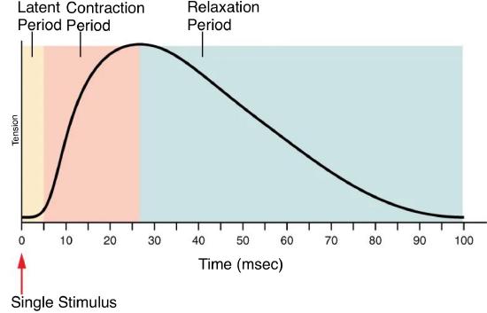
While a muscle twitch is possible, it is important to note that a single twitch does not lead to substantial muscle activity. A succession of action potentials must be directed toward the muscle fibers to induce a muscle contraction.
The subsequent twitch becomes more potent when muscle fibers are stimulated while a preceding twitch is still in progress. This effect is termed wave summation. The rationale lies in the concept that the excitation-contraction coupling effects stemming from consecutive motor neuron signals summate (Figure 21a). At a molecular level, summation transpires because the second stimulus releases increased calcium ions. These additional calcium ions become available to activate more sarcomeres while the muscle is still undergoing contraction from the initial stimulus. Consequently, summation leads to an amplified contraction of the motor unit.27

When the frequency of signaling from the motor neuron increases, summation ensues, leading to a progressive elevation in muscle tension within the motor unit until it attains a pinnacle. At this juncture, the tension achieved is approximately three to four times greater than the tension generated by a single twitch. This state is referred to as incomplete tetanus. During incomplete tetanus, the muscle undergoes rapid cycles of contraction, each punctuated by a brief relaxation phase (Figure 21a). Should the stimulus frequency be elevated so that the relaxation phase vanishes entirely, contractions become continuous, constituting complete tetanus (Figure 21b). Throughout tetanus, the concentration of calcium ions in the sarcoplasm is elevated to a level that permits nearly all sarcomeres to engage in cross-bridge formation and subsequent shortening, which allows for a sustained contraction to persist without interruption, albeit until the muscle succumbs to fatigue and can no longer sustain tension production.37
Treppe
Treppe, also known as the staircase effect or the Bowditch effect, refers to a physiological phenomenon in muscle contraction where each successive contraction of a muscle, when stimulated immediately after the previous contraction, is slightly stronger or more forceful than the previous one (Figure 22). This effect is characterized by a gradual increase in muscle tension with each contraction. Treppe can continue until the muscle reaches its maximum force of contraction. It is important to note that treppe is distinct from tetanus. Treppe occurs when stimuli are delivered at a frequency that allows partial relaxation between contractions. Treppe is often observed in skeletal muscles that have been at rest for some time and are then stimulated to contract repeatedly. It is attributed to the gradual increase in intracellular calcium concentration and improved muscle fiber recruitment with each successive contraction, resulting in enhanced muscle performance over time.

Muscle Tone
Skeletal muscles are seldom completely relaxed but maintain a slight, ongoing contraction even when not actively engaged in movement. This continuous, low-level contraction, known as muscle tone, serves important physiological functions. It plays a critical role in stabilizing joints and sustaining posture, ensuring that our bodies remain upright and stable. The mechanism behind muscle tone is intricate and involves the coordinated interaction between the nervous system and skeletal muscles. This interaction leads to the activation of a limited number of motor units at any given time, often cyclically, allowing muscles to sustain a constant level of tension without reaching complete exhaustion. While some motor units are active, others can recover and rest.
The absence of these subtle, ongoing contractions that contribute to muscle tone is termed hypotonia. Hypotonia can result from damage to specific parts of the CNS, such as the cerebellum, or from the loss of nerve innervation to a skeletal muscle, as seen in conditions like poliomyelitis. Muscles affected by hypotonia appear limp and may exhibit functional deficits, including weakened reflexes. Conversely, excessive muscle tone is referred to as hypertonia, often accompanied by hyperreflexia, which involves exaggerated reflex responses. Hypertonia can arise from damage to upper motor neurons within the CNS. It may manifest as muscle rigidity, as seen in conditions like Parkinson's disease, or as spasticity, characterized by a rhythmic alteration in muscle tone, causing a limb to return to its original position when passively stretched quickly. Spasticity is observed in some stroke patients and other neurological disorders.39
Nondepolarizing and Depolarizing Muscle Relaxants
Nondepolarizing muscle relaxants exert their neuromuscular blockade by binding to one or both α-subunits of the postjunctional ACh receptor (Figure 3), thereby preventing ACh from binding to these sites and opening the ion channel. Notable examples of nondepolarizing muscle relaxants include rocuronium, vecuronium, and cisatracurium. The blockade induced by nondepolarizing muscle relaxants can be reversed using cholinesterase inhibitors. Drugs like neostigmine act as anticholinesterase and counteract the neuromuscular block produced by nondepolarizing muscle relaxants. Sugammadex is another drug used to change certain types of nondepolarizing muscle relaxants. The choice of reversal agent depends on the specific pharmacological properties of the nondepolarizing muscle relaxant used. In contrast, depolarizing muscle relaxants like succinylcholine activate the motor end-plate similarly to ACh. Depolarizing muscle relaxants bind to the acetylcholine receptors on the motor end plate of the muscle cell, mimicking the action of ACh. This causes the muscle cell to depolarize, which initially leads to muscle contraction (fasciculations). Unlike ACh, which is quickly broken down by acetylcholinesterase, depolarizing muscle relaxants persist at the receptor. This sustained activation keeps the sodium channels open, preventing repolarization, and thus, the muscle remains in a depolarized, unresponsive state. Since the muscle cannot repolarize and generate new action potentials, it eventually leads to muscle paralysis.
Under normal physiological conditions, depolarizing muscle relaxants typically have a shorter action period than nondepolarizing muscle relaxants. However, a phenomenon known as desensitization may occur during the administration of a depolarizing block. Prolonged exposure to a depolarizing muscle relaxant can render the postjunctional membrane less responsive to ACh, resulting in the inability to elicit muscle contraction through nerve stimulation. This phenomenon is referred to as a Dual block, consisting of two stages: initially resembling a depolarizing block and then transitioning to a nondepolarizing block, akin to the actions of a nondepolarizing muscle relaxant. This latter stage is known as a Phase II Block.
Both depolarizing and nondepolarizing muscle relaxants play a vital role in interrupting nerve impulse transmission at the NMJ, resulting in partial paralysis (paresis) or complete paralysis of skeletal muscles. These pharmacological agents are indispensable for inducing muscle relaxation, facilitating tracheal intubation, and ensuring optimal conditions for surgical procedures. They are also employed in emergency departments, during cardiopulmonary resuscitation, and in intensive care units to assist with mechanical ventilation. Importantly, these agents lack analgesic or anesthetic properties and must never be used as a substitute for proper anesthesia, as they can render a patient paralyzed while still conscious.
Assessment of nondepolarizing and depolarizing blocks induced by muscle relaxants involves nerve stimulation and recording of action potentials from the muscle supplied by the selected nerve. Characteristics of a depolarizing block (Phase I Block) include fasciculations observed before paralysis, sustained tetanic and twitch rates upon nerve stimulation, and the absence of post-tetanic potentiation. In contrast, the features of a nondepolarizing block (Phase II Block) include a lack of fasciculations, a decrease in both tetanic and twitch rates during stimulation, and the presence of post-tetanic potentiation.
Certain antibiotics, particularly aminoglycosides, can inhibit ACh release from nerve terminals and intensify neuromuscular blockade when administered concurrently with therapeutic doses of neuromuscular blocking agents. Additionally, various pharmacological agents can either enhance or diminish the intensity of the muscle relaxant in use.
Curare, a term encompassing several South American arrow poisons traditionally utilized by indigenous peoples for immobilizing and paralyzing wildlife for sustenance, operates by inducing paralysis in skeletal muscles. This paralysis ultimately leads to fatality. Curare achieves its effects by binding to nicotinic ACh receptors at the motor end plate, effectively engaging in competitive inhibition with ACh. In modern medicine, curare is employed as a muscle relaxant during general anesthesia.40
This page titled 6.4: Skeletal Muscle was last modified on Wed, 19 Mar 2025 23:09:49 GMT and is shared under a CC BY-NC-SA 4.0 license and was authored, remixed, and/or curated by Chaya Gopalan (Consortium of Academic and Research Libraries in Illinois (CARLI)) .
The book could not be loaded just now.