Opening the book…
Smooth muscle, characterized by its lack of striations, is primarily found in the walls of hollow organs like the urinary bladder, uterus, stomach, and intestines, as well as within passageways such as the arteries and veins of the circulatory system. It also lines the respiratory, urinary, and reproductive tracts (Figure 23). Smooth muscle serves various functions within the body, including controlling the size of the iris and altering the shape of the lens in the eyes. Additionally, it plays a role in erecting hair in response to cold temperatures or fear within the skin, referred to as goosebumps. 41

Unlike skeletal muscle fibers, where stretching beyond the overlap of thin and thick filaments fails to generate tension, smooth muscle fibers can develop tension even when stretched to 2.5 times their resting length. This is because the resting length of smooth muscle fibers is significantly shorter than that of skeletal muscle fibers. Smooth muscle can be stretched considerably before reaching its optimal length. Moreover, in smooth muscle fibers, the thin filaments overlap with the thick filaments even in the stretched-out position, allowing cross-bridge interactions and tension development. This property of smooth muscle is advantageous, as it enables the accommodation of varying volumes of contents with minimal change in tension. For example, this feature is essential in structures like the bladder, which needs to adapt to variable fluid levels without significant tension alterations. Smooth muscle contraction, which relies on the presence of calcium ions, is characterized by several differences compared to skeletal muscle fibers, particularly in cellular structure and calcium handling. Smooth muscle fibers possess a much smaller diameter compared to skeletal muscle cells. Unlike skeletal muscle cells, smooth muscle fibers do not require T-tubules to penetrate deeply into the cell to transmit action potentials. This difference is due to the smaller size of smooth muscle fibers and the fact that they lack the highly organized sarcomere structure found in skeletal muscle.
Smooth muscle fibers have a limited sarcoplasmic reticulum for calcium storage but compensate with calcium ion channels in the sarcolemma. These calcium ion channels open during the action potential along the sarcolemma, allowing extracellular calcium ions to flow into the sarcoplasm and reach calmodulin, a regulatory protein crucial for smooth muscle contraction. Most of the calcium ions needed to trigger smooth muscle contraction come from this influx of extracellular calcium ions, which diffuse into the sarcoplasm.
Smooth muscle contraction continues until ATP-dependent calcium pumps actively transport calcium ions back into the sarcoplasmic reticulum and out of the cell. However, a low concentration of calcium ions remains in the sarcoplasm to maintain muscle tone. This residual calcium keeps the muscle in a slight state of contraction, which is particularly important in certain tracts and around blood vessels.
Because many smooth muscles need to function continuously without rest, they exhibit relatively low power output but can sustain contractions without significant energy expenditure. Some types of smooth muscle can maintain contractions even when calcium ion is removed and myosin kinase is inactivated or dephosphorylated. It is achieved through a subset of cross bridges, known as latch bridges, which link myosin heads and actin for extended periods without requiring ATP. This property allows for the maintenance of muscle tone in smooth muscle that lines arterioles and other visceral organs with minimal energy consumption.
Smooth muscle is considered involuntary muscle because it operates without conscious control. Various triggers can initiate smooth muscle contractions, including hormones, neural stimulation by the ANS, and local factors. In specific locations, such as the walls of visceral organs, stretching the muscle can also stimulate contraction, leading to the stretch-relaxation response.
The spindle-shaped smooth muscle fibers, which possess a single nucleus, typically measure 30 to 200 μm in length. These dimensions are significantly smaller than those of skeletal muscle fibers. While they lack the striations and sarcomeres observed in skeletal and cardiac muscle fibers, smooth muscle fibers contain actin and myosin contractile proteins and thick and thin filaments. Dense bodies are specialized structures found in the smooth muscle fibers. They are composed of proteins such as alpha-actinin, vinculin, and desmin, which provide structural support and stability to the smooth muscle cell. Dense bodies are interconnected by intermediate filaments, creating a lattice-like network throughout the cytoplasm of the smooth muscle cell. This network helps to transmit contractile forces generated by the action of myosin filaments across the cell, allowing for coordinated contraction and relaxation of the smooth muscle tissue. Dense bodies help hold thin filaments in place and anchor to the inner membrane of the sarcolemma. As the thick filaments pull thin filaments during the powerstroke action of muscle contraction, they tug on the dense bodies, which are linked to intermediate filament networks throughout the sarcoplasm. This coordinated action leads to the contraction of the entire muscle fiber, causing the ends to draw towards the center and generating a twisting motion in the middle (Figure 24). 41
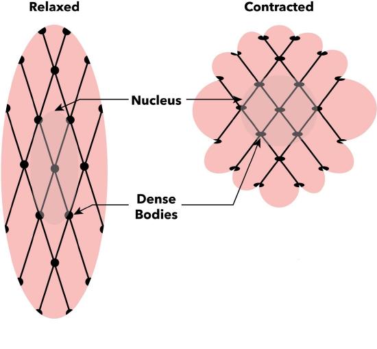
In contrast to skeletal and cardiac muscle cells, smooth muscle cells do not contain troponin to regulate cross-bridge formation. Instead, the regulatory protein calmodulin takes on this role. Calcium ions essential for muscle contraction are sourced from both the sarcoplasmic reticulum within the smooth muscle fibers and from the extracellular fluid through specialized membrane invaginations termed calveoli, which are comparable to T-tubules.
Smooth Muscle Contraction and Relaxation
A stimulus for smooth muscle contraction could be neural, humoral, or mechanical. Autonomic nerve stimulation, various hormones or neurotransmitters, and intrinsic changes causing stretch are examples of stimuli causing calcium entry into the sarcoplasm from multiple sources. These calcium ions bind to calmodulin, forming the calcium-calmodulin complex. Subsequently, this complex activates an enzyme known as myosin light chain kinase (MLCK). This enzyme activates myosin heads by phosphorylating them, which involves converting ATP to ADP and Pi, with Pi attaching to the head. Once activated, the myosin heads can attach to actin-binding sites and exert force on the thin filaments, pulling them towards thick filaments (Figure 25).44

In the absence of stimulus for muscle contraction, calcium ion levels decrease within the cell, and calmodulin no longer binds to calcium ions. This allows myosin light chain phosphatase (MLCP) to become active and catalyze the removal of phosphate groups from MLC. MLCP activity is controlled by various signaling pathways, such as cAMP and cGMP, serving as second messengers. They, in turn, activate protein kinases, which phosphorylate MLCP. In the presence of MLCP, the phosphorylation level of MLC decreases, inhibiting the interaction between myosin heads and actin filaments. This prevents cross-bridge formation and muscle contraction, ultimately leading to smooth muscle relaxation.46
Types of Smooth Muscle Fibers
Smooth muscle is organized into two primary types: single-unit smooth muscle and multiunit smooth muscle. These two types differ in their locations within the body and their characteristics.
Single-unit smooth muscle, also known as unitary muscle or visceral muscle, is a prevalent type of smooth muscle characterized by muscle fibers interconnected by gap junctions, enabling them to contract synchronously as a single unit. Found predominantly in the walls of visceral organs, excluding the heart, single-unit smooth muscle is abundant in structures like the digestive tract, urinary bladder, and uterus. It possesses self-excitable properties, capable of initiating contractions independently without nervous stimulation. These cells exhibit slow-wave potentials, manifesting as rhythmic fluctuations in membrane potential involving depolarization and hyperpolarization.
Visceral smooth muscle displays a stress-relaxation response, crucial for organs like the stomach and urinary bladder, which continually change size during filling and emptying. Initially contracting when stretched during filling, it quickly relaxes to prevent premature emptying, maintaining organ integrity. Additionally, visceral smooth muscle sustains muscle tone during emptying and contraction, preventing flaccidity. Its slow, steady contractions facilitate the movement of substances, such as food, in the digestive tract.
Multiunit Smooth Muscle, in contrast, lacks gap junctions and electrical coupling between cells. Contractions in one cell do not propagate to neighboring cells; they remain confined to the originally stimulated cell. Multiunit smooth muscle is neurogenic, with stimuli primarily generated by autonomic nerves. It surrounds structures like ciliary muscles of the eyes, large blood vessels, respiratory passageways, and the base of hair follicles. Contraction of multiunit smooth muscle, such as in hair follicles, can result in phenomena like goosebumps.
Unlike skeletal muscle, where motor neurons form highly organized NMJs with muscle fibers, smooth muscle lacks such organization. Instead, axons of neurons in the ANS course through smooth muscle, forming varicosities, bulges containing neurotransmitters. Varicosities release neurotransmitters into the synaptic cleft, activating smooth muscle cells (Figure 26). Additionally, visceral muscle in the walls of hollow organs (excluding the heart) contains pacesetter cells, which can spontaneously trigger action potentials and muscle contractions. These unique features contribute to the involuntary and often rhythmic nature of smooth muscle function.41

This page titled 6.5: Smooth Muscle was last modified on Wed, 19 Mar 2025 23:10:05 GMT and is shared under a CC BY-NC-SA 4.0 license and was authored, remixed, and/or curated by Chaya Gopalan (Consortium of Academic and Research Libraries in Illinois (CARLI)) .
The book could not be loaded just now.