Opening the book…
Mastication, or chewing, involves the adduction and lateral motions of the jaw bone. It is controlled by four muscles of the face.
- Differentiate between the actions of the masseter and the temporalis muscles in chewing
Key Points
- The masseter elevates the jaw, closing the mouth.
- The temporalis elevates and retracts the jaw.
- The lateral pterygoid is the only muscle of mastication that actively opens the jaw. Unilateral action of a lateral pterygoid produces lateral movement in the jaw, usually performed in concert with the medial pterygoids.
- The medial pterygoid elevates and closes the jaw, contributes to protrusion of the mandible, and assists in mastication.
Key Terms
- medial pterygoid: A muscle of mastication with two heads. It lies inferiorly to the medial pterygoid.
- temporalis: A broad muscle that fans out to cover much of the temporal bone on the side of the skull.
- lateral pterygoid: A muscle of mastication with two heads. It lies superiorly to the medial pterygoid.
- masseter: The large muscle which raises the lower jaw, and assists in mastication.
Mastication, or the act of chewing, involves adduction and lateral motion of the jaw bone. It is controlled by four bilateral muscles in the face. The lower jaw, or mandible, connects to the temporal bone of the skull via the temporomandibular joint, which allows movement in all planes.
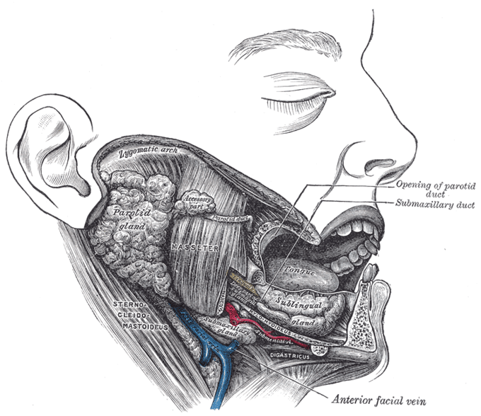
Location of the masseter muscle: The masseter muscle spans the opening of the mouth.
- Masseter: The most powerful muscle of mastication. It is quadrangular in shape and split into two regions, deep and superficial. It covers the other muscles of mastication.
- Attachments: The superficial region originates from the skull below the eye socket, while the deep part originates from the skull above the jaw. Both parts attach to the jaw.
- Actions: Elevates the jaw.
- Temporalis: A broad muscle that fans out to cover much of the temporal bone on the side of the skull.
- Attachments: The temporalis muscle has a wide, fan-shaped origin on the side of the skull and condenses into a tendon which attaches to the jaw.
- Actions: Elevates and retracts the jaw.
- Lateral Pterygoid: The lateral pterygoid muscle has a triangular shape with two head, superior and inferior. It is the major protractor (opener) of the jaw.
- Attachments: Both heads originate adjacent to the eye socket and converge into a tendon which attaches to the jaw.
- Actions: Together, the lateral pterygoids protract the jaw, working independently to produce lateral movement.
- Medial Pterygoid: The medial pterygoid muscle has a quadrangular shape with two heads, deep and superficial. It is located inferior to the lateral pterygoid.
- Attachments: The superficial head originates from the front of the skull and the deep head attaches to the skull adjacent to the eye socket. Both heads attach to the jaw.
- Actions: Elevates the jaw and assists in the production of lateral movement.

Location of the temporalis muscle and the lateral pterygoid: (a) Highlighted in orange, the temporalis muscle is a broad muscle extending from zygomatic bone. (b) Arrows indicate the location of the lateral pterygoid, highlighted with the medial pterygoid in orange.
KEY MOVEMENTS
- Elevation of the Jaw: Produced by the masseter, temporalis and medial pterygoid.
- Depression of the Jaw: Produced by the lateral pterygoid, assisted by the digastric, mylohyoid and geniohyoid muscles found in the neck.
- Protraction of the Jaw: Produced by the lateral pterygoid.
- Retraction of the Jaw: Produced by the temporalis.
- Lateral Movement of the Jaw: Produced by the lateral and medial pterygoid.
This page titled 9.7B: Chewing Muscles was last modified on Tue, 14 Oct 2025 23:13:00 GMT and is shared under a CC BY-SA 4.0 license and was authored, remixed, and/or curated by Boundless (now LumenLearning).
The book could not be loaded just now.