Opening the book…

- An image from a peripheral blood smear demonstrating two hairy cells with abundant pale blue cytoplasm, small cytoplasmic projections, and mature-looking nucleus giving the cell a “fried egg” appearance. 50x oil immersion. From MLS Collection, University of Alberta, https://doi.org/10.7939/R3ZG6GP2C
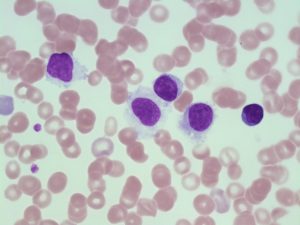
- An image from a peripheral blood smear demonstrating the different forms of hairy cells. 100x oil immersion. From MLS Collection, University of Alberta, https://doi.org/10.7939/R3610W71F
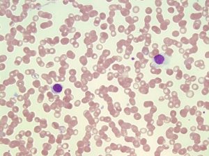
- An image from a peripheral blood smear showing a form of hairy cell with hairy cytoplasmic projections (left) and a different form of hairy cell with abundant pale blue cytoplasm (right). 50x oil immersion. From MLS Collection, University of Alberta, https://doi.org/10.7939/R32805D72
Cell Features:1-4
Two types of characteristic B lymphocyte morphologies can be seen in Hairy Cell Leukemia. Hairy cells are small to medium sized cells with either serrated cytoplasmic projections giving it a “hairy” appearance or more abundant pale blue cytoplasm giving it a “fried egg” appearance. The nucleus can be oval or indented, lacks nucleoli, and has an evenly stained mature chromatin patttern.
Cause:1,5,6
An indolent clonal mature B cell disorder has been associated with the BRAF -V600E mutation.
Age Group Affected:2,6
Middle-aged adults (median age of 55 years).
Laboratory Features for HCL:1,2,4,6
|
CBCD: Pancytopenia Relative lymphocytosis |
Peripheral Blood Smear: Hairy Cells (± Fried egg appearance) |
Bone Marrow: Dry Tap due to fibrosis (Aspirate) Hypocellular |
|
Immunologic markers: CD19, CD20, CD22, CD25, CD123, Annexin A1 |
Other useful tests: Tartrate-resistant acid phosphatase (TRAP) stain positive |
|
References:
1. Grever MR, Abdel-Wahab O, Andritsos LA, Banerji V, Barrientos J, Blachly JS, et al. Consensus guidelines for the diagnosis and management of patients with classic hairy cell leukemia. Blood [Internet]. 2016 Feb 2 [cited 2018 Jun 22];129(1):553–61. Available from: http://www.bloodjournal.org/cgi/doi/10.1182/blood-2016-01-689422
2. Turgeon ML. Malignant myeloid and monocytic disorders and plasma cell dyscrasias. In: Clinical hematology: theory and procedures. 4th ed. Philadelphia, PA: Lippincott Williams & Wilkins; 1999. p. 275-92.
3. Swerdlow SH, Campo E, Harris NL, Jaffe ES, Pileri SA, Stein H, et al. editors. WHO Classification of Tumours of Haematopoietic and Lymphoid Tissues Volume 2. 4th ed. International Agency for Research on Cancer (IARC); 2008.
4. Czader M. Mature lymphoid neoplasms. In: Rodak’s hematology clinical applications and principles. 5th ed. St. Louis, Missouri: Saunders; 2015. p. 619-41.
5. Troussard X, Cornet E. Hairy cell leukemia 2018: Update on diagnosis, risk-stratification, and treatment. Am J Hematol [Internet]. 2017 Dec 7 [cited 2018 Jun 25];92(12):1382–90. Available from: http://www.ncbi.nlm.nih.gov/pmc/articles/PMC5698705/
6. Jain P, Pemmaraju N, Ravandi F. Update on the biology and treatment options for hairy cell leukemia. Curr Treat Options Oncol [Internet]. 2014 Jun [cited 2018 Jun 27];15(2):187–209. Available from: http://www.ncbi.nlm.nih.gov/pmc/articles/PMC4198068/
This page titled 13.3: Hairy Cell Leukemia (HCL) was last modified on Fri, 11 Sep 2026 16:39:58 GMT and is shared under a CC BY-NC 4.0 license and was authored, remixed, and/or curated by Valentin Villatoro and Michelle To (Open Education Alberta) via source content that was edited to the style and standards of the LibreTexts platform.
The book could not be loaded just now.