Opening the book…
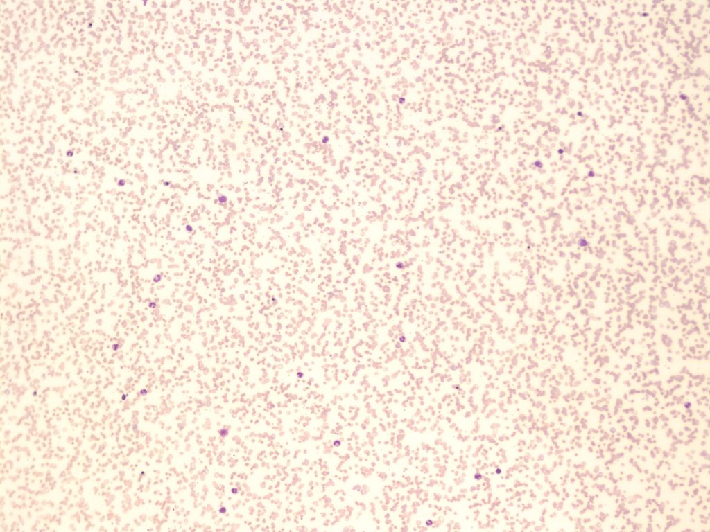
- A peripheral blood smear demonstrating agglutination of red blood cells. 10x magnification. From MLS Collection, University of Alberta, https://doi.org/10.7939/R3C824V90
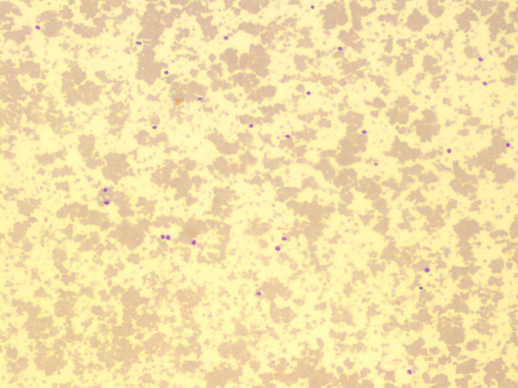
- A peripheral blood smear demonstrating severe agglutination of red blood cells. 10x magnification. From MLS Collection, University of Alberta, https://doi.org/10.7939/R34J0BC72
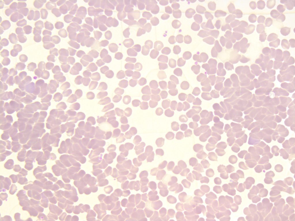
- A peripheral blood smear demonstrating autoagglutination of red blood cells. 50x oil immersion. From MLS Collection, University of Alberta, https://doi.org/10.7939/R3599ZH26
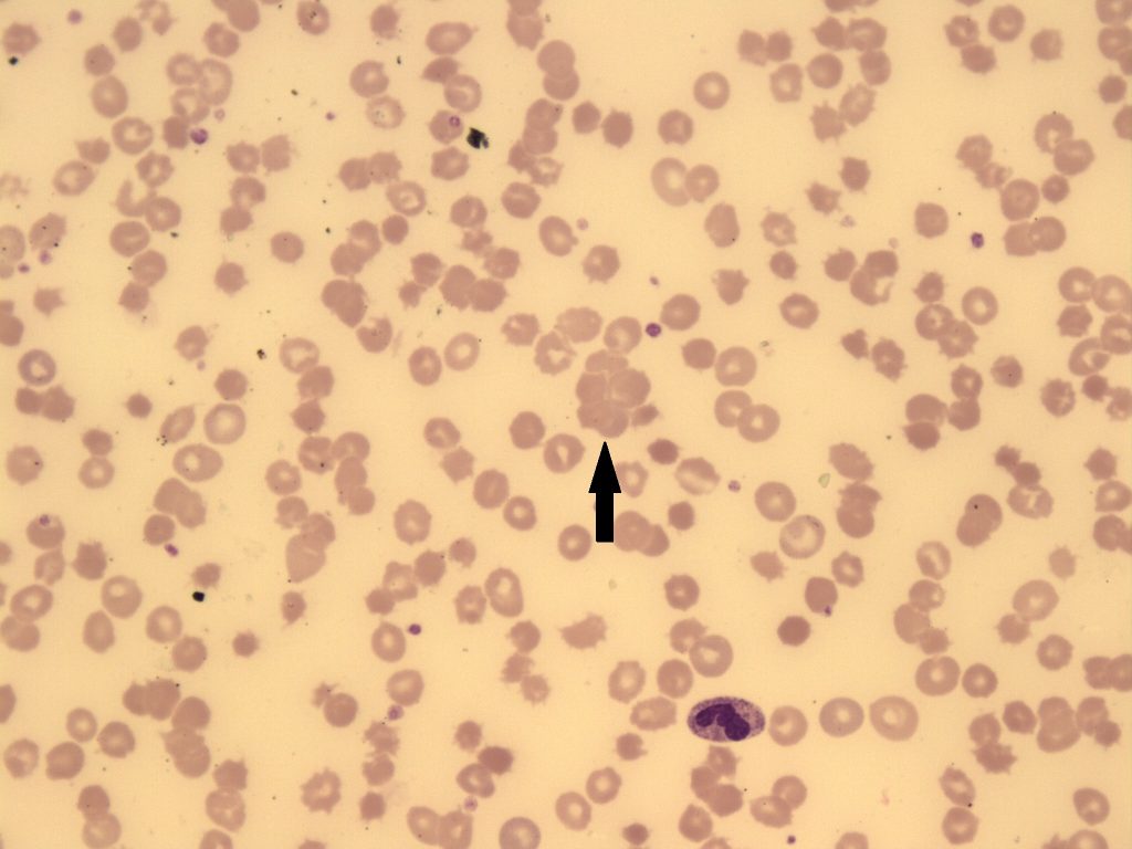
- An image from a peripheral blood smear showing agglutination of red blood cells. The arrow points to a cluster of red blood cells. 50x magnification. From MLS Collection, University of Alberta, https://doi.org/10.7939/R3SB3XD80
Cell Description:
This alteration of RBC distribution presents as irregular and random grape-like clusters or clumps. Agglutination is differentiated from Rouleaux by the lack of linear chains or “coin staking”. 1-4
The outlines of individual cells may not be evident.
Cell Formation:
Agglutination is caused by the formation of antibody-antigen complexes and occurs at room temperatures. Auto-agglutination is produced as a result of a complex formed between the patient’s own RBC antigens and antibodies, mediated by cold-reacting antibodies. Agglutination can be reversed when the blood sample is warmed to 37°C.1,2,5
Associated Disease/Clinical States: 2-4
Cold Hemagglutinin Disease
Paroxysmal Cold Hemoglobinuria
Cold Autoimmune Hemolytic Anemia
Note: Formation is NOT reversed with the addition of saline.5
References:
1. Hemolytic anemia: membrane defects. In: Clinical laboratory hematology. 3rd ed. New Jersey: Pearson; 2015. p. 317-33.
2. Jones KW. Evaluation of cell morphology and introduction to platelet and white blood cell morphology. In: Clinical hematology and fundamentals of hemostasis. 5th ed. Philadelphia: F.A. Davis Company; 2009. p. 93-116.
3. Ford J. Red blood cell morphology. Int J Lab Hematol [Internet]. 2013 Mar 9 [cited 2018 Jul 12];35(3):351–7. Available from: https://doi.org/10.1111/ijlh.12082
4. Turgeon ML. Normal erythrocyte lifecycle and physiology. In: Clinical hematology: theory and procedures. 4th ed. Philadelphia, PA: Lippincott Williams & Wilkins; 1999. p. 71-98.
5. Rodak BF, Carr JH. Variations in shape and distribution of erythrocytes. In: Clinical hematology atlas. 5th ed. St. Louis, Missouri: Elsevier Inc.; 2017. p. 93-106.
This page titled 2.3: Agglutination was last modified on Sat, 02 Sep 2023 08:16:26 GMT and is shared under a CC BY-NC 4.0 license and was authored, remixed, and/or curated by Valentin Villatoro and Michelle To (Open Education Alberta) via source content that was edited to the style and standards of the LibreTexts platform.
The book could not be loaded just now.