Opening the book…
Chemical digestion in the small intestine is facilitated by the coordinated efforts of three accessory digestive organs: the liver, pancreas, and gallbladder (refer to Figure 16). The liver plays a pivotal role in digestion by producing bile and transporting it to the duodenum. Meanwhile, the gallbladder serves as a reservoir, concentrating, storing, and releasing bile when needed. Additionally, the pancreas contributes to the digestive process by generating pancreatic juice, which includes digestive enzymes and bicarbonate ions. This pancreatic juice is then transported to the duodenum, which plays a crucial role in the breakdown of ingested nutrients. Some anesthetics can decrease the secretion of digestive enzymes and bile, slowing digestion. This is particularly relevant in postoperative care, where resumed digestive function is monitored.

Figure 16 | Accessory Organs | 21 The liver, pancreas, and gallbladder are considered accessory digestive organs, but their roles in the digestive system are vital. Externally, the liver is divided into the right, left, quadrate and caudate lobes. The right and left hepatic ducts merge into the common hepatic bile duct. The common hepatic duct then joins the cystic duct which extends from the gallbladder. This merger then forms the common bile duct. The pancreatic duct found along the pancreas joins the common bile duct.
The Pancreas
The pancreas, a soft and oblong gland, is situated transversely in the retroperitoneum behind the stomach. Its head is snugly positioned within the "c-shaped" curve of the duodenum. The body extends to the left for approximately 15.2 cm (6 in) before tapering into a tail that reaches the hilum of the spleen. This vital organ serves dual functions, encompassing both exocrine activities, involving the secretion of digestive enzymes, and endocrine functions, characterized by the release of hormones into the bloodstream (Figure 17).
The exocrine part of the pancreas arises from small, grape-like clusters of cells called acini. These acinar clusters are spread throughout the lobular structure of the pancreas. Acinar cells are crucial for producing enzyme-rich pancreatic juice, which is secreted into small ducts. These ducts merge to form the main pancreatic duct, which ultimately delivers the pancreatic juice into the duodenum to support digestion. Scattered among the acini are endocrine structures known as the islets of Langerhans. These cells release hormones pancreatic polypeptide, insulin, glucagon, and somatostatin, each playing a crucial role in the regulation of various physiological processes.22

Figure 17 | Exocrine and Endocrine Pancreas | 23 The pancreas is located to the right of the duodenum. The pancreatic duct runs along the pancreas from tail to the head. The acinar cells and pancreatic islet cells are structured into compartments known as lobules. Acinar cells produce digestive enzymes and pancreatic islet cells secrete hormones into the pancreatic duct.
Pancreatic Juice
The pancreas generates over a liter of pancreatic juice each day. This clear fluid primarily consists of water, salts, sodium bicarbonate, and digestive enzymes. The inclusion of sodium bicarbonate imparts slight alkalinity to pancreatic juice, maintaining a pH range between 7.1 to 8.2. This alkalinity serves several crucial functions, such as buffering the acidic gastric juice in chyme, inactivating pepsin from the stomach, and creating an optimal environment for the activity of pH-sensitive digestive enzymes in the small intestine.
Pancreatic enzymes play an active role in the digestion of carbohydrates, proteins, and fats. Notably, the pancreas produces protein-digesting enzymes in their inactive forms to prevent self-digestion. These enzymes undergo activation in the duodenum to avoid the digestion of the pancreas itself, a phenomenon observed in the disease pancreatitis. Enteropeptidase, an enzyme in the intestinal brush border, plays a key role in activating trypsin from trypsinogen of the pancreas. Subsequently, trypsin activates other pancreatic enzymes such as procarboxypeptidase and chymotrypsinogen, transforming them into their active forms, carboxypeptidase, and chymotrypsin. In contrast, enzymes responsible for digesting starch (amylase), fat (lipase), and nucleic acids (nuclease) are secreted in their active forms. This precautionary measure ensures that these enzymes do not pose a threat to the pancreas, unlike their protein-digesting counterparts.
Regulation of Pancreatic Secretion
The regulation of pancreatic secretion is a finely orchestrated process involving both hormonal signals and the parasympathetic nervous system. As acidic chyme enters the duodenum, it triggers the release of secretin, which, in turn, prompts duct cells to release bicarbonate-rich pancreatic juice, as depicted in Figure 18. Simultaneously, the presence of proteins and fats in the duodenum stimulates the secretion of CCK. CCK not only stimulates acini to secrete enzyme-rich pancreatic juice but also enhances the activity of secretin. Parasympathetic regulation primarily occurs during the cephalic and gastric phases of gastric secretion, wherein vagal stimulation prompts the secretion of pancreatic juice.24
Typically, the pancreas secretes just enough bicarbonate to counterbalance the amount of hydrochloric acid produced in the stomach—the secretion of bicarbonate by the pancreas results in the entry of hydrogen ions into the blood. Consequently, the acidic blood draining from the pancreas neutralizes the alkaline blood draining from the stomach. This intricate balancing act helps maintain the pH of the venous blood flowing to the liver, underscoring the pancreas's crucial role in acid-base homeostasis.

Figure 18 | Regulation of Pancreatic Secretion | 24 The secretion of pancreatic juice is regulated by the interaction of many factors such as acidic chyme, secretin, and cholecystokinin.
The Liver
The liver, weighing approximately three pounds in an adult, is the largest gland in the body. Beyond its role as an accessory digestive organ, the liver is a multifaceted organ with significant involvement in metabolism and regulation. Positioned inferior to the diaphragm in the right upper quadrant of the abdominal cavity, the liver benefits from the protection provided by the surrounding ribs. Its substantial size and diverse functions underscore its importance in maintaining various physiological processes within the body. The liver holds a paramount role in sustaining life by overseeing a wide spectrum of metabolic processes, crucial for eliminating substances that could be harmful if allowed to accumulate. Notably, the liver exhibits the remarkable ability to convert toxic ammonia into non-toxic waste, urea. Serving as the primary gateway for most nutrients absorbed by the digestive tract, the liver plays a pivotal role in processing and regulating the body's nutritional intake. Furthermore, the liver actively contributes to the synthesis of essential components such as plasma proteins, clotting agents, and bile. These products play vital roles in optimizing the absorption of fats, ensuring proper blood coagulation, and facilitating the digestive process.
Blood Supply to the Liver
The liver receives dual blood supplies, one from the hepatic artery and the other from the hepatic portal vein. The hepatic portal vein is a distinctive arrangement formed by the convergence of veins from the lower esophagus, stomach, pancreas, spleen, and intestines. The entry point for these vessels is the porta hepatis, meaning "gate to the liver." This unique vascular system connecting visceral organs to the liver elucidates why the liver is a common site for the metastasis of cancers originating in the alimentary canal.
The hepatic artery delivers oxygenated blood to the liver, while the hepatic portal vein transports partially deoxygenated blood along with nutrients, drugs, and toxins collected from the visceral organs. In addition to processing bloodborne nutrients and toxins, the liver releases necessary nutrients back into the bloodstream through the central vein, which converges with central veins from other lobules to form the hepatic vein. This hepatic vein, in turn, joins the inferior vena cava (Figure 19). This intricate network ensures that the liver efficiently regulates and manages the diverse array of substances transported through its dual blood supplies.25

Figure 19 | Hepatic Portal System | 25 The blood from gastrointestinal structures and the spleen flow into the hepatic portal vein. This is passed through the hepatic sinusoid and collected into the central vein. This continues into the hepatic vein and drains into the inferior vena cava.
Microscopic Anatomy of the Liver
Hepatocytes, constituting approximately 80 percent of the liver's volume, are the primary cell types in the liver, playing pivotal roles in its secretory, metabolic, and endocrine functions. Positioned around the fenestrated endothelium of hepatic sinusoids, which are open and porous blood capillaries formed by the branching of the nutrient-rich hepatic portal vein and oxygen-rich hepatic artery, hepatocytes have direct access to the blood (Figure 20). Their central location enables them to process nutrients, toxins, and waste materials carried by the blood. During this process, hepatocytes handle various substances, including bilirubin, a bile pigment, which is processed and excreted into the bile canaliculi, the smallest bile ducts. Additionally, proteins, lipids, and carbohydrates are either processed and secreted into the sinusoids or stored within the cells until needed. Within the hepatic sinusoids, there are also star-shaped reticuloendothelial cells known as Kupffer cells. These cells are phagocytic in nature, actively removing dead red and white blood cells, bacteria, and other foreign materials that may enter the sinusoids. This intricate cellular interplay in the liver ensures efficient processing, secretion, and detoxification, contributing to the organ's multifaceted functions.

Figure 20 | Microscopic Anatomy of the Liver | 26 At a microscopic level the liver is compartmentalized into lobules with a surrounding layer of connective tissue. Within these lobules is the central vein which drains into the hepatic vein by the interlobular vein. The plates of hepatocytes surround the central vein and between them run hepatic sinusoids. The sinusoids carry blood from the portal system into close contact with hepatocytes for nutrient processing and detoxification. At the periphery of the lobules is the portal triad which includes the portal venule, portal arteriole and bile duct.
Between adjacent hepatocytes, grooves in the cell membranes create space for each bile canaliculus (plural = canaliculi). These small ducts serve as conduits for the collection of bile produced by hepatocytes. Bile initially accumulates in these canaliculi and then proceeds into bile ductules, gradually advancing into larger bile ducts. The convergence of these bile ducts gives rise to the right and left hepatic ducts, which further amalgamate and exit the liver as the common hepatic duct. Subsequently, this duct joins forces with the cystic duct from the gallbladder, forming the common bile duct through which bile flows into the small intestine (Figure 21). A distinctive feature within the hepatic lobules is the portal triad, an arrangement encircling the lobular periphery. This triad comprises three fundamental structures: a bile duct, a branch of the hepatic artery, and a branch of the hepatic portal vein.1
Bile
Bile, a vital component for lipid digestion in the small intestine, is a yellowish, yellowish-brown, or yellowish-green alkaline solution with a pH ranging from 7.6 to 8.6. Secreted by hepatocytes in the liver, bile consists of bile salts, bile pigments (bilirubin), bile acids, phospholipids, cholesterol, triglycerides, water, and electrolytes. Its primary function is to emulsify lipids, a process critical for the efficient action of lipid-digesting enzymes. The key players in emulsification are bile salts and phospholipids, featuring both hydrophobic and hydrophilic regions. This molecular arrangement allows them to interact with large lipid molecules and watery chyme, breaking down large lipid globules into smaller fragments of about one μm in diameter, thereby significantly increasing the surface area for enzyme activity. This emulsification process mirrors the action of dish soap on fats mixed with water.
Hepatocytes contribute significantly to bile production, secreting approximately one liter each day. The gallbladder releases bile into the common bile duct as needed. The common bile duct, now carrying bile from both the liver and the gallbladder, joins the pancreatic duct near the duodenum to form the hepatopancreatic ampulla (or ampulla of Vater). The combined duct then empties its contents into the duodenum, the first part of the small intestine, through the major duodenal papilla.27
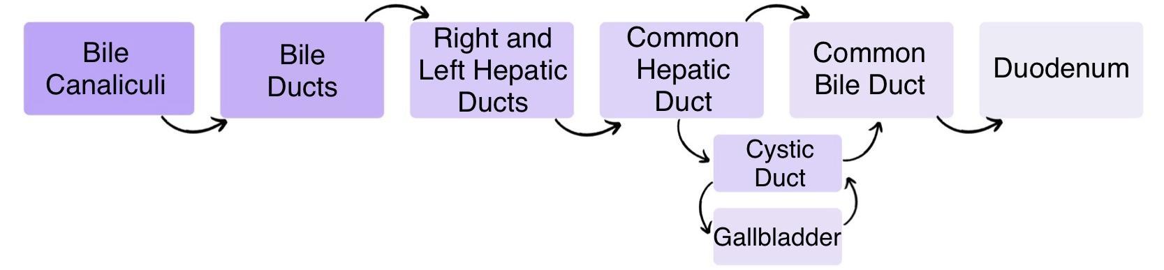
Figure 21 | Circulation of Bile | 27 Bile is secreted into bile canaliculi, drains into bile ducts, then into the right and left hepatic ducts, which form the common hepatic duct. Bile either flows directly to the common bile duct and duodenum, or is stored in the gallbladder via the cystic duct. During digestion, bile is released from the gallbladder back through the cystic duct into the common bile duct and then to the duodenum.
Despite eliminating most bile constituents in feces, bile salts undergo enterohepatic circulation, a unique blood vessel arrangement between the small intestine and the liver. In this recycling process, bile salts reach the ileum, are absorbed, and return to the liver through the hepatic portal blood, where hepatocytes excrete them into newly formed bile. Bilirubin, the primary bile pigment, is a byproduct generated when the spleen removes old or damaged red blood cells. Transported to the liver through the splenic vein of the hepatic portal system, breakdown products, including proteins, iron, and bilirubin, undergo recycling in the liver. Bilirubin, responsible for the green color of bile, is eventually transformed into stercobilin by intestinal bacteria, imparting a characteristic brown color to stool.
In certain disease states, the absence of bile entering the intestine results in white (‘acholic’) stool with high-fat content, as fats are not adequately broken down or absorbed. Bile production intensifies in response to fat-rich chyme entering the duodenum, triggered by the gut hormone secretin. Between meals, bile production persists, but it is conserved in concentrated form within the gallbladder. The release of bile from the liver to the gallbladder occurs when the hepatopancreatic ampulla, situated in the duodenum, acts like a valve.1
Metabolic Functions of the Liver
The liver stands as a metabolic powerhouse, orchestrating a vast array of biochemical processes crucial for maintaining systemic physiological homeostasis. In carbohydrate metabolism, it serves as the primary hub for blood glucose regulation. Following a meal, the liver takes up excess glucose from the portal circulation to store it as glycogen via glycogenesis. During periods of fasting, it maintains normoglycemia by releasing glucose back into the bloodstream, achieving this through both the breakdown of stored glycogen (glycogenolysis) and the de novo synthesis of glucose from non-carbohydrate precursors, such as lactate, glycerol, and glucogenic amino acids (gluconeogenesis).
Its role in lipid metabolism is equally vital, encompassing the de novo synthesis of cholesterol, phospholipids, and lipoproteins, as well as the oxidation of fatty acids via mitochondrial beta-oxidation to generate ATP. During prolonged fasting or starvation, the liver converts excess acetyl-CoA into ketone bodies (acetoacetate and beta-hydroxybutyrate), providing an essential alternative fuel source for extrahepatic tissues.
In protein metabolism, the liver is central to the processing of amino acids, utilizing transamination and deamination pathways. It synthesizes critical plasma proteins, including albumin—which maintains oncotic pressure—and essential coagulation factors. Furthermore, the liver plays a critical role in nitrogen homeostasis by detoxifying highly toxic ammonia, a byproduct of protein catabolism, into water-soluble urea through the urea cycle for subsequent renal excretion.
Beyond its synthetic and fuel-regulatory functions, the liver is the body's primary site of biotransformation and detoxification. It utilizes a two-phase enzymatic process (Phase I functionalization and Phase II conjugation) to metabolize endogenous substances, such as hormones and bilirubin, as well as exogenous xenobiotics and pharmaceuticals. This pathway converts lipophilic compounds into hydrophilic metabolites, facilitating their elimination. These processed substances, along with metabolic waste, are then excreted via bile into the gastrointestinal tract or released into the bloodstream for filtration by the kidneys.
Finally, the liver acts as a major reservoir for micronutrients, storing significant reserves of iron (complexed within the intracellular protein ferritin), fat-soluble vitamins (A, D, E, and K), and water-soluble vitamin B12. Additionally, it actively participates in endocrine activation, performing the essential first hydroxylation step required to convert cholecalciferol into biologically active vitamin D. This intricate, interconnected metabolic network underscores the liver's indispensable role in overall human physiology, as summarized in Table 5.
| Carbohydrate | Protein | Lipid |
|---|---|---|
| Glycogenesis | Production of plasma proteins | Synthesis of triglycerides and cholesterol |
| Glycogenolysis | Production of clotting factors | Lipolysis |
| Gluconeogenesis | Deamination | |
| Conversion of fructose and galactose to glucose | Transamination | |
| Urea cycle |
As blood, laden with substances from the visceral organs, including gut bacteria, is directed into the liver via the hepatic portal system, the liver employs a protective mechanism against enteric bacteria. This safeguard is facilitated by Kupffer cells. These specialized cells play a crucial role in the removal of enteric bacteria, ensuring the maintenance of a healthy and sterile environment within the liver. Additionally, Kupffer cells contribute to the breakdown of toxins through oxidation reactions, releasing free radicals such as reactive oxygen species. While the accumulation of free radicals can induce cell injury, the liver produces antioxidants, with glutathione being a major component as a defense mechanism to protect its cells from free radical-related damage.
Another noteworthy function of the liver is its involvement in calcium regulation. The liver excretes calcium from the body by secreting it into the bile, which then passes into the gut and is subsequently lost in the feces.1, 3, 9
The Gallbladder
The gallbladder, measuring approximately 8–10 cm (~3–4 in) in length, is situated in a shallow area on the posterior aspect of the right lobe of the liver. This muscular sac serves as a storage reservoir, concentration chamber, and, upon stimulation, a fuel for bile into the duodenum through the common bile duct. The inner lining of the gallbladder, known as the mucosa, plays a crucial role in absorbing water and ions from bile. This absorption mechanism results in the concentration of bile, often reaching up to 10-fold, before it is released into the duodenum.29

Figure 22 | Gallbladder | 30 The left hepatic duct and right hepatic duct collect bile from the liver and join to form the common hepatic duct.The gallbladder consists of three regions: the fundus (rounded end), body (main chamber), and neck (narrow region connecting to the cystic duct). The gallbladder stores and concentrates bile, and releases it into the two-way cystic duct when it is needed by the small intestine.
15.9: The Liver, Pancreas, and Gallbladder is shared under a not declared license and was authored, remixed, and/or curated by LibreTexts.
The book could not be loaded just now.