Opening the book…
Regulation of various electrolytes and substances is a vital function of the kidneys and related systems in the body. Although the handling of these ions is discussed in specific parts of the tubule, a summary of the regulation of key substances is as below.
Regulation of Sodium Ions
Sodium ion plays a significant role in the osmolarity of the plasma, and its concentration needs to be tightly regulated. An increase in sodium ion concentration can lead to increased blood volume and hypertension. The kidneys play a central role in regulating sodium ion balance. Sodium reabsorption occurs at multiple sites along the renal tubules, ensuring that the body maintains the appropriate sodium levels necessary for optimal physiological function. The reabsorption of sodium ions In the PCT is a crucial process. Sodium ions are cotransported alongside various solutes such as glucose, amino acids, phosphate, and lactate. Additionally, sodium ions are counter-transported via the sodium-hydrogen exchanger, a process that is intricately linked to the reabsorption of filtered bicarbonate ions. Sodium ions are reabsorbed along with chloride ions in the later part of PCT.
The thick ascending limb of the loop of Henle is responsible for the reabsorption of a substantial portion of filtered sodium ions. This is facilitated by the Na+/K+/2Cl-cotransporter. While sodium ions are primarily reabsorbed, some potassium ions diffuse back into the tubule lumen, contributing to sodium conservation. The early DCT plays a role in the reabsorption of sodium ions through the sodium-chloride cotransporter. In the late DCT and the collecting duct, sodium ions continue to be reabsorbed, accounting for approximately 3% of the total reabsorption. This final reabsorption step is predominantly orchestrated by principal cells, which simultaneously reabsorb water. Only a small fraction of filtered sodium ions, less than 1 percent, is excreted in the urine under normal conditions.
Regulation of Potassium Ions
The kidneys play a pivotal role in maintaining potassium balance, crucial for normal muscle and cardiac function. Potassium ions are primarily reabsorbed in the early proximal tubule through passive transport. However, sodium-potassium exchange pumps play a pivotal role in potassium reabsorption in the late proximal tubule. The thick ascending limb further contributes to potassium regulation. Here, the Na+/K+/2Cl- cotransporter is engaged, ensuring potassium movement. Within the distal tubule, potassium reabsorption continues, primarily through sodium-chloride cotransport mechanisms. The final stages of potassium handling occur in the late distal tubule and collecting ducts. Principal cells within these segments actively reabsorb sodium and water while controlling potassium secretion.
Figure 17 illustrates the homeostatic regulation of potassium levels in response to increased dietary K+ intake. When potassium intake rises, plasma K⁺ concentration increases, triggering the release of aldosterone from the adrenal cortex. It upregulates the expression and activity of Na+/K+ ATPase on the basolateral membrane, increasing sodium reabsorption and potassium secretion. Additionally, aldosterone enhances the activity of apical potassium channels, allowing more K⁺ to be secreted into the tubular lumen for excretion in the urine. This negative feedback mechanism helps maintain potassium homeostasis and prevents hyperkalemia, which could have detrimental effects on cellular function, particularly in excitable tissues like the heart and nervous system.

Figure 17 | Regulation of potassium homeostasis by aldosterone | An increase in potassium (K+) intake leads to elevated plasma K+ concentration, which stimulates the release of aldosterone. Aldosterone enhances K+ secretion at the cortical collecting tubules, promoting its excretion and maintaining potassium balance.
Sodium reabsorption and potassium secretion in principal cells are driven by the Na+/K+-ATPase pump, which creates a gradient that facilitates the passive movement of sodium ions into the cells via ENaC channels and the active secretion of potassium ions into the tubular lumen (Figure 18).
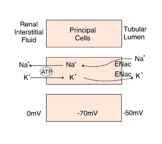
Figure 18 | Reabsorption of Sodium Ions and Secretion of Potassium Ions within the Principal Cells | This diagram illustrates the sodium (Na+) and potassium (K+) exchange that occurs in principal cells of the renal collecting duct. It shows the movement of ions between the tubular lumen and the renal interstitial fluid, facilitated by ENaC channels and the Na+/K+-ATPase pump.
Regulation of Chloride Ions
Chloride ion is closely associated with sodium in the extracellular environment. Its regulation closely mirrors that of sodium, particularly in maintaining acid-base balance in the extracellular space and its role in gastric acid production.
The kidneys play a central role in regulating the reabsorption of chloride ions, a pivotal component of overall electrolyte balance and renal function. In the early PCT, chloride ions are reabsorbed alongside sodium ions through the sodium-chloride cotransport mechanism. This process is highly efficient and contributes significantly to the reabsorption of chloride ions. In the late PCT, additional reabsorption of chloride ions occurs. Within the thick ascending limb of the Loop of Henle, chloride ions are reabsorbed by an active transport mechanism. This process, facilitated by the \(\ce{Na+}\)-\(\ce{K+}\)-\(\ce{2Cl-}\) cotransporter, contributes to the overall conservation of chloride ions. In addition to active transport, chloride ions are reabsorbed through the paracellular route in the thick ascending limb, enhancing chloride ion retention. The early DCT is involved in reabsorbing chloride ions through the sodium-chloride cotransporter. In the late DCT and collecting duct, the final phase of chloride ion reabsorption occurs, accounting for approximately 15% of the total reabsorption. Under normal conditions, only a small fraction of filtered chloride ions are excreted in the urine, which represents the minimal amount necessary to maintain chloride ion balance in the body.
Regulation of Calcium and Phosphate
The parathyroid glands monitor and respond to circulating calcium levels in the blood. When calcium levels drop too low, PTH is released to stimulate the DCT to reabsorb calcium from the filtrate. Conversely, less PTH is released when calcium levels are adequate or high, and more calcium remains in the filtrate to be excreted. Phosphate levels in the blood move in the opposite direction to calcium levels. PTH inhibits the reabsorption of phosphate when calcium levels are low, allowing calcium levels to rise. Additionally, PTH stimulates the renal conversion of calcidiol into calcitriol, the active form of vitamin D, which, in turn, stimulates the intestines to absorb more calcium from the diet. PTH increases calcium reabsorption through the activation of adenylate cyclase and cAMP-mediated mechanisms.
The bulk of calcium reabsorption occurs in the proximal tubule and the thick ascending limb of the loop of Henle. Over 90% of the filtered calcium is reabsorbed in these two segments, and this process is coupled with sodium reabsorption through passive transport mechanisms. The thick ascending limb of the loop of Henle plays a critical role in calcium reabsorption. This segment provides two routes for calcium to be reabsorbed: the paracellular route and the transcellular route. The distal tubule and the collecting duct are involved in the final stages of calcium reabsorption. Here, calcium reabsorption occurs through an active transport process, which can be modulated by PTH and vitamin D. Vitamin D increases the expression of calcium channels, calcium-binding proteins (calbindin), and ATP-dependent calcium pumps that enhance calcium uptake from the filtrate back into the bloodstream (Figure 19).

Figure 19 | The Role of Parathyroid Hormone (PTH) in Calcium Homeostasis | This illustration depicts the role of parathyroid hormone (PTH) and vitamin D in regulating calcium levels. PTH increases calcium reabsorption in the kidneys, stimulates calcium release from bones, and stimulates vitamin D activation. Activated vitamin D helps increase calcium absorption in the intestines.
The process of regulating phosphate levels within the body begins with the filtration of phosphate ions at the glomerulus. The primary site of reabsorption for phosphate ions is the proximal tubule. The reabsorption of phosphate in this segment occurs through the mechanism of sodium-phosphate cotransport. Approximately 85% of the filtered phosphate is reabsorbed in the proximal tubule. The remaining 15% of the filtered phosphate is excreted in the urine.22

Figure 20 | Calcium Transport in Principal Cells of the Renal Tubule | This illustration depicts the mechanisms of calcium ion (\(\ce{Ca2+}\)) transport in principal cells of the renal tubule, encompassing both transcellular and paracellular routes. ATP-dependent process such as \(\ce{Ca2+}\) ATPase activity is involved in the reabsorption of calcium. Water (\(\ce{H2O}\)) movement is driven by the osmotic gradient created by solute distribution.
In the principal cells of the collecting duct, the movement of sodium ions into the cell from the basolateral side is closely linked to calcium ion reabsorption through the action of the sodium-calcium exchanger (NCX1). This exchanger, located on the basolateral membrane, facilitates the movement of three \(\ce{Na+}\) ions into the cell in exchange for one calcium ion being transported out into the interstitial fluid. The driving force for this exchange is the electrochemical gradient of sodium, which is maintained by the sodium-potassium ATPase pump. This pump actively transports Na⁺ out of the cell and \(\ce{K+}\) into the cell, ensuring a low intracellular \(\ce{Na+}\) concentration. As a result, sodium passively enters the cell via NCX1, allowing for the extrusion of calcium ions against its concentration gradient. This secondary active transport mechanism is essential for calcium reabsorption into the bloodstream, as the removal of intracellular calcium facilitates further uptake from the tubular lumen. Thus, the coordinated action of the \(\ce{Na+}\)/\(\ce{K+}\) ATPase and NCX1 ensures efficient sodium and calcium transport in the collecting duct (Figure 20).
PTH is a critical regulator of phosphate balance. When the body senses an increase in phosphate levels, PTH secretion is stimulated. PTH, in turn, acts on the proximal tubule to decrease phosphate reabsorption. A cAMP-mediated mechanism controls the reabsorption of phosphate in the proximal tubule. The action of PTH increases cAMP levels, which, in turn, modulate the reabsorption of phosphate. Elevated levels of PTH lead to decreased phosphate reabsorption, enhancing its excretion in the urine.
Regulation of Magnesium Ions
The kidneys play a central role in regulating magnesium ion concentrations in the body. The renal regulation of magnesium ions begins with their filtration at the glomerulus. Approximately 25 percent of the filtered magnesium ions are reabsorbed in the proximal tubule. PTH, a key regulator of magnesium balance, controls this reabsorption. The majority of magnesium reabsorption, roughly 65 percent, occurs in the loop of Henle. It is essential to understand that magnesium reabsorption in the loop of Henle is competitive with calcium ions. This interaction between magnesium and calcium ions is pivotal. An increase in blood calcium levels, hypercalcemia, leads to an increased excretion of magnesium ions, while hypermagnesemia results in increased calcium excretion. This delicate balance ensures that the body maintains appropriate levels of both ions.
The distal and collecting tubules play a relatively minor role in the reabsorption of magnesium ions, accounting for less than 5 percent of total magnesium reabsorption. The extent of reabsorption in this segment is influenced by various factors, including the body's overall magnesium balance and the presence of certain hormones, such as aldosterone.
Regulation of Hydrogen Ions, Bicarbonate, and pH
Chemical buffers, the respiratory system, and the kidneys maintain the body's acid-base balance. The kidneys play a significant role in regulating blood pH by actively secreting hydrogen ions into the filtrate in the proximal tubule and by having intercalated cells secrete hydrogen ions into the urine in the collecting ducts, which are often buffered by bicarbonate, dihydrogen phosphate ions, or ammonia (Table 4). This process helps regulate blood pH within a normal range.
This page titled 11.10: Renal Electrolyte Regulation was last modified on Thu, 18 Jun 2026 02:08:02 GMT and is shared under a CC BY-NC-SA 4.0 license and was authored, remixed, and/or curated by Chaya Gopalan (Consortium of Academic and Research Libraries in Illinois (CARLI)) .
The book could not be loaded just now.