Opening the book…
After completing this section, you should be able to
-
- show, by means of a diagram, how two different amino acid residues can be combined to give two different dipeptides.
- draw the structure of a relatively simple peptide, given its full or abbreviated name and the structures of the appropriate amino acids.
- draw, or name, the six possible isomeric tripeptides that can be formed by combining three different amino acid residues (amino acid units) of given structure.
- account for the fact that there is restricted rotation about the C(O)-N bonds in peptides.
- illustrate the formation of a disulfide linkage between two cysteine residues, and show how such bonds can link together two separate peptide chains or can provide a bridge between two cysteine residues present in a single peptide molecule.
Make certain that you can define, and use in context, the key terms below.
- C‑terminal amino acid
- N‑terminal amino acid
- peptides
- residues
The general term peptide refers to an amino acid chain of unspecified length. However, chains of approximately 50 amino acids or more are typically referred to as proteins or polypeptides. In its physiologically active form, a protein may be composed of one or more polypeptide chains. A chain consisting of only two amino acid units is called a dipeptide; a chain consisting of three is a tripeptide. By convention, peptide and protein structures are depicted with the amino acid whose amino group is free (the N-terminal end) on the left and the amino acid with a free carboxyl group (the C-terminal end) to the right

Identify the C- and N-terminal amino acids for the peptide lys-val-phe-gly-arg-cys.
SolutionC-terminal amino acid: cysteine (cys or C); N-terminal amino acid: lysine (lys or K)
Peptide Bond Formation or Amide Synthesis
The formation of peptides is nothing more than the application of the amide synthesis reaction. By convention, the amide bond in the peptides should be made in the order that the amino acids are written. The amine end (N terminal) of an amino acid is always on the left, while the acid end (C terminal) is on the right.

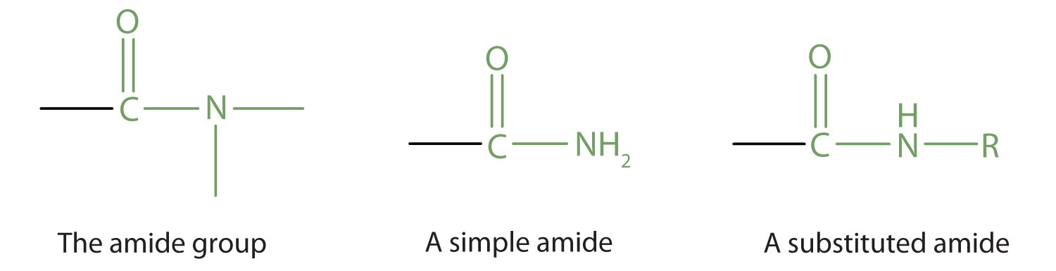
The amide functional group consists of a nitrogen atom bonded to a carbonyl carbon atom. If the two remaining bonds on the nitrogen atom are attached to hydrogen atoms, the compound is a simple amide. If one or both of the two remaining bonds on the atom are attached to alkyl or aryl groups, the compound is a substituted amide. Peptides are substituted amides.
For example, alanylserine is the dipeptide that results when an amide bond forms between the alanine carboxyl and the serine amino group.
Note that two dipeptides can result from reaction between alanine and serine, depending on which carboxyl group reacts with which amino group. If the alanine amino group reacts with the serine carboxyl, serylalanine results.


Resonance Contributors for the Peptide Bonds
A consideration of resonance contributors is crucial to any discussion of the amide functional group. One of the most important examples of amide groups in nature is the ‘peptide bond’ that links amino acids to form polypeptides and proteins.
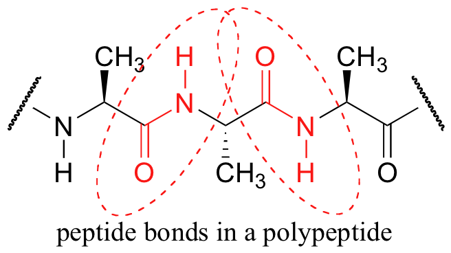
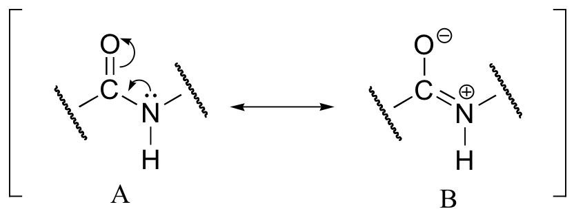
The amide bond that links amino acids together in peptides is no different from any other amide bond (Section 10.4). An amide nitrogen is nonbasic because its unshared electron pair is delocalized by resonance with the carbonyl group.
This overlap of the nitrogen p orbital with the p orbitals of the carbonyl group imparts a certain amount of double-bond character to the C–N bond and restricts rotation around it. The amide bond is therefore planar, and the N–H is oriented 180° to the C═O.
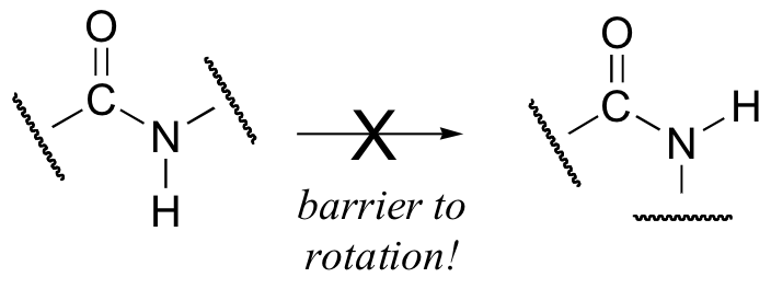
This, along with the observation that the bonding around the peptide nitrogen has trigonal planar geometry, strongly suggests that the nitrogen is sp2-hybridized. An important resonance contributor has a C=N double bond and a C-O single bond, with a separation of charge between the oxygen and the nitrogen. Although B is a minor contributor due to the separation of charges, it remains highly relevant in terms of peptide and protein structure – our proteins would not fold properly if there were free rotation about the peptide C-N bond.
Backbone Peptide or Protein Structure
The structure of a peptide can be written fairly easily without showing the complete amide synthesis reaction by learning the structure of the "backbone" for peptides and proteins.

After the backbone is written, go back and write the specific structure for the side chains as represented by the "R" as ser-ala-cys for this example. The amine end (N terminal) of an amino acid is always on the left (ser), while the acid end (C terminal) is on the right (cys).
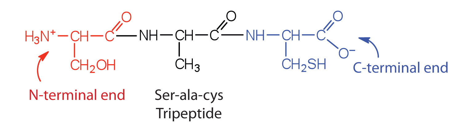
Write the structure for the following tripeptides. First, write the "backbone" and then add the specific side chains.
a. val-ser-cys b. glu-cys-gly c. phe-tyr-asn
Solutiona. Answer graphic b. Answer graphic c. Answer graphic


Six isomeric tripeptides contain valine, tyrosine, and glycine. Name them using both three- and one-letter abbreviations.
- Answer
-
Val-Tyr-Gly (VYG), Tyr-Gly-Val (YGV), Gly-Val-Tyr (GVY), Val-Gly-Tyr (VGY), Tyr-Val-Gly (YVG), Gly-Tyr-Val (GYV)
Draw the structure of M-P-V-G, and indicate its amide bonds.
- Answer
-

Disulfide Bridge
The amino acid cysteine undergoes oxidation and reduction reactions involving the -SH (sulfhydryl group). The oxidation of two sulfhydryl groups results in the formation of a disulfide bond by the removal of two hydrogen atoms. The oxidation of two cysteine amino acids is shown in the graphic. An unspecified oxidizing agent (O) provides oxygen, which reacts with the hydrogen (green) on the -SH group to form water. The sulfurs (green) join to make the disulfide bridge. This is an important bond to recognize in the tertiary structure of proteins. As we saw in Section 4.8, a disulfide is formed by mild oxidation of a thiol, RSH, and is cleaved by mild reduction. The reduction of a disulfide bond is the opposite reaction, which again leads to two separate cysteine molecules.
Cysteine residues in the peptide chain can form a loop by forming the disulfide bond (—S—S—), while cysteine residues in different peptide chains can actually link what were otherwise separate chains. Insulin was the first protein whose amino acid sequence was determined. This pioneering work, completed in 1953 after some 10 years of effort, earned a Nobel Prize for British biochemist Frederick Sanger (born 1918). He found that the primary structure consisted of two chains, totaling 51 amino acids, linked by two cysteine disulfide bridges. Also, note that the first peptide chain possesses an internal loop.
This page titled 12.4: Peptides and Proteins was last modified on Thu, 31 Jul 2025 00:06:38 GMT and is shared under a CC BY-SA 4.0 license and was authored, remixed, and/or curated by Sol Parajon Puenzo (Cañada College) .
The book could not be loaded just now.