Opening the book…
Search Fundamentals of Biochemistry
by William (Bill) W. Christie and Henry Jakubowski.
This section is an abbreviated and modified version of material from the Lipid Web, an introduction to the chemistry and biochemistry of individual lipid classes, written by William Christie.
(Learning goals written by Claude, Sonnet 4.6, Anthropic)
Sphingolipid Building Blocks: Long-Chain Bases, Ceramides, and Their Synthesis
- Describe the de novo sphingolipid biosynthetic pathway — tracing the PLP-dependent condensation of palmitoyl-CoA and L-serine by serine palmitoyltransferase (the rate-limiting, ORMDL-regulated heterotrimer) to 3-keto-sphinganine, NADPH-dependent reduction to sphinganine, N-acylation by one of six tissue-specific ceramide synthases (each with distinct acyl-CoA chain-length specificities), and finally Δ4-desaturation by DEGS1/2 to produce ceramide — explaining why the trans-Δ4 double bond is introduced only after N-acylation.
- Explain how the six mammalian ceramide synthases partition ceramide production into functionally distinct pools by their acyl-CoA specificity (CerS1: C18:0, brain; CerS2: C20–C26, lung/liver; CerS3: C26+, skin/testes; CerS5/6: C16, lung/intestine), and describe how the CERT protein transfers ceramide non-vesicularly from the cytoplasmic ER surface to the trans-Golgi for complex sphingolipid synthesis by recognizing PI(4)P via its PH domain and ceramide via its START domain.
- Describe the ceramide cycle — distinguishing de novo synthesis in the ER, the sphingomyelinase pathway (rapid hydrolysis of sphingomyelin at the plasma membrane to generate ceramide for signaling), and the salvage pathway (lysosomal ceramidase generates sphingosine that is re-N-acylated by ceramide synthases, contributing 50–90% of sphingolipid biosynthesis) — and explain the opposing biological functions of ceramide/sphingosine (pro-apoptotic) versus sphingosine-1-phosphate and ceramide-1-phosphate (pro-mitotic), establishing the "sphingolipid rheostat."
Complex Sphingolipids: Sphingomyelin, Glycosphingolipids, and Gangliosides
- Describe the synthesis of sphingomyelin by SMS1 (trans-Golgi lumen) and SMS2 (plasma membrane outer leaflet) — explaining the unique phosphocholine transfer from PC to ceramide (liberating DAG, linking sphingolipid and glycerolipid signaling), the proposed Asp→Arg→phosphocholine nucleophilic mechanism, and the CERT-dependence of SMS1 vs. local ceramide recycling by SMS2 — and describe the catabolism of sphingomyelin by acid, neutral, and alkaline sphingomyelinases in different organelles, connecting lysosomal acid sphingomyelinase deficiency to Niemann-Pick disease types A and B.
- Trace the biosynthesis of glucosylceramide (ceramide + UDP-Glc by GCS at the cis-Golgi cytoplasmic face, then P4-ATPase/FAPP2-mediated translocation to the trans-Golgi lumen) and galactosylceramide (ceramide + UDP-Gal in the ER lumen, by UDP-Gal:ceramide galactosyltransferase restricted to oligodendrocytes/Schwann cells/kidney/testes), and explain why glucosylceramide is the key precursor for all complex neutral oligoglycosphingolipids and gangliosides in animals while being the terminal product in plants and fungi.
- Describe ganglioside biosynthesis — explaining how lactosylceramide serves as the branch-point precursor from which GM3 synthase (cis-Golgi) initiates the 0-, a-, b-, and c-series gangliosides through sequential Golgi-localized sialyltransferases and glycosyltransferases — and describe ganglioside catabolism in the lysosome, explaining the roles of the saposin activator proteins (Sap A–D) and GM2-activator protein in enabling soluble hydrolases to access membrane-embedded substrates, and connecting deficiencies in specific hydrolases to gangliosidoses and Gaucher disease.
Membrane Organization, Disease, and Pharmacological Relevance
- Explain the "sphingoid motif" — describing how the 2-amino-1,3-dihydroxy arrangement and trans-Δ4 double bond of the sphingoid base enable an unusually large number of hydrogen bonds, charge-pairing, and van der Waals interactions with cholesterol and other sphingolipids — and explain how these interactions drive the spontaneous formation of liquid-ordered raft nanodomains in the outer leaflet of the plasma membrane, concentrating receptors, enzymes, and signaling proteins and distinguishing sphingomyelin/ceramide-rich domains from glycerophospholipid-cholesterol domains.
- Describe the pathophysiology of the major sphingolipidoses — including Gaucher disease (GBA1 deficiency → glucosylceramide accumulation in macrophage lysosomes), Niemann-Pick types A/B (acid sphingomyelinase deficiency → sphingomyelin accumulation), Farber disease (acid ceramidase/ASAH1 deficiency → ceramide accumulation), and Krabbe disease (galactosylceramidase deficiency → galactosylceramide accumulation and demyelination) — and describe how enzyme replacement therapy and substrate reduction therapy (glucosylceramide synthase inhibitors) address Gaucher disease.
Sphingolipids
The sphingolipids comprise a wide range of complex lipids. The defining component is a long-chain or sphingoid base, usually linked to a fatty acid via an amide bond. J.L.W. Thudichum, a German chemist working in London, first coined the root term “sphingo-” in 1884 following his discovery of the first glycosphingolipids because the enigmatic nature of the molecules reminded him of the riddle of the Sphinx. Regrettably, the importance of his work was not recognized until 25 years after his death, and the term “sphingolipide” was not introduced until 1947 by Herbert Carter and colleagues. Although they are much less enigmatic than they once were, sphingolipids remain extremely versatile molecules that continue to fascinate as new insights into their functions in healthy (and diseased) animal and plant tissues emerge. They are found in only a few bacterial genera, notably Sphingomonas, Sphingobacterium, and a few other species. Many pathogenic species utilize host sphingolipids to promote infections. Novel sphingolipid structures continue to be reported; for example, at the last count, 188 complex sphingolipids classified as gangliosides, varying only in the complex carbohydrate component, had been characterized in vertebrates.
Long-chain or sphingoid bases, of which sphingosine is typical, are the basic elements and are the simplest possible functional sphingolipids. They vary in chain length and in the presence of various functional groups, including double bonds in both cis- and trans- configurations at different locations along the aliphatic chain. Ceramides, which contain sphingoid bases linked to fatty acids by amide bonds, vary appreciably in the compositions of both aliphatic components, depending on their biological origins. The structures of sphingosine and ceramide, the sphingolipid building blocks, are shown in Figure \(\PageIndex{1}\).

Long-chain bases and ceramides have important biological properties in their own right, for example, in relation to intra- and intercellular molecular signaling, especially in animal cells. Another relatively simple sphingolipid, sphingosine-1-phosphate, is now recognized as a key factor in countless aspects of animal metabolism. The concentrations of these bioactive lipids respond rapidly to specific stimuli and then regulate downstream effectors and targets.
Ceramides are the precursors of many sphingophospho- and sphingoglycolipids with an immense range of functions in tissues. The properties and functions of these complex sphingolipids are quite distinct from those of the comparable glycerophospho- and glyceroglycolipids. For example, in animals, sphingomyelin has structural similarities to phosphatidylcholine but very different physical and biological properties, while the complex oligoglycosylceramides and gangliosides (glycosphingolipids, of which glucosylceramide is the precursor) have no true parallels among the glyceroglycolipids. Figure \(\PageIndex{2}\) shows the structures of the complex sphingolipids sphingomyelin and glucosylceramide.

Complex sphingolipids are synthesized in the endoplasmic reticulum and Golgi. They are located mainly in the plasma membrane of most mammalian cells, where they serve both structural and adhesive functions. The glycosphingolipids are especially important for myelin formation in the brain. However, sphingolipids have intracellular functions in all cellular compartments, including the nucleus. The first five carbon atoms of the sphingoid base in sphingolipids have a highly specific stereochemistry and constitute a key feature that has been termed the ‘sphingoid motif’, which, in comparison to other lipid species, facilitates a relatively large number of noncovalent interactions with other membrane lipids, via hydrogen-bonding, ion-ion interactions, and induced dipole-induced dipole interactions. A distinctive property of sphingolipids in membranes is that they spontaneously form transient nanodomains, termed 'rafts', usually in conjunction with cholesterol, where proteins, such as enzymes and receptors, congregate to carry out signaling and other functions. Thus, beyond their direct effects on metabolism, sphingolipids indirectly affect innumerable aspects of biochemistry through their physical properties.
While a well-balanced sphingolipid metabolism may seem important for animal health, growing evidence indicates that impaired sphingolipid metabolism and function are involved in the pathophysiology of many more common human diseases. These include diabetes, various cancers, microbial infections, Alzheimer's disease, and other neurological syndromes, and diseases of the cardiovascular and respiratory systems. In humans, many important genetic defects in sphingolipid metabolism, or sphingolipidoses, have been identified, especially storage diseases associated with the lysosomal compartment, where sphingolipids are catabolized. Sphingolipids and their metabolism are likely to prove of ever-increasing interest to scientists.
Sphingolipid composition and metabolism differ appreciably between animal and plant cells, particularly in the aliphatic components and, especially, the polar head groups. However, important similarities also exist. Sphingomyelin is the most abundant sphingolipid in animals, but it is absent in plants and fungi. Although less is known about their role in plants, complex sphingolipids are much more abundant in plant membranes than previously believed and are now recognized as key components of the plasma membrane and endomembrane system.
Overview of Sphingolipid Metabolism
Sphingolipid biosynthesis and catabolism involve many intermediate metabolites, each with distinctive biological activities. In animals, the relationships between these metabolites have been rationalized in terms of a ‘sphingomyelin, sphingolipid or ceramide cycle’, as shown in Figure \(\PageIndex{3}\).
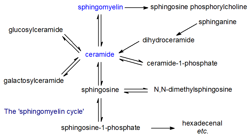
Many enzymes (and their isoforms) are involved, and their activities depend on factors such as intracellular location and activation mechanisms. Each compound in these pathways has characteristic metabolic properties. Thus, free sphingosine and other long-chain bases, which are the primary precursors of ceramides and thence of all the complex sphingolipids, function as mediators of many cellular events, for example by inhibiting the important enzyme protein kinase C. Ceramides are involved in cellular signaling, and especially in the regulation of apoptosis, cell differentiation, transformation, and proliferation, and most stress conditions. In contrast, sphingosine-1-phosphate and ceramide-1-phosphate promote cellular division (mitosis) rather than apoptosis, so the balance between these lipids and ceramide and/or sphingosine levels in cells is critical and requires exquisite control in each cellular compartment.
Similarly, the ‘structural’ sphingolipids, such as sphingomyelin, monoglycosylceramides, oligoglycosylceramides, and gangliosides, all have unique and characteristic biological functions, some of which stem from their physical properties and location within rafts, nanodomains of membranes. Most reactions in the sphingomyelin cycle are reversible, and the relevant enzymes are located in the endoplasmic reticulum, Golgi apparatus, plasma membrane, and mitochondria. The lysosomal compartment catabolizes more complex sphingolipids. Sphingolipids are especially important in providing the permeability barrier in the skin, where they are characterized by ultra-long fatty acyl components and fatty acyl groups linked to a hydroxyl group at the terminal end of the N-linked fatty acids (thereby generating a three-chain rather than a two-chain molecule).
Metabolic pathways comparable to the sphingomyelin cycle are believed to occur in plants, as shown in Figure \(\PageIndex{4}\). However, researchers have not studied them as extensively as in animals.
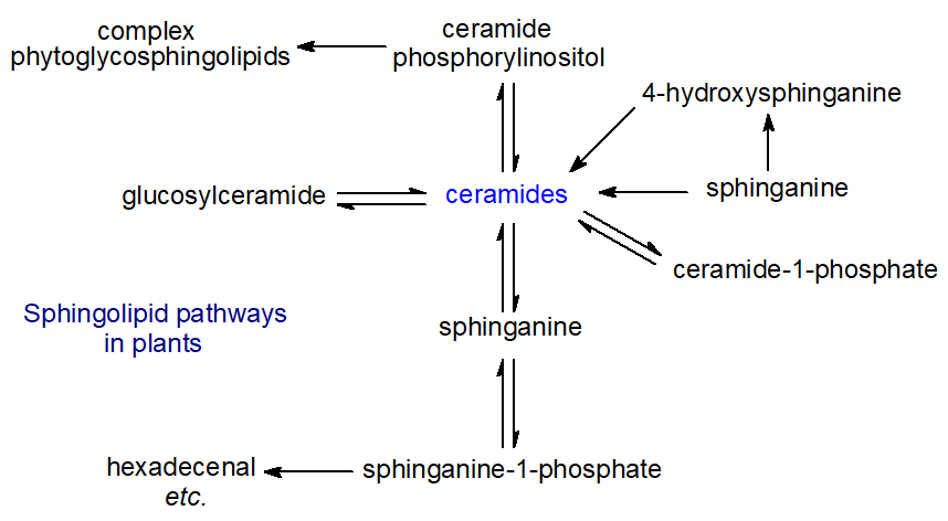
However, sphingolipid metabolites such as sphingosine-1-phosphate (or analogs) have been linked to programmed cell death, signal transduction, membrane stability, host-pathogen interactions, and stress responses. Plants also have a unique range of complex sphingolipids in their membranes, such as ceramide phosphorylinositol and phytoglycosphingolipids, and these now constitute a higher proportion of total lipids than previously thought. However, researchers have explored their functions only minimally. While relatively few bacterial species produce sphingolipids, some species produce sulfono-analogs of long-chain bases and ceramides (capnoids).
Fatty Acid Components of Sphingolipids
Sphingolipid fatty acids differ greatly from glycerolipid fatty acids, consisting of very-long-chain (up to C26) odd- and even-numbered saturated or monoenoic and related 2(R)-hydroxy components, while even longer fatty acids (C28 to C36) occur in spermatozoa and the epidermis. The dienoic acid 15,18‑tetracosadienoate (24:2(n‑6)), derived from the elongation of linoleic acid, is found in the ceramides and other sphingolipids of several tissues, but at relatively low levels. Polyunsaturated fatty acids are rarely present, although sphingomyelins of testes and spermatozoa are exceptions, as they contain even longer-chain fatty acids (up to 34 carbon atoms) and include 28:4(n‑6) and 30:5(n‑6). Skin ceramides also contain unusual very-long-chain fatty acids, while yeast sphingolipids are distinctive in containing mainly C26 fatty acids. In plants and yeasts, a similar range of chain lengths occurs as in animals, but 2-hydroxy acids predominate, sometimes accompanied by small amounts of 2,3‑dihydroxy acids; saturated fatty acids are most abundant, but monounsaturated fatty acids are present in higher proportions in the Brassica family (including Arabidopsis) and a few other species. Some fungal species contain monoenoic fatty acids with a trans-3 double bond and/or a hydroxyl group. Figure \(\PageIndex{5}\) shows typical sphingolipid fatty acids.

Very-long-chain saturated and monoenoic fatty acids for sphingolipid biosynthesis are produced from medium-chain precursors by elongases (ELOVL) in the endoplasmic reticulum of mammalian cells, and increasing evidence links specific isoforms to the biosynthesis of certain ceramides. For example, ELOVL1 has been linked to the production of ceramides containing C24 fatty acids (saturated and unsaturated), while ELOVL4 produces ultra-long-chain fatty acids in the skin. Yeasts possess three elongation enzymes: Elo1 (for medium- to long-chain fatty acids), Elo2 (up to C22), and Elo3 (up to C26).
The hydroxyl group is believed to enhance the hydrogen-bonding capacity of sphingolipids, helping stabilize membrane structures and strengthen interactions with membrane proteins. In mammals, a fatty acid 2-hydroxylase carries out hydroxylation; it is an NAD(P)H-dependent monooxygenase and an integral membrane protein of the endoplasmic reticulum. It converts unesterified long-chain fatty acids to 2‑hydroxy acids in vitro and probably in vivo. For example, experimental evidence is consistent with 2‑hydroxylation occurring at the fatty acid level before incorporation into ceramides in mouse brains, where the enzyme is highly expressed. A second enzyme of this kind is known to exist. It has yet to be characterized, and peroxisomal α-oxidation of 2-hydroxy acids may synthesize a proportion of the odd-chain fatty acids in the brain. Similarly, in skin, 2‑hydroxy and non-hydroxy fatty acids, as their CoA esters, are used equally readily for ceramide biosynthesis by ceramide synthases. Because mutations in the fatty acid 2‑hydroxylase in humans and mice cause demyelination disorders such as leukodystrophy, sphingolipids containing 2‑hydroxy acids likely have unique membrane functions that non-hydroxy analogs cannot replace.
In plants, 2‑hydroxyl groups appear to be inserted into fatty acyl chains while they are linked to ceramide, as ceramide synthase does not accept hydroxy fatty acids in vitro, at least. Two fatty acid 2‑hydroxylases (di-iron-oxo enzymes) have been identified in Arabidopsis, one specific for very-long-chain fatty acids and the other for palmitic acid. In fungi, a hydroxyl group is inserted at C2 of the fatty acid in a dihydroceramide intermediate.
Although fatty acids are only occasionally considered in the biological functions of sphingolipids, their influence is considerable, especially in relation to their physical properties and membrane function. For example, very-long-chain fatty acids may help stabilize highly curved membrane domains, as is required during cell division. The hydrophobic nature of the fatty acyl groups (together with the long-chain bases) enables the hydrogen bonding essential for forming raft nanodomains in membranes. As a general rule, lipid bilayers containing sphingolipids with 2-hydroxy-fatty acyl or 4-hydroxy-sphingoid base moieties tend to form condensed, more stable gel phases with higher melting temperatures than their non-hydroxylated equivalents because they have a more extended and stronger intermolecular hydrogen-bonding network. Fatty acid composition changes occur in some disease states; for example, increased concentrations of fatty acids >C24 are a feature of adrenoleukodystrophy, an X-linked genetic disorder.
Removal of very-long-chain fatty acids from sphingolipids in mutants of the model plant Arabidopsis inhibits the development of seedlings. For example, synthetic glycerolipids must contain very-long-chain fatty acids (C26) to allow growth in yeast mutants lacking sphingolipids, probably by stabilizing the proton-pumping enzyme H+-ATPase. Similarly, ceramides containing different fatty acids can be used in highly specific ways. Thus, in fungi, C16 or C18 hydroxy acids are used exclusively to synthesize glucosylceramide. Very-long-chain C24 and C26 hydroxy acids are used only to synthesize glycosyl inositol phosphorylceramide anchors for proteins. In plants, sphingolipids containing 2-hydroxy acids are protective against oxidative and other biotic stresses.
Links between Glycerolipid and Sphingolipid Metabolism
Sphingolipid and glycerolipid metabolism have been treated as separate sciences until relatively recently, partly for historical reasons and partly because analyzing the two lipid groups required different approaches and skills. However, the two overlap in many areas, not least because phosphatidylcholine is the biosynthetic precursor of sphingomyelin in animal cells. In plants and fungi, phosphatidylinositol is the biosynthetic precursor of ceramide phosphorylinositol. In contrast, ethanolamine phosphate derived from sphingolipid catabolism via sphingosine 1-phosphate is recycled for phosphatidylethanolamine biosynthesis, which is essential for survival in the protozoan parasite Trypanosoma brucei. In vitro studies show that sphingosine 1-phosphate activates phospholipase C, which hydrolyzes the lipid mediator phosphatidylinositol 4,5-bisphosphate to form diacylglycerols and inositol triphosphate. Sphingolipid-rich domains, or rafts, positively and negatively influence the location and functions of glycerophospholipids in membranes.
In addition, several examples of phosphoinositides and other complex lipids bind to enzymes of sphingolipid metabolism, either to regulate their activity or to facilitate their localization to various membranes. Thus, sphingosine kinase 2, one of the enzymes responsible for sphingosine 1-phosphate biosynthesis, binds to phosphatidylinositol monophosphate. The ceramide kinase responsible for ceramide 1-phosphate biosynthesis requires phosphatidylinositol 4,5-bisphosphate to function. Similarly, the CERT protein involved in ceramide transport has a binding site for phosphatidylinositol 4-phosphate. Sphingomyelin production at the trans-Golgi network triggers a signaling pathway that dephosphorylates phosphatidylinositol 4-phosphate, thereby interrupting cholesterol and sphingomyelin transport. Again, the interactions are not solely one-way, as ceramide 1‑phosphate (with phosphatidylinositol 4,5-bisphosphate) binds to the specific phospholipase A2 (cPLA2α) responsible for hydrolyzing phosphatidylinositol and thereby releasing arachidonic acid for eicosanoid production. In addition to phosphoinositides, phosphatidylserine activates neutral sphingomyelinase in the brain.
Long-Chain (Sphingoid) Bases
Long-chain/sphingoid bases are characteristic structural units of sphingolipids and are important structural and signaling lipids in animals, plants, and a few bacterial species. These are long-chain aliphatic amines, containing two or three hydroxyl groups and often a distinctive trans-double bond in position 4. More precisely, they are 2-amino-1,3-dihydroxy-alkanes or alkenes with (2S,3R)‑erythro stereochemistry, often with further structural modifications in the alkyl chain. They are important for the physical and biological properties of the more complex sphingolipids, but free sphingoid bases are also bioactive and interact with specific receptors and target molecules. As discussed below, the mechanisms for sphingoid base biosynthesis and for the N-acylated form (ceramides) are intimately linked.
Structures and Occurrence
In animal tissues, the most common or abundant sphingoid base is sphingosine ((2S,3R,4E)-2-amino-4-octadecene-1,3-diol) or sphing-4E-enine, i.e., with a C18 aliphatic chain, hydroxyl groups in positions 1 and 3, and an amine group in position 2; the double bond in position 4 has the trans (or E) configuration. Herbert Carter first characterized this in 1947 and also proposed the term “sphingolipides” for lipids containing sphingosine. It is usually accompanied by the saturated analog dihydrosphingosine (or sphinganine). Sphingoid bases are illustrated in Figure \(\PageIndex{6}\).

For shorthand purposes, a nomenclature similar to that for fatty acids can be used; the chain length and number of double bonds are denoted in the same manner with the prefix 'd' or 't' to designate di- and trihydroxy bases, respectively. Thus, sphingosine is denoted as d18:1 and phytosphingosine is t18:0. A superscript may indicate the position of the double bond. For example, 4-sphingenine can be abbreviated as d18:1Δ4t or 4E-d18:1. While alternative nomenclatures are occasionally used in publications, they are not recommended.
Long-chain bases found in animals, plants, and microorganisms now number over 100. Many of these may occur in a single tissue or organism, but almost always as part of a complex lipid containing an N-acyl-linked fatty acid, often with phosphate or carbohydrate functional groups. The aliphatic chains can contain from 14 to 28 carbon atoms, and most often they are saturated, monounsaturated, or diunsaturated, with double bonds in either the cis or trans configuration. For example, the main dienoic long-chain base (sphingadienine) in human plasma is D-erythro-1,3-dihydroxy-2-amino-4-trans,14-cis-octadecadiene, and it is especially abundant in the kidney, with higher levels in women than in men. It is not present in zebrafish, a widely used model species. Sphingolipids in some marine invertebrates and a dinoflagellate contain forms with three double bonds, such as sphinga-4E,8E,10E-trienine, sometimes with a methyl group at position 9. In addition, long-chain bases can have branched chains with methyl substituents at the omega‑1 (iso), omega‑2 (anteiso), or other positions; hydroxyl groups at positions 4, 5, or 6; ethoxy groups at position 3; and, in some organisms, a cyclopropane ring in the aliphatic chain. N-Methyl, N,N-dimethyl, and N,N,N-trimethyl derivatives of sphingoid bases have been detected in mouse brains.
The main C18 components of long-chain sphingomyelin bases in some animal tissues are accompanied by small amounts of C16 to C19 dihydroxy bases. The latter are more prevalent in ruminant tissues. In gangliosides from human brain and intestinal tissues, eicosasphingosine (2S,3R,4E-d20:1) occurs at appreciable concentrations, with amounts varying by region and membrane. However, human skin contains many isomers, including saturated, monoenoic, 6-hydroxy bases, and phytosphingosines with chain lengths ranging from C16 to C28. Many insect species contain shorter-chain bases. In the fruit fly, Drosophila melanogaster, a widely used model species in genetic and metabolic experiments, the main components are C14 bases. In contrast, nematodes such as Caenorhabditis elegans produce C17iso-methyl-branched sphingoid bases, which are essential for normal sphingolipid function.
The long-chain base composition of individual lipids can vary markedly between species, tissues, organelles, and even different membranes within a single organelle. For example, the data in Table \(\PageIndex{1}\) are perhaps from an extreme example. Still, it illustrates remarkable differences among lipids within a single part of the cell (rat liver mitochondria). Only part of the data from the paper cited is listed. It illustrates that 3-keto-sphinganine, produced in the first step of sphingosine biosynthesis (see below) and normally a minor component of sphingolipids - often not detectable- can vary from 28 to 100% of the sphingoid bases, depending on the lipid class and membrane within the organelle.
| Table \(\PageIndex{1}\): Long chain base composition of some lipid components of mitochondria from rat liver. | ||||
| Type | Base (%) | |||
|---|---|---|---|---|
| d18:1 | d18:0-3keto | t21:1 (phyto) | Unidentified | |
| Ceramidesa | 18 | 28 | 53 | - |
| Glucosylceramidesa | 3 | 95 | - | 3 |
| Lactosylceramidesb | 100 | |||
| a whole mitochondria; b mitochondrial inner membrane Data from Ardail, D. et al. FEBS Letts, 488, 160-164 (2001). |
||||
Phytosphingosine or 4D-hydroxy-sphinganine ((2S,3R,4R)-2-amino-octadecanetriol) is a common long-chain base of mainly plant origin. It is a saturated C18-trihydroxy compound, although unsaturated analogs, for example, with a trans (or occasionally a cis (Z)) double bond in position 8, i.e., dehydrophytosphingosine or 4D‑hydroxy-8-sphingenine, tend to be much more abundant. In many plant species, lipid class preferences also exist; for example, dihydroxy long-chain bases are more enriched in glucosylceramides than in glycosylinositolphosphoceramides. This is true in the model plant Arabidopsis thaliana, where the data listed for whole tissue are probably representative largely of the latter lipid, as shown in Table \(\PageIndex{2}\) below.
| Table \(\PageIndex{2}\): Sphingolipid long-chain base composition of whole tissue and glucosylceramides from Arabidopsis thaliana. | ||||||
| Base (%) | ||||||
|---|---|---|---|---|---|---|
| t18:1 (8Z) | t18:1 (8E) | t18:0 | d18:1 (8Z) | d18:1 (8E) | d18:0 | |
| Whole tissue | 12 | 70 | 13 | 4 | 1 | |
| Glucosylceramides | 44 | 22 | 5 | 28 | 2 | |
| Data from Sperling, P. et al. Plant Physiol. Biochem., 43, 1032-1038 (2005) | ||||||
Other plant long-chain bases have a double bond in position 4, which can be either cis or trans, although trans isomers are far more common. In contrast, the base d18:2Δ4E,8Z/E is relatively abundant in most plant species. In A. thaliana and related species, Δ4 long-chain bases are found mainly in the flowers and pollen and then exclusively as a component of the glucosylceramides. In general, as with Brassica species, composition depends on the species. Still, typically it is composed of up to eight different C18-sphingoid bases, with variable geometry of the double bond in position 8, i.e., (E/Z)-sphing-8-enine (d18:1Δ8), (4E,8E/Z)-sphinga-4,8-dienine (d18:2Δ4,8) and (8E/Z)-4-hydroxy-8-sphingenine (t18:1Δ8); d18:1Δ4, d18:0 and t18:0 tend to be present in small amounts only.
Phytosphingosine is not restricted to plants; it is found in significant amounts in intestinal cells and animal skin, with much lower relative proportions in the kidneys. Although non-mammalian sphingoid bases are generally poorly absorbed from the intestines, a small proportion of phytosphingosine and related sphingoid bases found in animal tissues may enter via the food chain.
Yeasts and fungi tend to have distinctive and characteristic long-chain base compositions. For example, filamentous fungi have 9-methyl-4E,8E-sphingadienine as the main sphingoid base in the glucosylceramides, as shown in Figure \(\PageIndex{7}\), but not in the ceramide phosphoinositol glycosides. At the same time, yeasts contain mainly the saturated C18 bases sphinganine and phytosphingosine, although some trans-4/8-unsaturated forms are usually present. Only a few bacterial species synthesize sphingolipids, but the family Bacteroidetes, abundant in the human gut, is an important exception; they usually contain saturated (and branched) long-chain bases. Other pathogenic bacteria may utilize sphingolipids and sphingoid bases from their hosts.

Sphingoid bases are surface-active amphiphiles with critical micellar concentrations of about 20 μM in aqueous solutions; they probably exist in the gel phase at physiological temperatures. They are unusual among lipids because they bear a small positive charge at neutral pH. However, their pKa (9.1) is lower than that of simple amines because of intramolecular hydrogen bonding. Together with their relatively high solubility (> 1μM), they can easily cross or move between membranes. In doing so, they increase membrane permeability to small solutes. In the esterified form, they form ordered lipid domains in membranes, such as rafts.
In complex sphingolipids, the sphingoid base links via its amine group to a fatty acid, including very-long-chain saturated or monoenoic and 2-hydroxy components, to form ceramides. These can be attached to a polar head group, such as phosphate or a carbohydrate, via the primary hydroxyl moiety. An important exception is sphingosine-1-phosphate, which is not acylated and has signaling functions in cells similar to those of lysophospholipids.
Biosynthesis and Metabolism
Sphinganine biosynthesis
The basic mechanism for sphinganine biosynthesis involves the condensation of palmitoyl-coenzyme A with L-serine, catalyzed by the membrane-bound enzyme serine palmitoyltransferase, which requires pyridoxal 5’-phosphate as a cofactor and binds to a specific lysine residue on the enzyme. The reaction occurs on the cytosolic side of the endoplasmic reticulum in animal, plant, and yeast cells with the formation of 3-keto-sphinganine, as illustrated in Figure \(\PageIndex{8}\).

This is believed to be the key regulatory or rate-limiting step in sphingolipid biosynthesis and is conserved in all organisms studied to date. Eliminating this enzyme is embryonically fatal in mammals and fruit flies. In mammals, serine palmitoyltransferase is a heterotrimer composed of two main subunits, designated SPTLC1 with either SPTLC2 or SPTLC3 (sometimes termed SPTLC2a and SPTLC2b, respectively). SPTLC1 is essential for activity and ubiquitously expressed, as is SPTLC2, whereas SPTLC3 is present in a relatively limited range of tissues and is most abundant in skin and placental tissue. In addition, two small subunits, ssSPTA and ssSPTB (other nomenclatures exist), differ by a single amino acid residue and may have regulatory functions; the active site lies at the interface between the two main subunits. ssSPTA is essential for serine palmitoyltransferase function during development and hematopoiesis.
A possible mechanism for the 1st step in the pathway, catalyzed by serine palmitoyltransferase, is shown in Figure \(\PageIndex{9}\).

Adding either small subunit to the complex substantially changes substrate preferences and enables synthesis of a wide range of natural homologs. In mammals, the SPTLC1-SPTLC2 complex forms C18 sphingoid bases specifically (with some C19 and C20), while SPTLC1 and SPTLC3 together give a broader product spectrum, including an anteiso-methylbranched-C18 isomer (from anteiso-methyl-palmitate as the precursor). Human skin synthesizes these branched bases only to a limited extent, but they are the main forms in lower invertebrates such as C. elegans. Serine palmitoyltransferase activity is governed by negative feedback and partly by orosomucoid (ORM-like or ORMDL) proteins: three in mammals (ORMDL1 to 3) and two in yeast (Orm1/2), which are ubiquitously expressed transmembrane proteins in the endoplasmic reticulum. Serine availability is also important.
Figure \(\PageIndex{10}\) shows an interactive iCn3D model of the human serine palmitoyltransferase complex (7K0M).

 Figure \(\PageIndex{10}\): Human serine palmitoyltransferase complex (7K0M).. Click the image for a popup or use this external link: https://structure.ncbi.nlm.nih.gov/i...tuGThTq9Vi4J58
- Gray and Plum (SPT1 A and E Chains
- Light Blue and Blue (SPT2 B and F Chains)
- Light Brown (small subunit A - ssA, C, and G chains)
- Yellow (ORM D and H)
The second step in sphinganine biosynthesis is the NADPH-dependent reduction of the keto group to a hydroxyl by a specific 3‑ketodihydrosphingosine reductase ('3KSR'), also on the cytosolic side of the endoplasmic reticulum. This step must occur rapidly as the intermediate is rarely encountered in tissues. The enzymes are presumed to be in similar subcellular locations in plant cells.
In plants, serine palmitoyltransferase is a heterodimer composed of LCB1 and LCB2 subunits with some homology to mammalian enzymes, while in the yeast Saccharomyces cerevisiae, it has three subunits: Lcb1, Lcb2, and Tsc3. Serine palmitoyltransferase is a water-soluble homodimer in a few bacteria that synthesize sphingoid bases. In the apicomplexan parasite Toxoplasma gondii, the enzyme is a homodimer located in the endoplasmic reticulum.
Free sphinganine formed in this way is rapidly N-acylated by acyl-CoA to form dihydroceramides by dihydroceramide synthases, which in animals are located primarily on the endoplasmic reticulum, presumably on the cytoplasmic surface. Animals and plants have multiple isoforms of this enzyme, for which the abbreviated term ‘ceramide synthase’ is now widely applied, as they utilize most other sphingoid bases, such as those produced by the hydrolysis of sphingolipids, as substrates. They are unique gene products, each located on a different chromosome, with considerable variation in enzyme expression across cell types within each tissue. Each isoenzyme has distinct specificities for the chain length of the fatty acyl-CoA moieties, but only to a limited extent for the base, suggesting that ceramides containing different fatty acids have differing roles in cellular physiology. These enzymes have six membrane-spanning regions, but the only substantial difference is in an 11-residue sequence in a loop between the last two putative transmembrane domains. Ceramides are central to all elements of sphingolipid biochemistry. These steps are illustrated in Figure \(\PageIndex{11}\).

Humans and mice have six ceramide synthases that use subsets of acyl-CoAs and thus produce ceramides with specific acyl chain lengths. Of these, ceramide synthase 2 is most abundant and is specific for CoA esters of very-long-chain fatty acids (C20 to C26); it is most active in the lungs, liver, and kidney. Ceramide synthase 1 is specific for 18:0 and is located mainly in the brain, with lower levels in skeletal muscle and testes. Ceramide synthase 3 produces the unusual ceramides of skin and testes. It uses C26-CoA and higher, including polyunsaturated-CoAs, in the latter tissue, while ceramide synthase 4 (skin, liver, heart, adipose tissue, and leukocytes) uses C18-C22-CoAs. Ceramide synthase 5 (lung epithelia and brain gray and white matter) generates C16 (mainly) and C18 ceramides, and ceramide synthase 6 (intestine, kidney, and lymph nodes) produces C14 and C16 ceramides. However, hydroxylation and the presence or otherwise of double bonds in the acyl-CoAs do not appear to influence the specificity of the ceramide synthases. Also, ceramide synthase mRNA expression does not always correlate with the fatty acid composition of sphingolipids in a given tissue, suggesting that other factors determine which molecular species are formed. One such is acyl-coenzyme A-binding protein (ACBP), which facilitates the synthesis of ceramides containing very-long fatty acids and stimulates ceramide synthases 2 and 3.
Insertion of the trans-double bond in position 4 to produce sphingosine occurs only after the sphinganine has been esterified in this way to form a ceramide, as illustrated in Figure \(\PageIndex{11}\), with desaturation occurring at the cytosolic surface of the endoplasmic reticulum also. The desaturases were first characterized in plants, simplifying the isolation of the appropriate enzymes in humans and other organisms. Two dihydroceramide desaturases have been identified in animals and designated 'DEGS1 and DEGS2'. Both enzymes insert a trans double bond at position 4, but DEGS2 is a dual-function enzyme that acts as a hydroxylase, adding a hydroxyl group at position 4 to generate phytoceramides. Enzyme distribution across tissues differs markedly: DEGS1 is expressed ubiquitously but is highest in the liver, Harderian gland, kidney, and lung. DEGS2 expression is largely restricted to the skin, intestine, and kidneys, where phytoceramides are more important. A considerable family of Δ4-sphingolipid desaturases has now been identified, and an early study by Stoffel and colleagues showed that Δ4-desaturation first involves syn-removal of the C(4)-HR and then the C(5)-HS hydrogens. This appears to be the first evidence that desaturases, in general, operate in this stepwise fashion.
The enzyme that inserts the cis-14 double bond into sphinga-4-trans,14-cis-dienine is fatty acid desaturase 3 (FADS3), which utilizes ceramides containing sphingosine as a precursor. The only other known activity of this enzyme is inserting a cis-double bond in position 13 of the CoA ester of vaccenic acid (11t-18:1) to produce the conjugated diene 11t,13c-18:2.
De novo synthesis of sphingoid bases is essential in most organisms, and inhibiting the biosynthetic pathways affects growth and viability. However, this can be tissue-specific: deletion of the liver-specific SPTLC2 in mice did not affect liver function, while a comparable deletion of the adipocyte-specific SPTLC1 caused major tissue defects. Presumably, the latter tissue cannot take up enough sphingolipid from the circulation to compensate. SPTLC3 deficiencies are associated with dermal pathologies, and genetic variants in SPTLC3 are associated with dyslipidemia and atherosclerosis. Studies of mutants lacking specific enzymes have similarly demonstrated the essentiality of sphingoid base synthesis in plants.
Click the link below.
- Link
-
Phytosphingosine and plant ceramides: Phytosphingosine is formed from sphinganine, produced as above, by hydroxylation at position 4, possibly via the free base in plants, although it can form from both sphinganine and a ceramide substrate in yeasts. Yeast has a single sphinganine C4‑hydroxylase, but Arabidopsis has two such enzymes (SBH1 and SBH2), which are critical for growth and viability. Much remains to be learned of the processes involved, but it is known that the enzyme responsible is closely related to a Δ4 desaturase. Indeed, bifunctional Δ4‑desaturase/Δ4-hydroxylases have been shown in Candida albicans and mammals, especially in keratinocytes (DEGS2 discussed above), in which either 4‑hydroxylation or Δ4‑desaturation is initiated by removal of the proR C-4 hydrogen. Sphinganine attached to ceramide is the substrate for 4-hydroxylation in intestinal cells.
In Arabidopsis thaliana leaves, 90% of the sphingoid bases are phytosphingosine with a Δ8‑double bond. In plants, in addition to Δ4‑desaturation, two distinct types (20 gene products) of sphingoid Δ8-desaturase have been characterized that catalyze the introduction of a double bond at positions 8 and 9 of phytosphingosine. These are evolutionarily distinct from the Δ4‑desaturases. One type produces mainly the trans (E)-8 isomer, and the other mostly the cis (Z)-8 isomer, with the trans isomer tending to predominate overall but varying by plant species. The trans isomer appears to form when the hydrogen on carbon 8 is removed first, and the cis isomer when carbon 9 is the point of attack. While most Δ8-desaturases require a 4‑hydroxysphinganine moiety as a substrate, the second does not.
In Arabidopsis, three different isoforms of ceramide synthase have been identified and denoted LOH1, LOH2, and LOH3. LOH1 and LOH3 (class II synthases) use phytosphingosine efficiently, but only LOH2 (class I synthase) uses sphinganine efficiently; LOH2 and LOH3 prefer unsaturated long-chain bases. LOH2 also shows marked fatty acid specificity, with near-complete specificity for palmitoyl-CoA and dihydroxy bases, while LOH1 shows greatest activity for 24:0- and 26:0-CoAs and trihydroxy bases; neither uses unsaturated acyl-CoA esters efficiently. In plants, fatty acid desaturases and hydroxylases are also closely related, and sphingolipid fatty acid α-hydroxylation is believed to occur on the ceramide rather than the free acyl chain. The Δ8-desaturase is believed to use ceramide as a substrate and direct products selectively toward complex sphingolipid synthesis, whereas Δ4‑desaturation directs ceramides toward glucosylceramide synthesis.
Long-chain bases with 4-hydroxyl groups are required for the viability of the filamentous fungus Aspergillus nidulans and for growth in plants such as A. thaliana. An 8E double bond confers aluminum tolerance to yeasts and plants and is important for chilling resistance in tomatoes. However, a trans-4 double bond in the sphingoid base does not appear essential for Arabidopsis growth and development.
Fungal sphingoid bases: Fungi produce trans Δ8-isomers only, but Δ4- and Δ8-desaturases do not occur in the widely studied yeast S. cerevisiae. In fungal sphingoid base biosynthesis, the double bonds at positions 4 and 8 and the methyl group at position 9 are inserted sequentially into the sphinganine portion of a ceramide, with the last step catalyzed by an S-adenosylmethionine-dependent methyltransferase similar to plant and bacterial cyclopropane fatty acid synthases. In S. cerevisiae, the ceramide synthase is a heteromeric protein complex containing three subunits, Lag1, Lac1, and Lip1, of which the first two are homologous proteins that feature eight transmembrane domains. The yeast Pichia pastoris has a distinct ceramide synthase that utilizes dihydroxy sphingoid bases and C16/C18 acyl-CoA as substrates to produce ceramides. The long-chain-base components of the ceramide are then desaturated in situ by a Δ4‑desaturase, and the fatty acid components are hydroxylated at position 2. Further desaturation of the long-chain base component by a Δ8-(trans)-desaturase occurs before an S-adenosylmethionine-dependent sphingolipid C-9 methyltransferase introduces the methyl group at position 9. As a final step, a trans-double bond may be introduced into position 3 of the fatty acid component. These ceramides are used exclusively to produce glucosylceramides, and a separate ceramide synthase, encoded by a different gene, is believed to produce the ceramide precursors for ceramide phosphorylinositol mannosides.
Viral sphingoid bases: The genome of an important marine virus (EhV) encodes for a novel serine palmitoyltransferase. This enzyme hijacks algal host metabolism to produce unusual hydroxylated C17 sphingoid bases, which accumulate in lytic cells of infected algae, including the important bloom-forming species Emiliania huxleyi. While this may seem esoteric, viruses constitute a significant proportion of the marine biome, and their control of algal bloom growth has global consequences.
Unesterified sphingosine: A cycle of reactions in tissues incorporates sphingoid bases into sphingolipids via ceramide intermediates, which serve innumerable functions before being broken down again into their component parts. All free sphingosine in tissues must arise this way, particularly through ceramidase action on ceramides. Five ceramidases are known, each with differing pH optima and subcellular locations. The levels of free sphingoids and their capacities to function as lipid mediators, as shown in Figure \(\PageIndex{12}\), are controlled mainly by enzymatic re‑acylation to form ceramides. However, sphingosine kinases act on some to produce sphingosine-1-phosphate.

Figure \(\PageIndex{12}\): Ceramide hydrolysis and resynthesis Free sphingoid bases are absorbed by enterocytes following the digestion of dietary sphingolipids in animals (including some from gut microorganisms). While some is converted to complex sphingolipids, much is catabolized, eventually forming palmitic acid.
Catabolism of sphingosine and other long-chain bases occurs after conversion to sphingosine-1-phosphate and analogs. Yeast has an alternative detoxification mechanism. Excess phytosphingosine is first acetylated, then converted to a vinyl ether, before export from the cell.
Biological Functions of Unesterified Sphingoid Bases
Sphingoid bases primarily serve as a basic component of ceramides and complex sphingolipids, where variations in their composition can influence the physical and biological properties of these lipids. Independent of this, in their free (unesterified) form, they are important mediators of many cellular events, even though they are present at low levels only in tissues (typically 25 and 50 nM in plasma), with intracellular levels determined by hydrolysis by ceramidases or by the action of sphingosine kinases (sphingosine-1-phosphate production). In animal cells, they indirectly inhibit protein kinase C, possibly by interfering with the binding of enzyme activators, such as diacylglycerols or phorbol esters. In addition, sphingoid bases potently inhibit cell growth, although they also stimulate cell proliferation and DNA synthesis. They are involved in apoptosis in a manner distinct from ceramides, by binding to specific proteins and regulating their phosphorylation. While sphingosine does not appear to participate in membrane raft formation, it may rigidify pre-existing gel domains in mixed bilayers. However, any such effects depend on local concentrations and pH. Some experimentally observed biological effects may result from conversion to sphingosine-1-phosphate.
Free sphingosine has been implicated in various pathological conditions; for example, plasma sphingosine levels are increased in hyperthyroidism and patients with type 2 diabetes. Lysosomal lipid storage is an initiating factor in Niemann-Pick type C disease. This neurodegenerative disorder disrupts calcium release, leading to cholesterol and sphingolipid buildup. In the human adrenal cortex, sphingosine, produced in situ by acid ceramidase, supports steroid production by serving as a ligand for steroidogenic factor 1 in the cell nucleus, which controls transcription of genes involved in cholesterol conversion to steroid hormones. Unesterified sphingoid bases may have a protective role against colon cancer in humans. Thus, N,N‑dimethylsphingosine and dihydrosphingosine, like the deoxysphingoid bases, are known to induce cell death in various malignant cells. Evidence suggests that sphingadienes of plant and animal origin inhibit colorectal cancer in mouse models by reducing sphingosine-1-phosphate levels. Consequently, researchers are testing synthetic analogs of long-chain bases for pharmaceutical potential.
Free sphingosine is believed to signal in plants by controlling pH gradients across membranes. In addition, free long-chain bases (and the balance with the 1-phosphate derivatives) are essential for regulating apoptosis in plants.
Ceramides
Structure and Occurrence
Figure \(\PageIndex{13}\) shows the ceramide structure again.

Figure \(\PageIndex{13}\): Structure of ceramides (with varying fatty acids in ester link)
Ceramides consist of a long-chain or sphingoid base linked to a fatty acid via an amide bond. They are essential intermediates in the biosynthesis and metabolism of all sphingolipids, including the complex sphingolipids in which the terminal primary hydroxyl group is linked to carbohydrate, phosphate, and so forth (sphingomyelin, glycosphingolipids, and gangliosides), as shown in Figure \(\PageIndex{14}\).
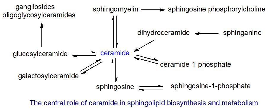
They are also the primary source of unesterified sphingoid bases and the important biological mediators sphingosine-1-phosphate and ceramide-1-phosphate. To date, researchers have identified 33 enzymes involved in ceramide metabolism. While ceramides are rarely found at greater than trace amounts in tissues other than skin, they can still exert important biological effects at these low levels. They are present in membranes where they participate in the formation of raft domains.
Each organism and tissue may synthesize ceramides, which contain a variety of di- and trihydroxy long-chain bases linked to fatty acids. As discussed previously, the fatty acids consist mainly of longer-chain (up to C24 or greater) saturated and monoenoic (mainly (n-9)) components, sometimes with a hydroxyl group in position 2. Other than in certain testicular cells, polyunsaturated fatty acids do not occur. Researchers have characterized more than 200 structurally distinct ceramide species in mammalian cells. In plants, 2-hydroxy acids predominate, sometimes accompanied by small amounts of 2,3-dihydroxy acids. Although small amounts of free ceramides are produced in all tissues as required for the specific biological functions described below, most are converted rapidly to more complex sphingolipids, including sphingomyelin (in animals) and the various glycosylceramides. The ceramides in skin are a remarkable exception to this rule and are discussed separately below.
A shorthand nomenclature combines those used conventionally for fatty acids and long-chain bases to denote ceramide species, including those as components of more complex lipids; e.g., N-palmitoyl-sphingosine is d18:1-16:0. Ceramides containing sphinganine are sometimes termed ‘dihydroceramides’.
Ceramide Biosynthesis
Ceramide production is complex and involves at least three pathways. De novo biosynthesis occurs in the endoplasmic reticulum, using palmitoyl-CoA and serine as precursors for the long-chain base component, which is converted to ceramide. Biosynthesis of the specific fatty acids in ceramides also involves various chain elongases (ELOVL). Alternative routes for ceramide production involve regeneration from complex sphingolipids. For example, in animals, the sphingomyelinase pathway converts sphingomyelin into ceramides (and vice versa) in the plasma membrane, Golgi, and mitochondria. Finally, hydrolytic enzymes in the lysosomal compartment can remove the polar moieties of complex glycosphingolipids, thereby recovering ceramides (or their fragments) through recycling/catabolism. Because these biosynthetic and metabolic pathways occur in different organelles, specific ceramide pools and sphingolipids have distinct biological properties and functions.
Ceramide synthesis de novo: This pathway is described mechanistically. In brief, in animals, sphinganine couples with a long-chain fatty acid to form dihydroceramide using one of six ceramide synthases in the endoplasmic reticulum, mainly before the double bond is introduced at position 4 of the sphingoid base. Of these, ceramide synthase 2 is most abundant and is specific for CoA esters of very-long-chain fatty acids (C20 to C26); it is most active in the central nervous system. Ceramide synthase 1 is specific for 18:0 and is located exclusively in the brain and skeletal muscle; ceramide synthases 5 and 6 generate 16:0-containing ceramides, and ceramide synthase 3 produces the unusual ceramides of skin and testes.
Figure \(\PageIndex{15}\) shows the synthesis of ceramide from sphinganine and palmitoyl-CoA (a repeat of Figure \(\PageIndex{11}\)
Each synthase has six membrane-spanning domains and contains a characteristic motif with the specific structures required for catalysis and substrate binding that are essential for its activity. They differ primarily in an 11-residue loop between the last two putative transmembrane domains. Beyond separate transcriptional regulation, many factors modulate ceramide synthase activity, including reversible dimerization. Ceramide synthase 2 has a sphingosine-1-phosphate binding motif, and this lipid may inhibit its activity. Acyl-coenzyme A-binding protein (ACBP) facilitates the synthesis of ceramides containing very-long fatty acids and stimulates ceramide synthases 2 and 3 in particular.
Most ceramides generated in this way are rapidly utilized to synthesize complex sphingolipids, especially sphingomyelin and hexosylceramides, thereby regulating cellular ceramide concentrations and controlling their biological activities. In mammalian cells, most complex glycerolipids are synthesized in the endoplasmic reticulum before transport to their final subcellular locations, whereas sphingolipids are synthesized differently. Ceramide is synthesized on the cytoplasmic leaflet of the endoplasmic reticulum. Complex sphingolipids form in the Golgi apparatus, and a key cytoplasmic protein, ceramide transporter, or 'CERT' (CERamide Trafficking), mediates non-vesicular transport of ceramide between these organelles. It has several distinct functional domains, including an N-terminal phosphatidylinositol-4-monophosphate (PI(4)P)-binding or Pleckstrin homology (PH) domain, which targets the Golgi apparatus, and a C-terminal ‘START’ domain, which can recognize ceramide species with the natural D-erythro stereochemistry, including dihydroceramide and phytoceramide (but not sphingosine). It holds them within a long amphiphilic cavity by hydrogen bonding with all three polar atoms of the sphingoid motif. A short peptide motif (FFAT) also recognizes a specific protein in the endoplasmic reticulum. The protein body is sufficiently flexible to allow ceramide to move from the endoplasmic reticulum to the Golgi without free movement through the cytosol.
Very-long-chain ceramides containing 24:0 or 24:1 fatty acids turn over much more rapidly in animal cells than those containing 16:0 or 18:0 fatty acids because the former convert more rapidly into complex sphingolipids, which may regulate the levels and perhaps the biological functions of the latter. In contrast, ceramides containing d16:1 and d18:1 sphingoid bases turn over at similar rates and do not affect ceramide flux through these pathways. CERT is a major factor in this specificity, as it extracts ceramides from membrane bilayers, preferring those required for complex sphingolipid synthesis. This removal establishes a gradient that sustains the process and prevents ceramide accumulation in the endoplasmic reticulum, which might otherwise disrupt the membrane and even cause cell death. Although the transfer itself is not ATP-dependent, the overall process requires ATP, possibly to maintain PI(4)P in a phosphorylated form, and the multiple factors that control this lipid's biosynthesis must also influence sphingolipid metabolism.
As a neutral lipid, ceramide can readily flip across membrane leaflets, which is also necessary for sphingomyelin synthesis in the Golgi lumen. A separate transport mechanism delivers the ceramide pool used to synthesize glycosylceramide to the Golgi and does not require ATP. In addition, some ceramide synthesis occurs in mitochondria, which can lead to cell death. Regulation of ceramide and subsequent sphingolipid biosynthesis is crucial, as excess sphingolipids can be toxic, while reduced synthesis can inhibit cell proliferation.
Some ceramides are transported from the liver to other tissues in plasma lipoproteins, especially the HDL2 and HDL3 subfractions, which contain apolipoprotein B. Some suggest that ceramide transport via lipoproteins could be a paracrine mechanism for regulating other cells' metabolism.
Ceramides are also produced during the catabolism of other complex sphingolipids, and especially by the action of one or other of the sphingomyelinases or phospholipase C on sphingomyelin in animal tissues as part of the 'sphingomyelin cycle' as shown in Figure \(\PageIndex{16}\).

Many agonists, including chemotherapeutic agents, tumor necrosis factor-alpha, 1,25-dihydroxyvitamin D3, endotoxin, gamma-interferon, interleukins, nerve growth factor, ionizing radiation, and heat, stimulate sphingomyelin hydrolysis to produce ceramide. In addition, reversal of the sphingomyelin synthesis reaction may generate ceramide, and some may be produced by the reverse operation of the enzyme ceramidase (see next section). These reactions are much faster than de novo synthesis, so they are especially relevant to ceramide signaling, particularly at the plasma membrane. For example, acid sphingomyelinase may be especially important in this context because it generates ceramides that initiate the cascade of events leading to apoptosis (see below).
Glycosidases can also hydrolyze glycosphingolipids to ceramides in tissues, but this process tends to be less important quantitatively (except in skin). The key enzymes of sphingolipid metabolism were first characterized in the yeast Saccharomyces cerevisiae, and they were found to be sufficiently similar to the corresponding enzymes in mammals to facilitate their study in mammals.
As discussed above, specific ceramide synthases use specific fatty acids for ceramide biosynthesis in animals, and we are slowly learning how these are compartmentalized and regulated within cells. Thus, ceramide synthesis and subsequent catabolism involve a complex web of at least 28 distinct enzymes, including six ceramide synthases and five sphingomyelinases, each encoded by distinct genes. Each enzyme may produce distinct ceramide species with characteristic biological properties. Ceramide species containing very-long-chain fatty acids (C24) turn over more rapidly than those containing C16/18 components.
Ceramide Catabolism
In animals, ceramidases control ceramide metabolism and function by hydrolyzing ceramide to form sphingoid bases and free fatty acids; this is the only route to unesterified sphingosine. This is illustrated in Figure \(\PageIndex{17}\).

Five such enzymes are known in humans, classified according to their pH optima, i.e., acid (‘ASAH1’), neutral (‘ASAH2’, which differs between humans and animals), and alkaline (three enzymes - ‘ACER1 to ACER3’), with differing cellular locations and fatty acid specificities and with the potential to affect distinct signaling and metabolic events. Acid ceramidase is particularly important, and aberrations in its synthesis or activity are involved in several human disease states, including the rare autosomal-recessive Farber disease, in which the enzyme is deficient, leading to ceramide accumulation; ceramide containing 26:0 in the blood is considered a biomarker for the disease. ASAH1 is located in lysosomes and hydrolyses ceramides containing small- to medium-chain fatty acids (C6-C18) most efficiently. Neutral ceramidase is located in the plasma membrane and the Golgi, especially in intestinal epithelial cells and colorectal tissues, and prefers long-chain components (C16 to C18); it also catalyzes the reverse reaction, which may support ceramide synthesis in mitochondria. ACER1 and ACER2 are found in the endoplasmic reticulum and Golgi, respectively, and they prefer species with very-long-chain acyl groups. ACER3 is present in both the endoplasmic reticulum and Golgi; in vitro, it has marked specificity for ceramides, dihydroceramides, and phytoceramides linked to unsaturated long-chain fatty acids (18:1, 20:1, or 20:4). Neutral/alkaline ceramidase activity has also been found in mitochondria and nuclei.
In Arabidopsis, an alkaline ceramidase (AtACER) can hydrolyze phytosphingosine-containing ceramides, and a related enzyme from rice prefers d18:1δ4-ceramide; the latter can function in reverse to increase C26- and C28-phytoceramide content. Several neutral ceramidases (AtNCERs) have been identified, but plants do not appear to have an equivalent to acid ceramidase. Ceramidases are also found in lower organisms, such as Pseudomonas aeruginosa and slime molds. The enzymes are secreted and are not integral membrane enzymes. Neutral ceramidase is found only in prokaryotes, including some pathogenic bacteria.
Sphingoid bases released by acid ceramidase can escape from lysosomes and be reused for ceramide biosynthesis by ceramide synthase. This has been termed the ‘salvage’ pathway and is important both quantitatively and biologically. For example, it has been estimated to contribute 50% to 90% of sphingolipid biosynthesis. The biological functions of ceramides are discussed below. Ceramides derived from the salvage pathway are likely spatially and functionally distinct from those synthesized de novo. In addition, sphingoid bases released in this way have biological functions, including serving as substrates for the synthesis of the biologically important metabolite sphingosine-1-phosphate. Therefore, regulating ceramidase action is central to biological processes in innumerable animals.
See the link below
- Link
-
Biological Functions of Ceramides
This text discusses the role of ceramides in the biosynthesis of complex glyco- and phospho-sphingolipids elsewhere. Ceramides, like other lipid second messengers in signal transduction, are produced rapidly and transiently in response to specific stimuli to target specific proteins, for example, to activate certain serine/threonine protein kinases or phosphatases. They may also regulate cellular processes by influencing membrane properties. While cells can produce them de novo for these functions, activating a sphingomyelinase under physiological stress or with other agents allows faster generation in animal tissues. Ceramides appear to form under all conditions of cellular stress through multiple activators in eukaryotic organisms. However, ceramides with different fatty acid and long-chain base (molecular species) compositions form in distinct cellular compartments or membranes through various mechanisms over different time scales, potentially with distinct functions. The biological functions of those ceramides containing medium-chain (up to C14), long-chain (C16 and C18), and very-long-chain (C20 and longer) fatty acids, in particular, may have to be considered separately.
Physical properties: Unsaturation in the sphingoid backbone augments intramolecular hydrogen bonding in the polar region, allowing close packing of ceramide molecules and tight intramolecular interactions in membranes. Another important factor is the length of the fatty acyl moiety, as shorter-chain ceramides tend to produce positive curvature in a lipid monolayer. Long-chain molecules have the opposite effect, with a marked intrinsic negative curvature that facilitates the formation of inverted hexagonal phases and increases acyl-chain order in bilayers. Through their interactions with ion channels, ceramides influence membrane permeability, rendering bilayers and cell membranes permeable to solutes ranging from small molecules to proteins.
Although ceramides are minor membrane components, their physical properties concentrate them into lateral liquid-ordered microdomains (a distinct form of 'raft' termed ‘ceramide-rich platforms’). However, these effects are again chain-length specific. These domains differ appreciably in composition from rafts enriched in sphingomyelin and cholesterol, and ceramides containing C12 to C18 fatty acids can displace cholesterol from rafts, modifying their physical properties. Ceramides are generated within rafts by acid sphingomyelinase, which causes small rafts to merge into larger units and modifies membrane structure, thereby permitting the oligomerization of specific proteins, such as cytokines and death receptors. Ceramides are also essential for exosome formation and/or secretion by facilitating or inducing membrane curvature. In contrast, sphingosine, sphingosine-1-phosphate, and ceramide-1-phosphate do not facilitate raft formation.
Ceramides can signal through these modified rafts. Specific receptor molecules and signaling proteins are recruited and clustered within such domains, excluding potential inhibitory signals while initiating and greatly amplifying primary signals. Ceramide-rich platforms are believed to amplify both receptor- and stress-mediated signaling events and may influence various disease states. Ceramide-enriched membrane domains formed in response to sphingomyelinase activity serve as sites for endocytic uptake of pathogens because they concentrate pathogen receptors and signaling complexes; in particular, these domains can enhance viral infections, including Norovirus, Japanese encephalitis virus, Ebola, and possibly SARS-CoV-2. However, elevated levels of ceramide inhibit the cellular uptake of HIV.
Although ceramides and diacylglycerols are structurally similar, they differ in occurrence, location, and membrane behavior. Ceramides cross synthetic lipid bilayers relatively quickly in vitro. Still, it is unclear whether they can flip across more complex biological membranes as readily, especially on ceramide-rich platforms. Restricted flipping could affect ceramides' signaling role, as those generated by distinct enzymes on opposite sides of a membrane could have distinct functions.
Enzyme activation: Ceramides generally modify intracellular signaling pathways to slow anabolism and promote catabolism. Among the wide range of biological functions in cellular signaling, ceramides are especially important in triggering apoptosis and have also been implicated in activating various protein kinase cascades, depending on the site of generation. The mechanism of these interactions is the subject of intensive study, but in this context, two intracellular targets have been identified as particularly important: at least two protein phosphatases (ceramide-activated protein phosphatases) and a family of protein kinases (ceramide-activated protein kinases). For example, phosphatases may regulate glycogen synthesis, insulin resistance, and responses to apoptotic stimuli. Ceramides generated by sphingomyelinase and by de novo synthesis are both important to the process. At the same time, ceramidases have contrasting effects on these and other biological effects of ceramides.
Apoptosis: The role of ceramides in regulating apoptosis, cell differentiation, transformation, and proliferation has received special attention. Apoptosis is a normal process that occurs in response to oxidative stress, in particular, in which a cell can be considered to commit suicide. It is essential for many aspects of normal development and maintaining tissue homeostasis. There are two pathways: 'extrinsic ', initiated in the plasma membrane by ligation of so-called 'death factors', such as tumor necrosis factor-α (TNF-α), and 'intrinsic ', induced by external actions in mitochondria, e.g., by DNA damage, oxidation, or radiation injury. Although the mechanism of ceramide interaction with these pathways is uncertain, a cascade of reactions is initiated, culminating in the release of intracellular caspase family proteases to promote apoptosis. In dysfunctional mitochondria, one mechanism involves forming membrane channels that release specific mitochondrial proteins, including caspases. Ceramides with fatty acids of different chain lengths are believed to function differently; for example, 16:0-ceramide generated by ceramide synthase 6 is especially pro-apoptotic. In contrast, ceramides with very-long-chain fatty acids accumulate in necroptosis, a form of regulated cell death. In keratinocytes, ceramides containing 2-hydroxy acids appear to protect against apoptosis. Ceramides also induce the related process of cellular senescence.
Failure to properly regulate apoptosis can have catastrophic consequences, and many disease states, including cancer, diabetes, neuropathies, Alzheimer's disease, Parkinson's disease, and atherosclerosis, are thought to arise from the deregulation of apoptosis. For example, ceramides have been implicated in TNF-α signaling and in cytotoxic responses to the amyloid Aβ peptide, both of which are involved in Alzheimer’s disease and neurodegeneration. In addition, ceramides appear to be involved in many aspects of aging biology and male and female fertility. These effects may hold implications for diseases associated with obesity and insulin resistance, including diabetes and cardiovascular disease.
Similarly, ceramides are intimately involved in the induction of autophagy, the 'maintenance' process by which cells remove proteins and excess or damaged organelles by engulfing them in a membrane-enclosed cellular compartment called the phagosome. In particular, maturing phagosomes are enriched in very-long-chain ceramides. While this process benefits recycling of cellular nutrients, excess ceramide can trigger unnecessary apoptosis.
Because animals and plants have multiple ceramide synthase isoforms specific to chain length and fatty acid, researchers suggest that ceramides with different fatty acids have distinct roles in cellular physiology. In particular, C16 ceramide appears to be especially important in apoptosis in non-neuronal tissues. In contrast, C18 ceramide has growth-arresting properties and may be involved in apoptosis in some carcinomas treated with chemotherapy agents. In addition, a transferase has been identified that transfers the acetyl group from platelet-activating factor to sphingosine with high specificity. The product, N-acetylsphingosine - the simplest of all ceramides- has signaling functions that are distinct from those of the parent lipids or other ceramides; it does not enter the salvage pathway in cancer cells in vitro and is cytotoxic.
In contrast, the ceramide metabolite, sphingosine-1-phosphate, has opposing effects on cell survival and proliferation. As ceramide and sphingosine-1-phosphate are interconvertible via sphingosine as an intermediate, which also has pro-apoptotic activity, the balance between these lipids and ceramide-1-phosphate is obviously of great metabolic importance. It has been termed the ‘sphingolipid rheostat ’, as illustrated in Figure \(\PageIndex{18}\).
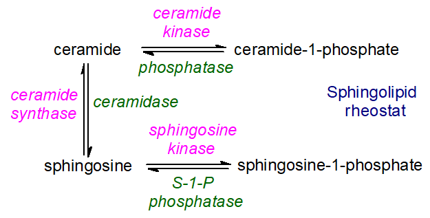
Figure \(\PageIndex{18}\): Sphingolipid rheostat Plants: Relatively little information is available on the role of ceramides in cell signaling, but suggestions link sphingolipid catabolic products to programmed cell death, signal transduction, membrane stability, host-pathogen interactions, and stress responses. For example, there is evidence that enhanced synthesis of ceramides with very-long-chain fatty acids and trihydroxy sphingoid bases by ceramide synthases LOH1 and LOH3 promotes cell division and growth, while, in contrast, accumulation of the ceramide species C16 fatty acid with a dihydroxy sphingoid base, due to LOH2 overexpression, leads to plant dwarfing and programmed cell death. Ceramides aggregate in rafts in plant membranes with other sphingolipids and sterols, as in animal tissues. Similarly, in the yeast S. cerevisiae, a widely used model organism, ceramide species with different N-acyl chains and sphingoid bases regulate different sets of functionally related genes.
Skin Ceramides
The mammalian skin forms a protective barrier between the host's internal tissues and the hostile external environment, which can include chemicals, ultraviolet light, mechanical damage, and pathogenic microorganisms, while preventing the loss of water and electrolytes. It consists of stratified layers of increasingly differentiated cells, or keratinocytes, in which the basal layer renews tissue and then migrates upward and differentiates, accumulating specific lipids and proteins that alter cellular architecture. Eventually, the keratinocytes lose their nucleus and become flattened structures of insoluble protein surrounded by lipids termed ‘corneocytes’ in the outermost impermeable layer, or stratum corneum. By secreting antimicrobial peptides and proteins, keratinocytes strengthen the skin's defense against commensal microorganisms and opportunistic pathogens; lipid mediators such as free sphingoid bases and eicosanoids in the stratum corneum, and free fatty acids in sebum, reinforce this defense.
The stratum corneum contains high levels of ceramides (as much as 50% of the total lipids), including O-acylceramides, which exist in the free form and are linked by ester bonds to structural proteins. They are present mainly in the extracellular domains (interstices). They are accompanied by nearly equimolar amounts of cholesterol and free fatty acids. This ratio is believed to be essential for normal tissue organization into the membrane structures that support the epidermal barrier's function. Unlike other biological membranes, skin membranes consist mainly of two lamellar phases, which form crystalline lateral phases with repeat distances of approximately 6 and 13 nm. Small lipid subdomains in a liquid phase may also exist.
Some of these skin ceramides have distinctive structures not seen in other tissues; many forms are commonly recognized. They can contain the normal range of longer-chain fatty acids (a), e.g., formula 1 in the figure, some of which have a hydroxyl group in position 2 (a*), e.g., formula 2, linked to dihydroxy bases with a trans-double bond in position 4 or to trihydroxy bases. This is illustrated in Figure \(\PageIndex{18}\).

Figure \(\PageIndex{18}\): Skin ceramides In addition, there are O‑acyl ceramides in which a unique very-long-chain fatty acid component (typically C30 or C32) has a terminal hydroxyl group, and this may be in the free form or esterified with linoleate (c), e.g., formulae 3 and 4; the sphingoid base can be either di- (b) or trihydroxy (b*), e.g., formula 4; the latter is not a common feature in sphingolipids of animal origin, and can include both phytosphingosine and the unique 6‑hydroxy-4-sphingenine in human epidermis. Ceramides of type 1 (with the 1-O-hydroxyl group of the sphingoid base acylated by a very-long-chain fatty acid) are also present (1‑O‑acylceramides - illustrated above). These account for 5% of the total ceramides in the epidermis of mice and humans and represent as many as 700 molecular species. In total, researchers have identified 15 classes of free ceramides and three classes of covalently bound ceramides, with up to 1700 distinct molecular species. Researchers first studied these lipids in detail in pig skin as a convenient experimental model, but they have also characterized them in humans and rats. In addition, researchers have characterized several molecular forms of glucosylceramide, based on similar ceramide structures, in skin, and these are also essential for its proper function.
Lipid composition can vary depending on the skin layer (keratinocytes, stratum corneum, etc.). These lipids play a key role in the skin's barrier properties, limiting water and solute loss while preventing the ingress of harmful substances. Because the aliphatic chains in ceramides and fatty acids are mainly non-branched, long-chain saturated compounds with a high melting point and a small polar head group, the lipid chains are mostly in a solid crystalline or gel state, which exhibits low lateral diffusion and low permeability at physiological temperatures. For example, the stratum corneum layer of the skin has a water permeability of only one-thousandth that of other biomembranes. Natural and synthetic ceramides are now commonly added to cosmetics and other skin care preparations.
Most steps in ceramide biosynthesis linked to ω-O-acylated fatty acids occur in the endoplasmic reticulum of keratinocytes. First, de novo fatty acid synthesis of very-long-chain (and ultra-long-chain, ≥C26) acyl-CoA requires the chain-elongation enzymes ELOVL1 and ELOVL4. Desaturation and oxidation can occur in the 2 (α) and terminal (ω) positions. The ω‑hydroxylation step requires a cytochrome P450 enzyme, designated CYP4F22, involved in synthesizing hydroxy-eicosatetraenoic acids (HETE). Mutations are a cause of lamellar ichthyosis, and knockout mice deficient in the equivalent enzyme were found to die within 8 hours of birth.

Ceramides are first synthesized by ceramide synthase 3 (CERS3), which has high specificity for very-long-chain fatty acids (>C26) and incorporates the ω‑hydroxy fatty acid. This is acylated with linoleate by an unusual enzyme related to the phospholipase A family, PNPLA1, which catalyzes esterification by first releasing linoleate from triacylglycerols in the skin and then acting as an acyltransferase to link linoleate directly to the ω-hydroxyl moiety of the ultra-long-chain fatty acid. PNPLA1 is unique among phospholipases because it is involved in sphingolipid metabolism rather than glycerophospholipid metabolism and catalyzes transacylation rather than hydrolysis. In addition, adipose tissue lipase releases some linoleate from triacylglycerols, aided by the protein ABHD5. This process is vital for proper skin barrier function and keratinocyte differentiation, as mice with defective triacylglycerol biosynthesis and metabolism, including a deficiency of the acyl-CoA synthase ACSL1, cannot synthesize ω‑O‑acylceramides and have an impaired skin barrier. Mutations in the human PNPLA1 gene are believed to cause the autosomal recessive disease, congenital ichthyosis.
The resulting ceramides are converted to the complex sphingolipids sphingomyelin and especially glucosylceramide, which are transferred with the aid of ATP-binding cassette (ABC) transporters together with degradative enzymes into the stratum corneum via specific organelles termed 'lamellar bodies.' These organelles must fuse with the apical plasma membrane of the outermost cell layer of the epidermis so that their contents can be secreted. Only then does the final step of lipid-precursor hydrolysis occur in the extracellular spaces of the stratum corneum: acid sphingomyelinase generates ceramides from sphingomyelin, and β-glucocerebrosidase generates them from glucosylceramides. This mechanism ensures that ceramides, with their potentially harmful biological activities, never accumulate within nucleated cells.
Eventually, ceramides bearing a terminal ω-hydroxyl group on the fatty acyl moiety are covalently bound to proteins of the cornified envelope, especially involucrin (a structural protein and critical precursor to the cornified cell envelope), which forms the outer, protective barrier of human skin. This is illustrated in Figure \(\PageIndex{19}\).
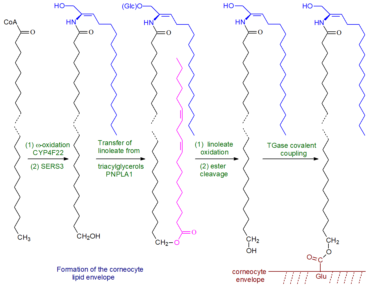
Figure \(\PageIndex{19}\): Formation of the corneocyte lipid envelope
Sphingomyelin and Related Sphingophospholipids
Structure and Occurrence of Sphingomyelin
Sphingomyelin, or ceramide 1-phosphocholine, consists of a ceramide unit with a phosphorylcholine moiety attached at position 1 of the sphingoid base. It is thus the sphingolipid analog of phosphatidylcholine and, like that lipid, is zwitterionic. The d18:1/16:0 molecular species is illustrated as an example in Figure \(\PageIndex{20}\).

Sphingomyelin is primarily of animal origin and is a ubiquitous component of all animal cell membranes, from mammals to nematodes (and in a few protozoa), where it is, by far, the most abundant sphingolipid. Indeed, it can comprise 50% or more of the lipids in certain tissues, though it is usually less abundant than phosphatidylcholine. For example, it makes up about 10% of the brain's lipids (especially in myelin) but 70% of the phospholipids of the human lens. Like phosphatidylcholine, sphingomyelin is most concentrated in the plasma membrane of cells (up to 20%), the endocytic recycling compartment, and the trans Golgi network. It is also abundant in the nucleus, where it is the main phospholipid associated with chromatin. However, it is present in very low amounts in the endoplasmic reticulum (2 to 4%) and even less in mitochondria. All the sphingomyelin in human erythrocyte membranes is in the outer leaflet, and ~90% of that in the plasma membrane of nucleated cells is in the outer leaflet. All lipoprotein fractions in plasma contain appreciable amounts of sphingomyelin, with a higher proportion in the VLDL/LDL. Sphingomyelin is the single most abundant lipid in the erythrocytes of most ruminant animals, where it replaces phosphatidylcholine entirely. In this instance, a highly active phospholipase A breaks down the glycerophospholipids but not sphingomyelin.
Sphingomyelin is not synthesized in plants or fungi, which produce the sphingophospholipid ceramide phosphoinositol and related lipids instead, or in bacteria, and its evolutionary significance remains speculative. However, many bacteria and viruses utilize sphingomyelin or its metabolism in their hosts for growth and viability.
Sphingosine is usually the most abundant long-chain base constituent (along with sphinganine and C20 homologs), although other bases can be present, especially in ruminant animals. In contrast, sphinganine is the major sphingoid base in the sphingomyelin of human lens membranes, linked mainly to 16:0. Typically, the fatty acids are very-long-chain saturated and monounsaturated, including odd-numbered components. Compared with glycosphingolipids, 2‑hydroxy acids are rarely detected and occur in small amounts. They are found in testes, spermatozoa, the kidney, and the skin sphingomyelin. The absolute proportions of each fatty acid and sphingoid base can vary markedly between tissues and species, and some of the variability in compositions can be seen from the data in Table \(\PageIndex{3}\) and Table \(\PageIndex{4}\) below.
| Table \(\PageIndex{3}\): Fatty acid compositions of sphingomyelin (wt % of the total) in some animal tissues. | ||||||||||
| Source | Fatty acids | |||||||||
|---|---|---|---|---|---|---|---|---|---|---|
| 16:0 | 18:0 | 18:1 | 20:0 | 22:0 | 22:1 | 23:0 | 23:1 | 24:0 | 24:1 | |
| Egg | 66 | 10 | 1 | 4 | 6 | 1 | 2 | - | 5 | 3 |
| Bovine brain | 3 | 42 | - | 6 | 7 | 3 | 3 | 3 | 6 | 27 |
| Cow's milk | 14 | 3 | 1 | 1 | 22 | - | 32 | - | 19 | 5 |
| Adapted from Ramstedt, B. et al. Analysis of natural and synthetic sphingomyelins using high-performance thin-layer chromatography. Eur. J. Biochem., 266, 997-1002 (1999); DOI. | ||||||||||
| Table \(\PageIndex{4}\): Long-chain base compositions of sphingomyelin (wt% of the total) in some animal tissues. | |||||||
| Source | Sphingoid base | ||||||
|---|---|---|---|---|---|---|---|
| d16:0* | d17:0 | d17:1 | d17:1-methyl | d18:0 | d18:1 | d19:0 | |
| Egg | 7 | 93 | |||||
| Bovine brain | 19 | 81 | |||||
| Cow's milk | 9 | 15 | 8 | 11 | 10 | 44 | 3 |
| Also from Ramstedt, B. et al. Eur. J. Biochem., 266, 997-1002 (1999); DOI. * d = dihydroxy base |
|||||||
Palmitic acid (16:0) is the most common fatty acid component of sphingomyelin in the peripheral cells of mammals. In contrast, stearic acid (18:0) is more abundant in neural tissue, but this only hints at the potential complexity, as variability can occur within tissues. For example, about 60% of the fatty acids in sphingomyelin in the grey matter of the human brain are stearic acid (18:0). In comparison, lignoceric (24:0) and nervonic (24:1) acids make up 60% of the corresponding lipids in white matter. However, this is dependent on the stage of development. During the first two years of life, the 18:0 concentration in white matter sphingomyelin decreases from 82% to 33%, while the proportions of 24:0 and 24:1 increase. This pronounced shift from long-chain to very-long-chain sphingomyelins does not apply to the cerebral cortex. Researchers have detected approximately 100 molecular species of sphingomyelin in human plasma. Although polyunsaturated fatty acids such as arachidonic acid are rarely present, the literature has sometimes mistakenly identified them. Exceptions include the sphingomyelins of testes and spermatozoa, which contain very-long-chain polyunsaturated fatty acids (up to 34 carbon atoms); the major components are 28:4(n-6) and 30:5(n-6), with some having hydroxyl groups at position 2.
Biosynthesis, Metabolism, and Function of Sphingomyelin
Sphingomyelin biosynthesis differs from that of phosphatidylcholine. It indeed depends upon it, as it involves the transfer of phosphorylcholine from phosphatidylcholine to ceramide, synthesized in the endoplasmic reticulum, with the liberation of 1,2-diacyl-sn-glycerols, as illustrated in Figure \(\PageIndex{21}\).

A ceramide choline phosphotransferase (sphingomyelin synthase, SMS) catalyzes the reaction. It occurs primarily on the luminal side of the trans-Golgi and also at the plasma membrane, catalyzed by two related enzymes, each with six transmembrane domains and N- and C-termini facing the cytosol: SMS1 and SMS2. Both enzymes are present in the Golgi, but only SMS2 is in the plasma membrane (facing the extracellular space in this instance) and may be necessary to form raft domains (see below). SMS2 is also present in rat liver nuclear membranes. Notably, in the absence of ceramide, both SMS1 and 2 have phospholipase C activity, which may regulate the steady-state levels of phosphatidylcholine, diacylglycerol, and sphingomyelin. The reaction does not use free phosphorylcholine or CDP-choline as a donor.
Figure \(\PageIndex{22}\) shows an interactive iCn3D model of the AlphaFold model of human Golgi membrane phosphatidylcholine:ceramide cholinephosphotransferase 1, also called sphingomyelin synthase 1 (Q86VZ5).

Figure \(\PageIndex{22}\): . Click the image for a popup or use this external link: https://structure.ncbi.nlm.nih.gov/i...v6s5wHRgPXKJC6
The gray helices are the transmembrane helices. A cytoplasmic protein:protein interaction domain called SAM (sterile alpha motif) is shown in magenta. The other cytoplasmic C-terminal domain is shown in cyan. Much of the cytoplasmic domain is disordered in this computational structure. Side chains involved in binding phosphatidylcholine are shown as CPK-colored sticks. Note that D95 and S97 are located in a disordered section in this model but would close in the active site in the actual structure.
It has been proposed that Asp101 (95 in the AlphaFold structure) deprotonates Arg220 (214 in the model), which then acts as a nucleophile that attacks the phosphate group of phosphatidylcholine. Given arginine's very high pKa, this mechanism, if true, is somewhat unique. Phosphocholine is linked to ceramide, which produces sphingomyelin. Three key, highly conserved amino acids in the active site are Asp101, Arg220, and Asn358.
A specific ceramide transport molecule (CERT) is important to the reaction with SMS1 because it transfers ceramide from the cytosolic surface of the endoplasmic reticulum to the trans-Golgi in an ATP-dependent, non-vesicular manner. Vesicular transport delivers much of the sphingomyelin produced in the Golgi to the apical plasma membrane. Sphingomyelin synthesis is partially regulated by phosphatidylinositol metabolism and is linked to sterol homeostasis through the oxysterol-binding protein (OSBP).
SMS2 in the plasma membrane is not dependent on CERT-mediated ceramide delivery but is believed to convert locally produced ceramide back to sphingomyelin; this may be an important protective mechanism for the cell. The location of the enzymes explains the enrichment of sphingomyelin in specific membranes and their sidedness (i.e., the luminal trans-Golgi and the outer leaflet of the plasma membrane), while ceramide reaching the cis-Golgi is used to synthesize glucosylceramide. Because sphingomyelin differs appreciably from its ceramide precursors, sphingomyelin synthases must have considerable substrate specificity. The reaction can be reversible, using sphingomyelin to generate ceramide for specific signaling functions. Sphingomyelin biosynthesis links the sphingolipid signaling pathway (pro-apoptotic - see below) and that of glycerolipids via the mitogenic diacylglycerol by‑products. Although the importance of this production relative to that via phosphatidylinositol is unknown, it may be significant locally at the external leaflet of the plasma membrane.
An alternative pathway of sphingomyelin synthesis has been demonstrated in the endoplasmic reticulum. In this pathway, ceramide is first converted to ceramide phosphoethanolamine via transfer of the head group from phosphatidylethanolamine, followed by stepwise methylation of the ethanolamine moiety. However, the physiological significance of this pathway remains unclear.
It was long thought that sphingomyelin served only as a substitute for phosphatidylcholine as a membrane building block, i.e., by forming a stable, chemically resistant outer leaflet of the plasma membrane lipid bilayer. For example, it may limit oxygen ingress and, hence, the oxidation of adjacent unsaturated acyl chains. While this is certainly one function, the apparent similarity between PC and SM is superficial, and they differ significantly in hydrogen-bonding capacity and physical properties. For example, sphingomyelin has an amide bond at position 2 and a hydroxyl at position 3 of the sphingoid base, both of which can participate in hydrogen bonding. At the same time, the trans double bond also appears to assist intermolecular interactions in membranes. Indeed, the first five carbon atoms of the sphingoid base in sphingolipids form a key feature termed the ‘sphingoid motif’, which facilitates many molecular interactions with other membrane lipids via hydrogen bonding, charge pairing, hydrophobic, and van der Waals forces. In contrast, the two ester carbonyl groups can act only as hydrogen acceptors with phosphatidylcholine. The degree of unsaturation of the alkyl moieties differs between lipids, giving them dissimilar membrane-packing properties.
Sphingomyelin and other sphingolipids now show a strong tendency to interact with proteins and cholesterol, often via strong van der Waals interactions and hydrogen bonding, forming transient nano-domains in membranes known as 'rafts' and on the surface of lipoprotein particles. Initially, it was thought that saturated sphingomyelin formed a liquid-ordered phase with cholesterol, or a gel phase with saturated ceramides, leading to lateral segregation within the membrane, and that sphingomyelin and cholesterol metabolism were closely integrated; some even suggested that sphingomyelin concentration might control cholesterol distribution in cells. Meanwhile, our understanding of raft formation in membranes has changed substantially in recent years. While cholesterol interactions are important, they may not be the major factor in vivo. Ceramide can displace cholesterol from sphingomyelin when it forms in membranes through sphingomyelin hydrolysis.
Other functions: Sphingomyelin per se is generally considered relatively inert, although modern molecular biology methods are uncovering potential regulatory functions through interactions with specific proteins. For example, it has been shown to inhibit phospholipase A2α, a key enzyme in eicosanoid production. Sphingomyelin in the plasma membrane may be essential for transferrin internalization and, in turn, iron uptake, and it appears to be required for the activity of many membrane-bound proteins, including certain ion channels and receptors. As the most abundant sphingolipid in the nucleus, it is intimately involved in chromatin assembly and dynamics and is an integral component of the nuclear matrix. A single molecular species of sphingomyelin with a C18 acyl chain binds specifically to a coat protein designated 'p24', enabling it to form membrane vesicles. In addition, sphingomyelin is selectively recognized and acts as a receptor for actinoporins, pore-forming toxins produced by sea anemones.
Sphingomyelin has a specific binding site on the amyloid beta peptide (Aβ) in the brain, and in vitro studies suggest this may promote aggregation of these proteins in Alzheimer's disease. In turn, this depletes brain sphingomyelin by activating acid sphingomyelinase, disrupting many protein–lipid interactions and downstream signaling pathways. In contrast, ganglioside GM1 may protect against Aβ aggregation.
Beyond its role in membranes, it serves as a precursor for ceramides, long-chain bases, sphingosine-1-phosphate, and many other biologically important sphingolipids as part of the 'sphingomyelin cycle' (also termed the ‘sphingolipid’ or ‘ceramide’ cycles depending on the context). Some metabolites are intracellular and intercellular messengers, while others are essential membrane constituents. The sphingomyelin cycle extends to other sphingolipids through sphingomyelinases and enzymes such as glycosylhydrolases and glycosyltransferases, producing innumerable new oligoglycosylceramides. It can also produce sn-1,2-diacylglycerols, which are central to many metabolic and signaling pathways. These molecular relationships are illustrated briefly in Figure \(\PageIndex{23}\).
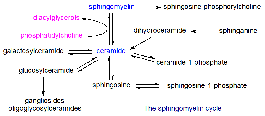
In particular, sphingomyelin is a major source of ceramides in most cellular organelles, including the nucleus and even mitochondria, via the action of sphingomyelinases (see next section). In addition to serving as a source of other sphingolipids, ceramides are required to trigger apoptosis and other metabolic changes. As ceramides do not mix well with glycerophospholipids and cholesterol, this conversion forms new membrane domains enriched in ceramide that exclude cholesterol and differ in composition from other sphingolipid rafts. This profoundly affects membrane function, especially that of the plasma membrane, as different proteins may be recruited or excluded depending on their relative affinities for cholesterol and ceramides. It may also influence disease states such as cancer.
Chlamydiae and other bacterial pathogens acquire sphingomyelin from their hosts' Golgi apparatus and plasma membrane, which is essential for their viability and growth. Other pathogenic bacteria, notably Pseudomonas aeruginosa and Neisseria gonorrhoeae, can hijack sphingomyelin-catabolic enzymes, with deleterious effects on the host. Likewise, human immunodeficiency virus (HIV) and the hepatitis C virus utilize host sphingomyelin for their nefarious purposes.
Nutrition: Although there is no known nutritional requirement for sphingomyelin or other sphingolipids, they are components of any diet that includes egg, meat, or dairy products. Thus, per capita sphingolipid consumption in the United States, for example, is estimated at approximately 0.3-0.4 g/d. Because sphingolipids constitute an appreciable proportion of the polar lipid constituents of milk, they may be significant if they are minor nutrients for infants and benefit their development. Animal experiments show that dietary sphingolipids can reduce intestinal cholesterol and lipid absorption, thereby lowering serum lipid concentrations. Feeding sphingolipids inhibits colon carcinogenesis and may alleviate some symptoms of inflammatory bowel disease. 2-Hydroxyoleic acid suppresses growth and induces autophagy in cancer cells by stimulating sphingomyelin synthesis and increasing its levels in the plasma membrane. On the other hand, plasma sphingomyelin levels are considered an independent risk factor for atherosclerosis, possibly because it retains cholesterol within cells and the arterial wall, thereby diminishing reverse cholesterol transfer via HDL.
Sphingomyelin Catabolism
Unlike glycerolipids, dietary sphingolipids are not hydrolyzed by pancreatic enzymes alone. Rather, most of the sphingomyelin in the diet is hydrolyzed in the brush border of the intestines by an alkaline sphingomyelinase (at a pH of 8.5–9 optimally) to ceramide and thence by a neutral ceramidase to free fatty acids and sphingosine. Some of this enzyme is also present in the liver, from which it is secreted into bile, which is then secreted into the intestinal lumen, where it can hydrolyze sphingomyelin and other phospholipids in the presence of bile salts. The sphingosine released at the brush border is absorbed; some is re-N-acylated to form ceramides, and the remainder is converted via sphingosine-1-phosphate to palmitic acid, which is esterified into the triacylglycerol component of chylomicrons. Some of these sphingolipid intermediates may have signaling functions and anti-inflammatory properties in intestinal cells. Alkaline sphingomyelinase is unusual because its structure and other properties differ significantly from those of intracellular enzymes with a related function; it belongs to the (ecto) nucleotide pyrophosphatase/phosphodiesterase (NPP) family, which includes autotaxin. The enzyme is believed to help produce sphingolipid metabolites in the intestines and colon, which may influence certain disease states. For example, it appears to inhibit colon cancer by increasing ceramide levels. In addition, alkaline sphingomyelinase has phospholipase C activity towards the pro-inflammatory metabolites platelet-activating factor and lysophosphatidylcholine, potentially conferring further beneficial effects. Reducing endogenous sphingomyelin levels and increasing ceramide levels in intestinal cell membranes may reduce dietary cholesterol uptake.
Catabolism in other tissues: The key enzymes for the degradation of sphingomyelin to ceramides in most tissues are also sphingomyelinases (phosphodiesterases), as shown in Figure \(\PageIndex{24}\).

These function similarly to phospholipase C, generating ceramides, with ceramide signaling as their main product. Many such enzymes have distinct pH optima and metal-ion requirements, operate in different cell regions, and may have distinct biochemical roles. Thus, acid sphingomyelinase is found in endo-lysosomes, while neutral sphingomyelinases are found in the plasma membrane, endoplasmic reticulum, Golgi, and mitochondria, and alkaline sphingomyelinase is found in the intestines. It should not be forgotten that the other reaction product is phosphocholine, which is an important nutrient. Bacterial sphingomyelinases are known to lyse red blood cells. Intriguingly, there is a sphingomyelinase in the bacterium Pseudomonas aeruginosa that can also act as a sphingomyelin synthase in vitro at least.
The lysosomal acid sphingomyelinase (pH optimum ca. 5) is expressed ubiquitously. It plays a key housekeeping role in maintaining normal membrane turnover and sphingolipid remodeling, especially in lipoproteins. While other lysosomal sphingolipid hydrolases require a saposin activator protein for full activity, the acid sphingomyelinase has an N-terminal saposin domain. It also has the unique lysosomal phospholipid bis(monoacylglycero)phosphate.
A related secreted acid sphingomyelinase (Zn2+-dependent) can be transported to the cell's outer membrane and is especially important in endothelial cells of the human coronary artery. The same gene encodes this enzyme, but it differs from the lysosomal enzyme by requiring Zn2+ ions for activation and having a different glycosylation pattern. It can also operate at neutral pH and has multiple functions, including involvement in many aspects of cellular signaling and membrane sphingomyelin turnover. At the plasma membrane, it produces ceramides and regulates the trafficking of palmitoylated proteins from the Golgi to their new location.
Neutral sphingomyelinases (pH optima 7.4), of which four distinct enzymes are known, are located in the membranes of the endoplasmic reticulum, Golgi, and plasma membrane, with one in mitochondria (MA-NSM), where they signal by generating ceramides and, in turn, other biologically active sphingolipids. Human NSM-1 has 423 amino acid residues and a molecular weight of 47.6 kDa; it has two putative transmembrane domains in the C-terminus and resides mainly in the nucleus and endoplasmic reticulum. It has broad specificity for choline phospholipids, but is most active with sphingomyelin and may not play a significant role in cellular signaling. In contrast, NSM-2, which is located in the Golgi apparatus and plasma membrane, is activated by phosphatidylserine and is important for ceramide signaling. It is especially important in the brain and nervous tissue, where it is required for the secretion of hypothalamic-releasing hormones. However, it is also involved in cellular functions and physiological processes in many other tissues. Dysregulation of NMS-2 has been reported as a factor in many inflammation-related pathologies. Neutral sphingomyelinase-3 is found mainly in the plasma membrane of bone and cartilage, where it is vital for mineralization; it is also important in striated and cardiac muscle. Little is known about the mitochondrial enzyme's function. Losses, mutations, and poor expression of the gene encoding neutral sphingomyelinase have been observed in several cancers. Still, exposure to ionizing irradiation led to rapid hydrolysis of sphingomyelin to ceramide by this enzyme, and thence to cancer cell death.
A diverse range of factors activate the enzymes, including chemotherapeutic agents, tumor necrosis factor-alpha, 1,25-dihydroxy-vitamin D3, endotoxin, gamma-interferon, interleukins, nerve growth factor, and most conditions known to induce cellular stress, especially in relation to inflammation. Because they use the most abundant sphingolipid in animal tissues to generate ceramides and other sphingolipid metabolites with important signaling functions, sphingomyelinases are believed to regulate signaling mechanisms, especially in the cell nucleus. Thus, they have a much broader metabolic role than simply catabolizing sphingomyelin.
The type A and B forms of Niemann-Pick disease are lysosomal lipid storage disorders that are a consequence of a deficiency of acid sphingomyelinase, with a resulting accumulation of sphingomyelin and smaller amounts of other sphingolipids, including gangliosides, in cells and tissues, especially in the monocyte/macrophage system, to form the so-called “foam cells” that characterize the disease. A resulting lack of ceramide production may contribute to disease pathology. Increasing sphingomyelin levels, in turn, elevate cholesterol concentrations. Membranes containing ceramides have a much lower capacity to bind cholesterol, so sphingomyelin degradation may play a role in cholesterol homeostasis. Type C Niemann-Pick disease differs from the A and B forms and is caused by defects in two distinct cholesterol-binding proteins (NPC1 and NPC2).
Glucosyl- and Galactosylceramides (Cerebrosides)
Two natural monoglycosylceramides are especially important in animals: glucosylceramide and galactosylceramide. Both have biological functions in their own right, especially as structural membrane components, as in the brain, where galactosylceramide is required to maintain the structure and stability of myelin and to support oligodendrocyte differentiation. Glucosylceramide is a vital component of all cell types and is most abundant in human skin; it is the key intermediate in the biosynthesis of lactosylceramide and, hence, complex oligoglycosphingolipids, including gangliosides. This monoglycosylceramide is also a major component of plant and fungal membranes. Although the two lipids have very similar structures in that D-galactose is an epimer of D-glucose and differ only in the configuration at C4, they have very different biological properties. A few other monoglycosylceramides are produced in nature, for example, by some bacteria of the order Sphingomonadales of α‑proteobacteria.
Structure and Occurrence
β-D-Galactosylceramide (Galβ1-1'Cer) is the principal glycosphingolipid in brain tissue; hence the trivial name "cerebroside", first conferred on it in 1874. Galactosylceramides are found in all nervous tissues, and indeed at low levels in all organs, but in the brain they can amount to 2% of the dry weight of grey matter and 12% of white matter or 23% of myelin lipids, where they insulate the axons of neuronal cells and constitute a substantial component of the extended plasma membrane of oligodendrocytes. It is also present in some fungal species. While galactosylceramide can be sulfated to form a sulfatide or sialylated to form ganglioside GM4, only a small proportion undergoes further galactosylation to form Gal2Cer, the precursor for the limited galactosyl-series of oligoglycosphingolipids.
β-D-Glucosylceramide (Glcβ1-1'Cer), with the trivial name "glucocerebroside", is a major constituent of skin lipids and is essential for maintaining the skin's water permeability barrier. It is also abundant in animal tissues such as the spleen and erythrocytes, and in nervous tissues, especially neurons, at low levels; it is also found in plants. Higher-than-normal concentrations of this glycosphingolipid have been reported in the apical plasma membrane domain of intestinal epithelial cells (especially the absorptive villous cells) and in the urinary bladder. The d18:1/16:0 molecular species of the two lipids are illustrated in Figure \(\PageIndex{25}\).

However, of equal or greater importance to the natural occurrence of glucosylceramide per se is its role as the biosynthetic precursor of lactosylceramide in animals and, hence, of most of the complex neutral oligoglycolipids and gangliosides. In contrast, glucosylceramide is the end-product of the biosynthetic pathway in plants and fungi.
Interestingly, the proportion of galactosylceramides relative to glucosylceramides in myelin glycolipids increases greatly in the ascending phylogenetic tree, and the ratio of hydroxy- to nonhydroxy fatty acids in cerebrosides increases with the complexity of the central nervous system. There is also an intriguing sex difference in the kidney: galactosylceramide, rather than glucosylceramide, is present only in male mice (or in androgen-treated adult females). Only glucosylceramide is present in the nerves of the most primitive animals (protostomes).
In the brain, the galactosylceramides are enriched in very-long-chain fatty acids (C22–C26). The fatty acid and long-chain base compositions of cerebrosides from the intestines of the Japanese quail are listed in Table \(\PageIndex{5}\) for illustrative purposes. The fatty acid components resemble those of other sphingolipids, although the percentage of 2-hydroxy acids is higher than that in sphingomyelin. In this case, they are exclusively saturated, though a small proportion of monoenoic components may also be found in other tissues. Glucosylceramides tend to contain mainly non-hydroxylated fatty acids that are of relatively shorter chain length. The proportion of trihydroxy bases listed is perhaps higher than in many other tissues or species studied, probably reflecting the diet. Usually, sphingosine is the main long-chain base in cerebrosides of animal tissues.
| Table \(\PageIndex{5}\): Composition of fatty acids and long-chain bases (wt % of the total) in cerebrosides of intestines from the Japanese quail.* | ||||
| Long‑chain bases | Fatty acids | Non-hydroxy acids |
2-Hydroxy acids |
|
|---|---|---|---|---|
| Type | % | % | % | |
| t18:0 | 43 | 16:0 | 5 | 6 |
| d18:0 | 9 | 18:0 | 3 | trace |
| d18:1 | 27 | 20:0 | 2 | 4 |
| t20:0 | 6 | 21:0 | trace | 2 |
| d20:0 | 3 | 22:0 | 4 | 43 |
| d20:1 | 11 | 23:0 | 1 | 13 |
| 24:0 | 3 | 12 | ||
| * The cerebrosides comprised 81% galactosylceramide and 19% glucosylceramide. From Hirabayashi, Y. et al., Lipids, 21, 710-714 (1986); DOI. |
||||
Plants: Glucosylceramide is the only glycosphingolipid common to plants, fungi, and animals. It has often been described incorrectly as the main sphingolipid in plants. However, this has been because the more polar complex glycosylinositol phosphoceramides are not easily extracted and, until relatively recently, were missed in conventional analyses. Nonetheless, glucosylceramide is abundant in photosynthetic tissues and constitutes approximately a third of the total sphingolipids, where the main long-chain bases are C18 4,8‑diunsaturated (Z/Z and E/Z) (not sphingosine as illustrated above); it is a major component of the outer layer of the plasma membrane and is also enriched in the late endosomes and plant tonoplast. Small amounts of monoglycosylceramides containing a β‑D‑mannopyranosyl unit may be present in non-photosynthetic tissues, but plants have not been found to contain galactosylceramides. Glucosylceramide is a common lipid component of yeast and other fungi, including most fungal pathogens. However, it does not occur in the yeast Saccharomyces cerevisiae, a widely used experimental model, although trace levels of galactosylceramide have been detected.
The fatty acid and long-chain base compositions of glucosylceramides from two plant sources are listed in Table \(\PageIndex{6}\). Perhaps surprisingly, the fatty acid components are not very different from those in animal tissues, comprising mainly longer-chain saturated and monoenoic acids, with a high proportion being saturated and having a hydroxyl group in position 2. In the examples selected for the table, di- and tri-hydroxy long-chain bases were found, mainly diunsaturated (Z/Z and E/Z) and almost entirely C18 in chain length. Pollen contains much higher concentrations of glucosylceramides than leaves, with substantial compositional differences. For example, the long-chain bases in Arabidopsis leaves consist mainly of t18:1, with relatively little d18:1, t18:0, and d18:0 (with 16:0, 24:0, and 24:1 hydroxy fatty acids mainly), but no d18:2 base. However, this is 50% of those in pollen. While saturated 2-hydroxy acids predominate in most plants, some cereal glucosylceramides contain high proportions of monounsaturated, very-long-chain fatty acids of the n-9 family. Glucosylceramides from algae tend to resemble those from higher plants, although microalgae have also been reported to contain novel structures.
| Table \(\PageIndex{6}\): Composition of fatty acids and long-chain bases (wt % of the total) in glucosylceramides of seeds from scarlet runner beans and kidney beans. | |||||
| Fatty acidsa | Long-chain basesb | ||||
|---|---|---|---|---|---|
| Type | Runner beans | Kidney beans | Runner beans | Kidney beans | |
| % | % | % | % | ||
| 16:0 | 4 | 5 | t18:0 | trace | trace |
| Other non-hydroxy |
1 | 2 | t18:1-8t | 13 | 11 |
| 14:0-OH | 1 | 1 | t18:1-8c | 10 | 9 |
| 15:0-OH | 1 | 1 | d18:0 | trace | trace |
| 16:0-OH | 58 | 58 | d18:1-8c/t | 1 | 3 |
| 18:0-OH | trace | trace | d18:1-4t | trace | trace |
| 20:0-OH | trace | trace | d18:2-4t,8t | 45 | 60 |
| 22:0-OH | 7 | 6 | d18:2-4t,8c | 31 | 17 |
| 23:0-OH | 2 | 1 | |||
| 24:0-OH | 23 | 23 | |||
| 25:0-OH | 1 | 1 | |||
| 26:0-OH | 1 | 1 | |||
| From Kojima, M. et al., J. Agric. Food. Chem., 39, 1709-1714 (1991); DOI, but see also Yamashita, S. et al. for further data: DOI a including 2-hydroxy acids; b di- and tri-hydroxy bases with cis or trans double bonds in the positions indicated. |
|||||
Biosynthesis
Ceramides synthesized both de novo and by sphingomyelin catabolism are used to synthesize monoglycosylceramides in animal tissues. The biosynthetic mechanism resembles that of glycosyldiacylglycerols: the carbohydrate moiety is directly transferred from a sugar-nucleotide (e.g., uridine 5-diphosphate(UDP)-galactose, UDP-glucose, etc.) to a ceramide unit synthesized in the endoplasmic reticulum. This is illustrated in Figure \(\PageIndex{26}\).

During transfer, specific glycosyltransferases invert the glycosidic bond from alpha to beta. Synthesis of β-D-galactosylceramide takes place on the luminal surface of the endoplasmic reticulum. However, it can access the cytosolic surface via an energy-independent flip-flop process. Expression of UDP-galactose:ceramide galactosyltransferase (galactosylceramide synthase) is restricted to oligodendrocytes, Schwann cells, kidneys, and testes. Before sulfation, galactosylceramide is transported to the trans-Golgi compartment.
In contrast, after CERT transfers the precursor ceramides from the endoplasmic reticulum to the cytosolic side of early Golgi membranes, glucosylceramide is produced by a glucosylceramide synthase present in this membrane (with the possible exception of neuronal tissues). It is then converted to more complex oligoglycosylceramides. This must be translocated to the luminal leaflet of the trans-Golgi network via both vesicular and non-vesicular transport. The latter is mediated by a conserved clade of integral membrane proteins, i.e., phospholipid flippases (P4-ATPases) designated ATP10A and ATP10D, together with the four phosphate adapter protein-2 (FAPP2) and glycolipid transfer protein (GLTP) in humans, with related enzymes in fungi, which utilize the energy from ATP catalysis to translocate lipids across cellular membranes. Human enzymes are entirely specific for glucosylceramide, not galactosylceramide. Indeed, the galactosyl- and glycosylceramide synthases have no significant sequence homology, indicating different evolutionary origins.
Both galactosyl- and glucosylceramide must cross the plasma membrane to function in protein interactions and signaling. Some glucosylceramide is carried by lipoproteins (VLDL, LDL, and HDL) in circulation and presumably requires active transport for absorption and distribution across target-tissue membranes.
In plants, glucosylceramides are also formed by an evolutionarily conserved glucosylceramide synthase involving UDP-glucose in the endoplasmic reticulum. However, an alternative mechanism has been described that uses sterol glucoside as the immediate glucose donor to ceramide. Evidence also suggests that, in some plant and fungal tissues, glucosylceramide synthesis requires ceramides containing Δ4 trans-double bonds, but not other sphingolipids. However, there is a distinct ceramide synthase in the yeast Pichia pastoris, which produces ceramides of defined composition exclusively for the production of glucosylceramides. A separate ceramide synthase with different specificities produces the ceramide precursors for phosphorylinositol, which only contains phytosphingosine as the long-chain base. In fungi, researchers have characterized glucosylceramide synthases, but have not yet identified a galactosylceramide synthase. Researchers have characterized enzymes responsible for glucuronosylceramide and α-galactosylceramide biosynthesis in some bacterial species.
Function
Galactosylceramides: A remarkable property of cerebrosides is that their melting point is well above physiological body temperature, so glycolipids have a paracrystalline structure at this temperature. Each cerebroside molecule may form up to eight inter- or intramolecular hydrogen bonds by lateral interaction between the polar hydrogens of the sugar and the hydroxyl and amide groups of the sphingosine base of the ceramide moiety, and this dense network of hydrogen bonds is believed to contribute to the high transition temperature and the compact alignment of cerebrosides in membranes. As with sphingomyelin, monoglycosylceramides tend to concentrate in the outer leaflet of the plasma membrane with cholesterol, and thus in myelin within specific membrane domains termed rafts. Indeed, the latter appear to segregate more strongly than sphingomyelin via hydrogen bonds and hydrophobic interactions, and these forces also help bind a wide range of proteins, including enzymes and receptors, found in raft domains.
Galactosylceramide is essential to myelin structure and function and is involved in oligodendrocyte differentiation. While molecular species with 2’-hydroxy fatty acid constituents are not essential for myelin formation, they are critical for long-term myelin stability, presumably because increased hydrogen bonding with neighboring lipids in membranes stabilizes the phase structure. Galactosylceramide is important as a precursor of 3’-sulfo-galactosylceramide, which is also essential to brain development and numerous functions in other tissues. By interacting with sulfatide in the membranes of opposing layers in the myelin sheath via carbohydrate-carbohydrate interactions, it forms a glycosynapse that contributes to myelin's long-term stability.
Glucosylceramides: Glucosylceramides have similar physical properties in membranes to the galactose analog, and they are also concentrated in raft domains in the outer leaflet of the plasma membrane. As mentioned briefly above, they are the primary precursor for most complex oligoglycosphingolipids in animal tissues, especially in the brain, where synthesis is vital for producing most neuronal oligoglycosphingolipids. Glucosylceramide, per se, is essential for axonal growth. They are major constituents of skin lipids, essential for lamellar body formation in the stratum corneum and for maintaining the skin's water-permeability barrier. In addition, epidermal glucosylceramides and sphingomyelin are sources of the unusual complex ceramides found in the stratum corneum, including those with terminal hydroxyl groups and estolide-linked fatty acids. Some glucosylceramides in the skin are covalently linked to proteins via their terminal hydroxyl groups, presumably to strengthen the epidermal barrier.
Much of the evidence for the function of glycosylceramides in animals comes from cell lines in which lipid synthesis has been suppressed in vitro by various means. Glucosylceramide does not appear to be essential for the viability of certain cell lines in culture. However, disruption of the global synthase gene in mice results in embryonic death. It is essential for cancer cell survival, and deletion in other cell types can lead to abnormalities. In addition to serving as an intermediate in the biosynthesis of more complex glycosphingolipids and playing a role in the skin's permeability barrier (discussed above), glucosylceramide is believed to be required for intracellular membrane transport, cell proliferation and survival, and various immune functions. In contrast, evidence suggests it may have adverse implications for various disease states. For example, the overexpression of glucosylceramide synthase in cancer cells has been linked to tumor progression, leading to reduced ceramide levels and increased resistance to chemotherapy. The lipid has also been associated with drug resistance in a wider context. In the nematode Caenorhabditis elegans, glucosylceramide containing the fatty acid 22:0 is reported to be a longevity metabolite that functions through clathrin membrane localization, a protein that regulates membrane budding.
In Arabidopsis, glucosylceramides are critical for cell differentiation and organogenesis but not necessarily for cell viability. Glycosphingolipids may impart positive membrane curvature, facilitating vesicle fusion. There is evidence that glycosylceramides (but not glycosyldiacylglycerols), together with sterols, are located in rafts in plant membranes in an analogous manner to sphingolipids in animal tissues and that they are associated with specific proteins. Correlative studies suggest that glucosylceramides help the plant plasma membrane withstand cold and drought stress. For example, glycosylceramides containing 2-hydroxy monounsaturated very-long-chain fatty acids and long-chain bases with 4-cis double bonds appear to be present at higher concentrations in plants tolerant of chilling and freezing. While fungal glucosylceramides with a 9-methyl group within the sphingosine backbone elicit defense responses in rice, cerebrosides with double bonds in positions 4 and/or 8 of the long-chain base appear to be involved in the defense of some plant species against fungal attack.
Less is known about the function of glucosylceramides in fungi, although they are major constituents of the plasma membrane and cell wall. They are believed to be involved in cell wall assembly, cell division and differentiation, and signaling. The methyl branch on the long-chain base is essential for cell division and alkali tolerance. In fungal pathogens, the host immune system recognizes glucosylceramides, which regulate virulence, often after export into the external environment as extracellular vesicles. In contrast to animals, ceramide monohexosides are not precursors for oligoglycosylceramides in fungi. Some plant molecular species of this lipid (a Δ8 double bond in the long-chain base is essential) show fruiting-inducing activity in the fungus Schizophyllum commune.
α-D-Galactosylceramides: Cerebrosides linked to an α-D- rather than a β-D-galactosyl unit, such as that found in the marine sponge Agelas mauritianus, in human gut microflora, and even in cow's milk, are potent stimulators of mammalian immune systems by binding to the protein CD1d on the surface of antigen-presenting cells and activating invariant natural killer T cells. Indeed, this was one of the first pieces of evidence showing that glycolipids, like glycoproteins, could elicit an immune response. Subsequently, it was shown that α-galactosylceramide with a 24:1 fatty acid, though present in very small amounts, is loaded onto the CD1d or CD40 protein and presented as the natural endogenous ligand for NKT cells in the thymus and periphery. Once activated, NKT cells secrete a range of pro- and anti-inflammatory cytokines to modulate innate and adaptive immune responses. The α‑glucosyl and α‑psychosine analogs show similar activity.
It is unclear whether animal tissues synthesize α-galactosylceramide, and it is likely derived primarily from members of the gut microbiome, such as Bacteroides fragilis and related species (although, in general, few bacterial species produce sphingolipids). Researchers have identified ceramide-galactosyltransferases that synthesize this lipid in two intestinal microbiome bacteria. In the mouse gut, the main molecular form consisted of a 2‑(R)‑hydroxylated hexadecanoyl chain linked to C18-sphinganine, while in B. fragilis it contained longer-chain components with iso-methyl branches in the sphingoid base and often fatty acid moieties. The sphinganine chain branching is a critical determinant of NKT cell activation by the bacterial enzyme. A decrease in the production of this lipid was observed in mice exposed to stress conditions that alter the gut microbiota, including a Western-type diet, colitis, and influenza A virus infection, with potential consequences for systemic immune responses. Catabolic enzymes control its concentration in animal tissues through a two-step mechanism: removal of the acyl chain by an acid ceramidase and hydrolysis of the sugar residue by an α-glycosidase. Initial studies with animal models suggest that treatment with α‑D‑galactosylceramides is effective against lung and colorectal cancers, melanomas, and leukemia. Preclinical trials of this lipid and its synthetic analogs have shown that they are safe and effective as anti-tumor immunotherapeutic agents and vaccine adjuvants. Indeed, a phase I trial in patients with high-risk melanoma has given promising preliminary results.
Catabolism of Glycosphingolipids
In animal tissues, lysosomes are the main sites for degrading all glycosphingolipids, including monoglycosylceramides, oligoglycosphingolipids, and gangliosides. These membrane-bound organelles have a limiting external membrane and internal lysosomal vesicles that contain soluble digestive enzymes active at the organelle's acidic pH. Lysosomes transport all membrane components and break them down into their primary components. For glycosphingolipids, this means fatty acids, sphingoid bases, and monosaccharides, which can be recovered for reuse or further degraded. Thus, sections of the plasma membrane are endocytosed and transported through the endosomal compartment to the lysosomes. The compositional and physical arrangement of lysosomal membranes makes them resistant to digestion, with bis(monoacylglycero)phosphate (lysobisphosphatidic acid) as a characteristic component of the inner membrane. A glycocalyx of highly N-glycosylated integral membrane proteins protects the perimeter membrane, aided by the ganglioside GM3, which resists degradation. This glycocalyx forms an efficient hydrophilic barrier at the luminal surface of the lysosomal perimeter membrane, protecting it from protease and hydrolase degradation and preventing lipids and their hydrolysis products from escaping from the lysosomal lumen.
Oligoglycosylceramides and gangliosides are degraded by sequential removal of monosaccharide units by specific exohydrolases from the non-reducing end until a monoglycosylceramide unit is reached, when glucosylceramide β-glucosidases or an analogous β-galactosidase (one isoform) removes the final carbohydrate moiety. Several glucosylceramidases are known; GBA1 is a lysosomal hydrolase, GBA2 is a ubiquitous non-lysosomal enzyme, and GBA3 is a cytosolic β-glucosidase. The last is found in the kidney, liver, spleen, and a few other mammalian tissues, but its function is unclear.
Because glycolipids with fewer than four carbohydrate residues are embedded in intralysosomal membranes, while the degradative enzymes are soluble, the process requires negatively charged lipids and specific activator proteins, which are water-soluble, low-molecular-weight glycoproteins. These are not catalytically active themselves but act as cofactors, either by directing the enzyme to the substrate or by activating it through binding. Five such proteins are known: the GM2-activator protein (specific for gangliosides) and the Sphingolipid Activator Proteins, or saposins A, B, C, and D, which perturb membranes enough to let degradative enzymes reach glycolipid substrates. The four saposins are derived by proteolytic processing from a single precursor protein, prosaposin, which is synthesized in the endoplasmic reticulum, transported to the Golgi for glycosylation, and then to the lysosomes. Of these, saposin C is essential for degrading galactosyl- and glucosylceramide, while saposin B is required for hydrolyzing sulfatide, globotriaosylceramide, and digalactosylceramide. The hydrolysis products of monoglycosylceramides are ceramides and monosaccharides, with net retention of the latter's stereochemistry. This is illustrated in Figure \(\PageIndex{26}\).
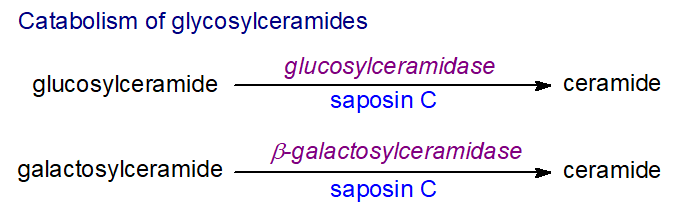
Anionic lipids such as bis(monoacylglycero)phosphate aid the reactions. In particular, this increases the GM2-activator's ability to solubilize lipids and stimulates the hydrolysis of membrane-bound GM1, GM2, and some kidney sulfatides. Saposin D stimulates lysosomal ceramide degradation by acid ceramidase and also helps solubilize negatively charged lipids at an appropriate pH. Eventually, acid ceramidase hydrolyzes ceramides into fatty acids and sphingoid bases.
β-Glucosylceramidase and saposin C are also required to generate structural ceramides from glucosylceramide in the outer skin layers, a process essential for optimal skin barrier function and survival. Some glucosylceramide is hydrolyzed by the enzyme GBA2 at the plasma membrane, where the resulting ceramide is rapidly converted to sphingomyelin by sphingomyelinase 2, which may be co-localized with the glucosidase. In addition, cellular β-glucosidases can transfer the glucose moiety between glucosylceramide and other lipids, as in the formation of cholesterol glucoside.
Small but significant amounts of glucosyl- and galactosylceramides are ingested in the human diet. Pancreatic enzymes do not hydrolyze them, but the intestinal brush border degrades them with lactase-phlorizin hydrolase (which also hydrolyses lactose in milk) to ceramides and thence to sphingosine.
An Arabidopsis homolog of human glucosylceramidase (AtGCD3) preferentially hydrolyses glucosylceramides that contain long acyl chains, and three further isoforms may exist based on sequence homology.
Genetic Disorders and Disease
Harmful quantities of glucosylceramide accumulate in the spleen, liver, lungs, bone marrow, and, in rare cases, the brain of patients with Gaucher disease, the most common inherited metabolic disorder (autosomal recessive) involving storage of excessive amounts of complex sphingolipids. Three clinical forms (phenotypes) are commonly recognized, and the most dangerous involve the brain (Types 2 and 3). All patients have a deficiency in lysosomal glucosylceramide-β-glucosidase (GBA1), which catalyzes the first step in glucosylceramide catabolism. The enzyme may be present, but a mutation prevents it from forming its correct conformation. However, other factors may be involved, as patients with defective saposin C, the lysosomal activator protein, develop similar symptoms.
In the brain, glucosylceramide accumulates when complex lipids turn over during brain development and myelin sheath formation. The excess glucosylceramide arises mainly from the biodegradation of old red and white blood cells. As a result, glucosylceramide remains stored within the lysosomes of macrophages, i.e., specialized cells that remove worn-out cells by degrading them into simple molecules for recycling, thereby preventing them from functioning normally and often leading to chronic inflammation. The enlarged macrophages containing undigested glucosylceramide are termed Gaucher cells. They overexpress and secrete certain proteins into the circulation, and some of these are used as biomarkers. In addition, these cells convert glucosylceramide to gangliosides more rapidly, increasing plasma and spleen ganglioside GM3 levels in patients with Gaucher disease. Fortunately, effective enzyme replacement therapies now exist for patients with the milder (non-neurological or Type 1) form of Gaucher disease that successfully reverse most manifestations of the disorder, including decreasing liver and spleen size and reducing skeletal abnormalities. Two oral drugs that inhibit glucosylceramide synthesis have also been approved.
Defective GBA1 enzyme activity in humans has been implicated in an increased risk of multiple myeloma and other cancers. Oligoglycosylceramides and gangliosides, in particular, are involved in the pathology of many cancers, and glucosylceramide is an important precursor of these. Inhibition of glucosylceramide synthase, which is overexpressed in many human tumors, results in a marked arrest of cell growth in vitro. This may have the potential to treat colorectal and other cancers.
A deficiency in glucocerebrosidase activity may predispose individuals to more common disorders such as Parkinson's disease and Lewy body dementia. Excess glucosylceramide production and the formation of more complex glycosphingolipids are factors in polycystic kidney disease. Accumulating lysosomal substrates in all lysosomal storage disorders appears to impair lysosomal integrity and cause broader disruptions to lipid metabolism, membrane structure, and function. In contrast, inhibiting glucosylceramidases may benefit cystic fibrosis. The next section discusses Krabbe disease.
Galactosylceramide is believed to function as an initial receptor for the human immunodeficiency virus (HIV) in mucosal epithelial cells. It controls the early infection-independent phase of HIV transfer to T cells. Glucosylceramide levels regulate the uptake of viruses that rely upon the late endosomal compartment for fusion, including the influenza A and Ebola viruses.
Gangliosides
The German scientist Ernst Klenk first applied the name ganglioside in 1942 to a mixture of complex glycosphingolipids newly isolated from ganglion cells of the brain. Subsequently, he demonstrated that, as part of an oligosaccharide chain, they contained an acidic carbohydrate component, which he named "neuraminic acid" - later termed "sialic acid" from the Greek "sialon" for saliva, from which they were first isolated. However, researchers did not fully characterize the first ganglioside species until 1963. Innumerable sphingolipids are now known that differ in both the glycan (glucose, galactose, N-acetylgalactosamine, and sialic acid residues) and ceramide structures. They occur throughout the animal kingdom, from echinoderms to higher animals, but not in plants. These highly polar, acidic, and relatively hydrophilic molecules have distinctive physical properties essential to the vital functions of gangliosides in the membranes of the central nervous system and other tissues.
Sialic acids and Gangliosides
Sialic acids: Gangliosides are oligoglycosylceramides derived as a first step from lactosylceramide, and they are defined by the presence of one to as many as five sialic acid residues, i.e., carbohydrate molecules with a nine-carbon backbone and a carboxylic acid group, a subclass of the superfamily of naturally occurring non‑2‑ulosonic acids. Of the many forms that have been characterized, only a few are relevant to gangliosides, and the most important of these is N-acetylneuraminic acid (‘NANA’ or ‘SA’ or 'Neu5Ac' or 'NeuAc'). Less often, the sialic acid component is N-glycolylneuraminic acid (Neu5Gc), which differs by only one oxygen atom at the C-5 N-acetyl group, or it can be a Neu5Ac analog in which the amide group is replaced by a hydroxyl group, i.e., 3-deoxy-D-glycero-D-galacto-nonulosonic acid (ketodeoxynonulosonic acid or ‘KDN’). The sialic acids are joined via α-glycosidic linkages to one or more monosaccharide units, e.g., via the hydroxyl group on position 2 or to another sialic acid residue. The polar head groups of lipids carry a net negative charge at pH 7.0 and are acidic. Their structures are shown in Figure \(\PageIndex{27}\).

Humans lack Neu5Gc: Neu5Ac is the biosynthetic precursor of Neu5Gc, a component of gangliosides in most animal species, including mice, horses, sheep, and goats, via the enzyme CMP-N-acetylneuraminic acid hydroxylase (CMAH). However, NeuGc is not synthesized in humans (or in birds and New World monkeys), although it is present in other primates such as the great apes. Because it is a xenogeneic antigen, healthy humans normally produce anti-NeuGc antibodies (especially after injection of NeuGc-containing glycoconjugates). The absence or irreversible inactivation of many relevant genes, but especially a critical exon in the CMAH gene, both for sialolipids and peptides in humans, suggests that this may have been a major biochemical branch-point in human evolution that occurred ~2 to 3 million years ago after the divergence of humans and chimpanzees from a common ancestor. It may also contribute to the superior performance of the human brain, as overexpression of Neu5Gc in transgenic mouse brains has been shown to cause abnormal development. It could also mean there was a fertility barrier between us and other hominids during evolution.
While these are speculations, some evidence suggests that the loss of Neu5Gc in humans had complex effects on immunity, providing greater ability to clear sublethal bacterial challenges. Some NeuGc may be obtained from the diet, for example, in meat and milk, and may be incorporated into human gangliosides to a limited extent, especially in fetal tissues and some cancers. In the latter, the preferential expression of dietary Neu5Gc has been ascribed to their higher metabolic rate.
2. Structure and Occurrence of Gangliosides
Most common gangliosides are derived from the ganglio- and neolacto-series of neutral oligoglycosphingolipids (Table 1) and should be named systematically in the same way, with the position of the sialic acid residue(s) indicated as for branched structures. However, they are more conveniently defined by a short-hand nomenclature system proposed by Svennerholm in which M, D, T, and Q refer to mono-, di-, tri, and tetrasialogangliosides, respectively, and the numbers 1, 2, 3, etc refer to the order of migration of the gangliosides on thin-layer chromatography. For example, the order of migration of monosialogangliosides is GM3 > GM2 > GM1 (sometimes defined by subscripts, e.g., GM1 or GM1). Further terms indicate variations within the basic structures, e.g., GM1a, GD1b, etc. Although more systematic alternatives have been proposed, IUPAC-IUB has approved the Svennerholm nomenclature. Ganglio-series glycosphingolipids having 0, 1, 2, and 3 sialic acid residues linked to the inner galactose unit are termed asialo- (or 0-), a-, b-, and c-series gangliosides, respectively, while gangliosides having sialic acid residues linked to the inner N-galactosamine residue are classified as α-series gangliosides. The structures for these groups are illustrated in the section on ganglioside biosynthesis below for practical convenience.
As of 2020, more than 200 gangliosides with variations in the carbohydrate chain had been characterized in vertebrates alone. One of the most studied monosialo-gangliosides and the first to be fully characterized is ganglioside GM1a (Neu5Acα2-3(Galβ1-3GalNAcβ1-4)Galβ1-4Glcβ1Cer), a major brain ganglioside of mammals and the preferred ligand of cholera toxin, illustrated in Figure \(\PageIndex{28}\).

It can also be depicted using the abbreviated structure in Figure \(\PageIndex{29}\).
IUPAC-IUB recommends a less-used alternative nomenclature based upon the ganglion (Gg) root structure. This nomenclature uses Roman numerals to designate each hexose unit and the location of Neu5Ac along the carbohydrate chain, with Arabic superscripts to designate the hydroxyl group to which it is linked. By this system, GM1a is defined as II3-α-Neu5Ac-Gg4Cer.
Brain gangliosides: Gangliosides can account for 6% of brain lipid weight (20 to 500 times more than in other tissues), and they constitute 10 to 12% of the total lipid content (20-25% of the outer layer) of neuronal membranes, for example. They are also synthesized and present at low levels (1 to 2% of total lipids) in all animal tissues, where, like neutral oligoglycosphingolipids, they concentrate in the outer leaflet of the plasma membrane in nanodomains known as 'rafts' or related structures. Mammalian neurons actively synthesize gangliosides of the ganglio-series. Still, oligodendrocytes in the brain produce myelin-forming glycosphingolipids, such as galactosylceramide and sulfatide, together with a minor amount of ganglioside GM4.
The brain contains 20 to 500 times more gangliosides than most non-neural tissues, with three times as much gray matter as white matter. As the brain develops, ganglioside content and sialylation increase. Species and tissue show large differences. For example, during embryogenesis and the postnatal period in the human central nervous system, the total amount of gangliosides increases approximately threefold, while that of GM1 and GD1a increases 12 to 15-fold. During the same period, the hemato-series gangliosides GM3, GD3, and 9-OAc-GD3, which lack a hexosamine residue, predominate. Still, they are present in much lower amounts in adults and only in some areas of the brain. In the mouse brain, total ganglioside levels are almost 8-fold higher in adults than in embryos, with a similar shift in composition from simple (GM3 and GD3) to more complex gangliosides. Ganglioside changes during brain maturation correlate with many neurodevelopmental milestones, and gangliosides play a crucial role in neuronal function and brain development, especially during infancy, when nutrient demand is high as the brain undergoes rapid restructuring.
The main gangliosides (~95%) of the adult mammalian brain are the ganglio series GM1, GD1a, GD1b, and GQ1b, while lactosyl series gangliosides such as GM3 (sialyllactosylceramide) are found mainly in the extra-neural tissues. The remaining ~5% consists of minor components in the brain, including gangliosides GM4, GM3, GD3, GM2, GD2, Fuc-GM1, Fuc-GD1b, GT1a, and GP1c, with proportions varying across species. In contrast, modern mass spectrometric methodology (electrospray ionization ion mobility MS) has revealed a much higher degree of sialylation than previously recognized, including a complete series of mono- to octasialylated gangliosides in the fetal frontal lobe. This methodology later identified many previously unknown acetylated gangliosides in the fetal hippocampus. Ganglioside content and composition in the brain also change with aging, with a substantial decrease in lipid-bound sialic acid and an increase in the proportion of more complex carbohydrate forms in the elderly.
Gangliosides in other tissues and species: Among extraneural tissues, lactosyl series gangliosides such as GM3 (sialyllactosylceramide) and monosialogangliosides generally predominate. Erythrocytes, bone marrow, testis, spleen, and liver contain relatively high concentrations of ganglioside GD1a. At the same time, GM4 is more abundant in the kidney, GM2 in the bone marrow, GM1 in erythrocytes, and GM3 in the intestine. In mouse germ cells, gangliosides switch from the a- to the 0-series upon differentiation as they cross the blood-testis barrier. Skin fibroblasts and many cells of visceral organs mainly generate globo-series gangliosides. Similarly, the globo- and lacto-series gangliosides are characteristic components of the stage-specific embryonic antigens (SSEA), which underlie the development and differentiation of human embryonic stem cells. Sialyl-lactotetraosylceramide is present in the latter and in the brains of children under two, but not in adult human tissues. Gangliosides can cross the placental barrier into the fetus, and those in milk, derived from the apical plasma membrane of secretory cells in the mammary gland, may be nutritionally important for the newborn. GD3 is the main ganglioside in human breast milk early in lactation, whereas GM3 is more abundant in later stages (and in bovine milk). Unfortunately, gangliosides are poorly characterized and quantified in foods in general.
A 5-N-deacetylated form of ganglioside GM3 has been detected in human melanoma tumors. In addition, O-acetylation or lactonization of the sialic acid residue can increase potential complexity. Gangliosides containing O-acetylated sialic acids, such as 9-OAc-GD3, are expressed during embryonic development and in the retina and cerebellum of adult rats but not in other brain regions. They also occur in certain tumors and may protect them from apoptosis. Such gangliosides may even be more widespread, but they are missed during isolation after treatment with mild alkali, a common analytical practice. Another complexity is the presence of gangliosides with sulfate groups, which have been isolated from human, mouse, and monkey kidney cells. KDN-containing gangliosides are minor components of fish egg, ovarian fluid, sperm, and testis, as well as some mammalian tissues.
Gangliosides from marine invertebrates (echinoderms), such as starfish and sea cucumbers, differ greatly in structure from those in vertebrates and lack a shorthand nomenclature. They include forms with distinctive ceramide compositions, atypical carbohydrate residues, sialic acids within the oligosaccharide chain, or glycosyl inositol-phosphoceramide structures. The mollusk Aplysia kurodai lacks gangliosides but produces complex oligoglycosylceramides with attached 2-aminoethylphosphonic acids and/or phosphoethanolamine groups that may serve as ganglioside surrogates.
Ceramide structures: In general, ganglioside ceramide structures are relatively simple. Sphingosine is usually the main sphingoid base, with the C20 analog present in central nervous system gangliosides. Stearic acid (18:0) can be 80 to 90% of the fatty acid constituents in the brain, accompanied by small amounts of 16:0, 20:0, and 22:0, with little or no polyunsaturated or 2-hydroxy acids, except in some exceptional circumstances (e.g., some carcinomas). Palmitic acid is more abundant in the intestines and liver gangliosides, while 2-hydroxylated fatty acids are relatively abundant in the latter and the kidney. The composition of the base and fatty acid components also differs across cells and brain regions. During development, the nature and concentrations of these constituents change markedly, and, for example, the ratio of C20/C18-sphingosine in ganglioside GD1a of the cerebellum increases 16-fold from 8-day-old to 2-year-old rats. In gangliosides outside the nervous system, C20-sphingosine is barely detectable, and fatty acid constituents often span a much wider range (C14 to C24).
The ceramide component matters for ganglioside biological function, and synthetically replacing the fatty acid with α-linolenic acid dramatically alters ganglioside activity in vitro. However, the carbohydrate moiety is of primary importance for most of its functions, and a detailed discussion of these structures would take us into the realms of chemistry best left to carbohydrate experts (see the reading list below). The number of different gangliosides may be relatively small in any given cell type. Still, their nature and composition may be characteristic and, in some way, related to the cell's function. Notably, some terminal ganglioside glycan structures are also present in membrane glycoproteins.
3. Biosynthesis
Evidence shows that the pool of glucosylceramide and lactosylceramide utilized for ganglioside biosynthesis differs from that of other neutral oligoglycosylceramides. This may explain differences between the two groups in the fatty acid and sphingoid base components, which will also depend on cell type. It remains unclear how ganglioside precursors enter the Golgi and trans-Golgi network, where synthesis occurs at the luminal leaflet. Still, regulating intracellular sphingolipid traffic may be as important as controlling enzyme expression and activity in determining the final compositions of the various glycosphingolipid types.
In humans, sialic acid biosynthesis occurs through a series of reactions in the cytosol. The Neu5Ac produced is transferred to the nucleus and activated by the cytosine 5'-monophosphate N-acetylneuraminic acid synthetase (CMAS) to form CMP-Neu5Ac. This is transported to the Golgi apparatus by a family of sialyltransferases specific for particular glycosidic linkages (α2,3, α2,6, α2,8, and α2,9).
Thereafter, the pathways for the biosynthesis of the common series of gangliosides of the ganglio-series, for example, involve sequential activities of distinct membrane-spanning sialyltransferases and glycosyltransferases as illustrated in Figure \(\PageIndex{29}\) for the four main 0-, a-, b- and c-series of gangliosides.
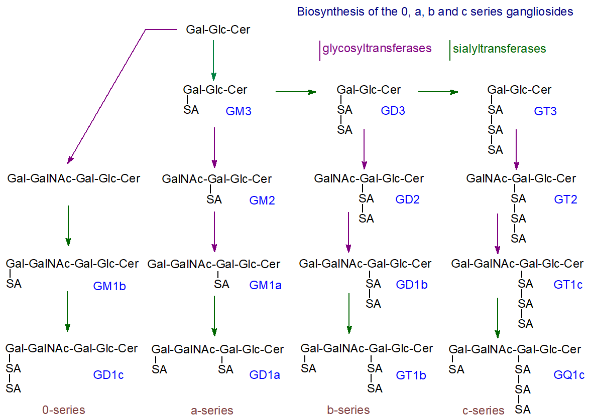
The required enzymes are bound to Golgi membranes in the order corresponding to the addition of the carbohydrate components. Thus, the sialyltransferase that catalyzes synthesis of the relatively simple ganglioside GM3 is located in the cis-Golgi. In contrast, those that catalyze the terminal steps of ganglioside synthesis are located in the distal, or trans-, Golgi region. GM3 synthase, in particular, catalyzes the transfer of Neu5Ac from cytidine monophosphate (CMP)-Neu5Ac to the terminal galactose residue of lactosylceramide and exhibits unique specificity.
The simple ganglioside GM3 is synthesized by adding sialic acid to lactosylceramide via CMP:LacCer α2-3 sialyltransferase (or GM3 synthase), before appropriate synthases produce GD3 and GT3. Subsequently, GM3, GD3, and GT3 serve as precursors of more complex gangliosides through additional glycosyl- and sialyltransferases. An alternative theory, supported by some evidence, proposes that a multiglycosyltransferase complex synthesizes each ganglioside rather than a series of individual enzymes. Further sialylation of the a, b, and c series, and at different positions in the carbohydrate chain, can produce an increasingly complex and heterogeneous range of products, such as α-series gangliosides with sialic acid residue(s) linked to the inner N-acetylgalactosamine residue (not illustrated). GM4, or NeuAcα2,3Gal-Ceramide, a minor brain component present in a few other tissues at low levels, is an exception because galactosylceramide is its precursor. Finally, the newly synthesized gangliosides are transferred to the external leaflet of the plasma membrane via the luminal surface of transport vesicles. Gangliosides are also important constituents of nuclear membranes.
Changes in ganglioside composition in the brain and other tissues during embryonic and postnatal development are mainly governed by changes in the expression level and activity of glycosyl- and sialyl-transferases, although the latter can also be regulated by glycosylation and phosphorylation.
The presence of distinctive sialidases that differ from the catabolic lysosomal enzymes (see below) in raft-like regions of the plasma membrane bring about further changes in the composition of the cell surface gangliosides that can be specific to particular cell types, causing a shift from poly-sialylated species involving a decrease of GM3 and formation of GM2 then GM1 by hydrolysis of terminal sialosyl residues linked either α2‑8 on another sialic acid or α2‑3 on galactose. Because GM1 resists most sialidases, its concentration tends to increase relative to oligosialo species as developmental and other GM1-requiring processes take hold. This may affect important cellular events, such as neuronal differentiation and apoptosis. Conversely, sialylation may occur in some neuronal membranes, increasing the proportions of poly-sialylated species. In particular, a CMP-NeuAc:GM3 sialyltransferase can sialylate GM3. Researchers have identified gangliosides GM1 and GD1a in both nuclear envelope membranes, along with two neuraminidases.
Ganglioside lactones, where ester linkages connect the sialic acids, have been detected as minor components in brain tissues. Lactonization occurs at the plasma membrane. As lactonization profoundly influences the shape and biological properties of the original ganglioside, lactonization-delactonization in a membrane might trigger specific cellular reactions. Similarly, GD3 ganglioside can undergo O-acetylation at C9 of the outer sialic acid with important metabolic implications.
Gangliosides added to many cell preparations in vitro are rapidly taken up by cells, while gangliosides injected into animals in vivo are rapidly internalized by tissues. They can cross the blood-brain barrier and enter the fetus via the placenta. Similarly, intestinal cells absorb dietary gangliosides intact. Intestinal cells can break them down into their lipid and carbohydrate constituents for reuse. The sialic acids released by intestinal sialidase are transported from the plasma to the brain and other tissues, influencing ganglioside expression. Some experimental evidence suggests that dietary gangliosides may improve cognitive function in animals and humans.
Catabolism
After endocytosis, ganglioside degradation occurs at the surface of intralysosomal luminal vesicles generated by inward budding of the endosomal membrane. In brief, for gangliosides, soluble sialidases (neuraminidases) and exoglycohydrolases remove sialic acid and sugar residues sequentially from the non-reducing terminal unit, as illustrated for ganglioside GM1, eventually forming ceramide, which ceramidases then split into a long-chain base and fatty acids. This degradation occurs through the endocytosis-endosome-lysosome pathway, which requires an acidic pH inside the organelle. In addition to sialidases and exoglycohydrolases, these reactions require effector molecules called sphingolipid activator proteins, including saposins (Sap) and the specific GM2-activator protein (GM2-AP). Ganglioside GM3 is a component of the lysosomal membrane but is protected from degradation by a glycocalyx of the membrane facing the lysosome. Anionic lipids, especially bis(monoacylglycero)phosphate, stimulate ganglioside degradation in membranes, while cholesterol inhibits it. The catabolic pathway is shown in Figure \(\PageIndex{30}\).
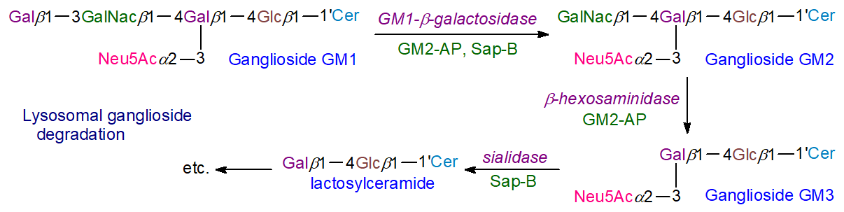
This process is a salvage mechanism important to the overall cellular economy, since a high proportion of the hydrolysis products are recycled for glycolipid biosynthesis. Generating ceramide and sphingosine may also support these lipids' regulatory and signaling functions. In addition, some partial hydrolysis of gangliosides occurs in the plasma membrane as part of the biosynthetic remodeling process discussed above. Defects in catabolism lead to the gangliosidoses, which will be discussed later.
Click the link below
- Link
-
Ganglioside Function
Cell surface effects: In their natural biological environment, gangliosides carry a negative charge due to sialic acids, which also enhance the hydrophilicity of the polysaccharide constituent. This is somewhat balanced by the hydrophobic ceramide moiety, so overall the molecules are amphiphilic but very different from glycerophospholipids, which are essential for forming membrane bilayers. Indeed, a ganglioside such as GM1 is water-soluble (unique among lipids) and can form large aggregates due to its hydrophilic properties. The ceramide unit, with its capacity to form hydrogen bonds with glycerophospholipids, helps ensure that gangliosides insert stably into the outer leaflet of the plasma membrane.
Thus, gangliosides are anchored in membranes by their ceramide units, with the double-tailed sialoglycan components extending from the cell surface, where they can participate in intermolecular interactions through a network of hydrogen bonds and hydrophobic interactions. For example, the glucose-ceramide bond of GM1 is oriented in the outer leaflet of the plasma membrane such that the glycan extends perpendicularly to the plane of the lipid bilayer. All gangliosides, but especially the simplest GM3 or Neu5Acα2-3Galβ1-4Glcβ1Cer, have a structural role, and they have a natural propensity to segregate laterally and to associate with each other and with other sphingolipids, phospholipids, and cholesterol into raft nano-domains or in related structures, such as the caveolae, where the very large surface area occupied by the oligosaccharide chain imparts a strong positive curvature to the membrane. In this environment, gangliosides can interact with one another through side-by-side hydrogen bonds, mediated by water molecules that bridge the chains.
Further, GM3 and other gangliosides self-aggregate into clusters on the surface of human peripheral blood lymphocytes, and evidence suggests that the density of these clusters in membranes governs their antigen reactivity. In addition, gangliosides and other oligoglycosylceramides are believed to cluster via hydrogen-bond donor-acceptor (cis) interactions, facilitated by hydroxyl and acetamide groups, to form glycosynaptic domains related to, but functionally distinct from, raft signaling platforms (with lower cholesterol concentrations). Many of the biological functions of gangliosides are mediated by their localization in these nanodomains, where they may have specialized roles in cell adhesion, growth, and motility through interactions with specific proteins and signal transduction pathways. However, not all gangliosides are present in such raft-like structures.
Receptor/signaling functions: Gangliosides can bind membrane proteins directly via carbohydrate-carbohydrate or carbohydrate-amino acid interactions, often involving specific ganglioside head groups, thereby altering protein localization within membrane microdomains to recruit signaling partners or altering receptor dimerization and other effects. In rafts and caveolae, especially, gangliosides can modulate cell signaling processes by interacting with specific receptors, adhesion molecules, and ion channels. Cell–cell (trans) interactions occur when sialoglycans on one cell bind to complementary binding proteins (lectins) on adjacent cells, promoting cell adhesion and enabling regulation of intracellular signaling pathways; e.g., myelin-associated glycoprotein on myelin sheaths binds to gangliosides on axonal membranes.
In addition, gangliosides act as receptors for interferon, epidermal growth factor, nerve growth factor, and insulin, and may regulate cell signaling and control cell growth and differentiation. Intact gangliosides inhibit growth by making cells less sensitive to epidermal growth factor stimulation, while removing the N-acetyl group of sialic acid enhances this effect and stimulates growth. Gangliosides function as antigens or receptors by recognizing specific molecules (lectins), including bacterial toxins, at the cell surface and by modulating the charge density at the membrane surface (see the section on Gangliosides and Disease below). They also regulate protein activity within the plasma membrane, especially receptor-type tyrosine kinases. For example, the phosphorylation state and activity of insulin receptors in caveolae, and hence the insulin resistance of cells, are controlled by the concentration of GM3, the main ganglioside in plasma and other extraneural tissues. GM3 also interacts with the epidermal growth factor receptor, inhibiting cell growth. GM1 strongly influences specific neuronal functions by interacting with specific receptors, such as the tropomyosin receptor kinase (Trk) A (TrkA) receptor, by altering its conformation to enable interaction with the nerve growth factor (NGF) ligand.
GM3 (SA-Gal-Glc-Cer) is a serum ganglioside highly enriched in a membrane microdomain termed a glycosynapse, and it forms complexes with co-localized cell signaling molecules. It has a function in the innate immune function of macrophages, and it has been demonstrated that molecular species of GM3 with differing acyl-chain structures and modifications can operate as pro- and anti-inflammatory modulators of Toll-like receptor 4 (TLR4); very-long-chain and α-hydroxy GM3 species increase TLR4 activation, while long-chain and unsaturated GM3 species have the opposite effect. In addition, gangliosides are cell-type-specific antigens with key roles in immune defense. For example, a major immunological function of gangliosides and sialic acids is to protect cells from immune attack and autoimmunity. They recognize and protect host organs and tissues from complement attack by binding to the complement regulatory protein factor H, which can exert strong cytotoxic and inflammatory activity. In particular, sialic acids protect autologous cells from complement killing by binding to this protein via the α2–3-linked sialic acid glycans of the GD3 ganglioside. Conversely, breakdown of this system can lead to autoimmune diseases.
Brain function: One of the first examples of a ganglioside influencing a signaling event studied in detail concerns the simple ganglioside GD3, which plays a central role in early neurogenesis. GD3 binds to the epidermal growth factor receptor (EGFR) via a protein-carbohydrate interaction involving its terminal N-acetylneuraminic acid and a lysine residue in the transmembrane domain of the receptor and also by a carbohydrate-carbohydrate interaction, thereby maintaining the latter in its inactive monomeric state. EGFR then binds epidermal growth factor and transitions from an inactive monomer to an active homodimer, triggering receptor autophosphorylation and activating a signaling cascade that promotes cell proliferation. This has proven essential for regulating stem cell self-renewal capacity in the brain. In contrast, the neutral oligosaccharide Gb4 exerts the opposite effect on EGFR by directly interacting with it, potentiating its autophosphorylation and activating the downstream cascade.
Molecular biology techniques, such as targeted gene deletion, enable the elimination of specific enzymes in experimental animals and are improving understanding of each ganglioside's function. They are essential to central myelination, maintaining the integrity of axons and myelin, and transmitting nervous impulses. These effects may be mediated by interactions of gangliosides' negatively charged sialic acid residues with calcium ions, which are critical for neuronal responses. For example, a variant of GD3, 9-O-acetyl GD3, appears to be involved in glial-guided neuronal migration during brain development in the rat, while GM1 may have a similar function in humans; it determines which growth cone of unpolarized neurons becomes the axon. By stabilizing neuronal circuits, gangliosides support memory; conversely, disturbances in ganglioside synthesis can lead to neurodegenerative disorders (see below). Ganglioside GM3 in raft domains has been shown to play an indispensable role in the development, function, and viability of cochlear hair cells, and, by extension, it is essential for hearing. In contrast, mice that express GM3 primarily and lack typical brain complex gangliosides exhibit weight loss, progressive motor and sensory dysfunction, and deterioration in spatial learning and memory with aging. GD3 is important for retinal structure and visual function in mice.
Nerve stimulation, environmental factors, or drug treatments can alter ganglioside composition. The various interconvertible ganglioside types in the plasma membrane of neurons are particularly important for neuronal development, as they regulate processes such as axonal determination and growth, signaling, and repair. In addition, gangliosides are believed to act as ligands that maintain myelin stability and control nerve regeneration by binding to a specific myelin-associated glycoprotein. The presence of gangliosides in cell nuclei suggests a possible role in expressing genes relevant to neuronal function. For example, the monosialoganglioside GM1 has been shown to promote the differentiation of various neuronal cell lines in culture. It protects the nervous system by promoting neural stem cell survival and proliferation, facilitating axon stability and regeneration, and inhibiting neurodegeneration through autophagy, for example, after ischemic stroke. Within membrane rafts, this ganglioside plays key roles in several signaling systems through its association with specific proteins that have glycolipid-binding domains, including those that modulate ion transport, neuronal differentiation, G protein-coupled receptors (GPCRs), immune responses, and neuroprotection. It is important for Ca2+ and Na+ homeostasis in the nucleus and plasma membrane, and in regulating the effects of platelet-derived growth factor. However, there have been unpleasant complications when GM1 has been administered for therapeutic purposes. GD1a is sometimes considered to be a reserve pool for GM1.
After nerve injury, toll-like receptor 2 (TLR2) signaling is important for inducing neuropathic pain; ganglioside GT1b acts as a TLR2 agonist, inducing mechanical and thermal hypersensitivity.
Other functions: The ganglioside GD3 is essential for apoptosis by blocking the activation of specific transcription factors and preventing the induction of antiapoptotic genes. 9-O-Acetylation of the GD3 molecule prevents ganglioside oxidation and blocks its pro-apoptotic effects. Similarly, GD3 regulates autophagy, i.e., the degradation and/or recycling of cellular components. Gangliosides are also important in reproduction; in mice, GD1a is important for oocyte maturation, monospermic fertilization, and embryonic development, while GM1 is important for sperm-oocyte interactions and sperm maturation. Deletion of the GM2/GD2 synthase leads to infertility in male mice and the production of a novel fucosylated ganglioside containing very-long-chain polyunsaturated fatty acids. Related studies in gene-knockout mice show that b-series gangliosides are important for adipocyte leptin secretion. In contrast, a-series gangliosides interact with the leptin receptor in the hypothalamus to influence the energy balance.
Gangliosides and Disease
Bacterial toxins and viruses: In adaptive immunity, a-series and o-series gangliosides in the plasma membrane support receptor function and stimulation on certain T-cell subsets by acting as pattern-recognition receptors for invading pathogens. In particular, certain gangliosides bind specifically to viruses, various bacterial toxins (such as those from botulinum, tetanus, and cholera), and blood merozoites of the deadliest malaria parasite, Plasmodium falciparum, and mediate interactions between microbes and host cells during infections, with NeuAc as the main recognition module. The best-known example is cholera toxin, an enterotoxin produced by Vibrio cholerae, which binds to the cell-surface receptor ganglioside GM1; each of the five B-chains binds one GM1 molecule. Interestingly, the subsequent metabolism of the ganglioside-toxin complex depends on the fatty acid components of the ganglioside. Toxins are believed to use gangliosides to hijack an existing retrograde transport pathway from the plasma membrane to the endoplasmic reticulum. For example, cholera toxin crosses the intestinal epithelial barrier via GM1, possibly through endocytosis of the toxin-GM1 complex via caveolae into the apical endosome and then into the Golgi/endoplasmic reticulum, where the complex dissociates. As a result, the toxin persistently activates adenylate cyclase and continuously produces cAMP, leading to the severe fluid loss typical of cholera infections. Another example is botulinum toxin, which binds to a complex of a polysialoganglioside and the protein synaptotagmin that together act as a high-affinity receptor complex enabling its neurotoxic effects. Similarly, ganglioside GM2 binds to a toxin secreted by Clostridium perfringens.
Influenza viruses have two glycoproteins in their envelope membranes: hemagglutinin and neuraminidase. Hemagglutinin binds to cellular receptors such as gangliosides. After entering respiratory epithelial cells, the virus's sialidase (neuraminidase) cleaves sialic acid from receptors, preventing additional viruses from entering. Variations in these proteins drive the development of new vaccines. The carbohydrate moiety of gangliosides is essential for initial viral binding, but the lipid moiety is believed to control intracellular transport.
Some gangliosides, especially GD1a, have anti-inflammatory properties, as they inhibit the effects of bacterial lipopolysaccharides by preventing the activation of tumor necrosis factor (TNF) and other cytokines. In contrast, GM2 may increase cytokine production in similar circumstances, while the heat-labile toxins of Escherichia coli bind to several gangliosides in macrophages, thus activating an inflammatory response.
Gangliosidoses and other neurodegenerative diseases: It appears to be a general rule that accumulating lysosomal substrates in all lysosomal storage disorders impair lysosomal integrity and disrupt lipid metabolism and membrane structure and function, inevitably triggering pathologic mechanisms. Endogenous antibody generation against gangliosides is often a factor, and some argue that gangliosides and their sialic acid components lie at the border of immune tolerance.
As with neutral oligoglycosylceramides and ceramide monohexosides, many unpleasant lipidoses involve storing excessive amounts of gangliosides in tissues because of failures in catabolism. The most important of these are the GM2 gangliosidoses, i.e., Tay-Sachs disease (and the similar Sandhoff disease), a fatal genetic disorder found mainly in Jewish populations in which harmful quantities of the ganglioside GM2 accumulate in nerve cells of the brain and other tissues. Lyso-GM2 (non-acylated) in plasma may serve as a marker. Researchers have identified a modified GM2 derivative containing taurine linked to the sialic acid carboxyl group in the brains of such patients. As infants with the most common form of the disease develop, the nerve cells become distended, and a relentless deterioration of mental and physical abilities occurs. The condition is caused by insufficient activity of specific enzymes, i.e., β‑N‑acetylhexosaminidase, which catalyzes the degradation of gangliosides by removing the terminal N-acetylgalactosamine residue from GM2 or the GM2 activator protein.
In addition, a generalized GM1 gangliosidosis (an autosomal recessive and neurodegenerative disease) has been characterized in which ganglioside GM1 accumulates in the nervous system, leading to mental retardation and enlargement of the liver. The condition results from a deficiency of the lysosomal β-galactosidase enzyme, which hydrolyses the terminal β-galactosyl residues from GM1 ganglioside to produce GM2. Storing substantial amounts of unwanted lipids in the lysosomal system appears to cause cellular starvation, depleting essential elements such as iron in brain tissue. Lyso-GM1 in plasma is now seen as a useful diagnostic aid. Small amounts of some gangliosides accumulate as secondary storage compounds in Niemann–Pick disease. The Guillain–Barré syndrome is an acute inflammatory disorder, usually triggered by a severe infection, that affects the peripheral nervous system. The immune system produces antibodies to gangliosides, leading to axonal damage and, in some cases, paralysis. Huntington’s disease is believed to involve disruption of the metabolic pathways between glycosylceramides and gangliosides, and a mutation in a sialyltransferase causes a human autosomal recessive infantile-onset epilepsy syndrome. Impaired ganglioside metabolism is also relevant to Alzheimer’s disease because complexation with ganglioside GM1 may promote aggregation of amyloid β-protein deposits that characteristically accumulate in the brain in this condition (this explanation is not universally accepted). In general, in ganglioside deficiencies, natural or induced, progressive inflammatory reactions appear to occur, leading to neurodegeneration, in part because of deterioration in the architecture of lipid rafts.
In contrast, at normal tissue concentrations, gangliosides such as GM1 are believed to have anti-inflammatory and neuroprotective effects in certain types of neuronal injury, Parkinson's disease, and related diseases. For example, in Parkinson's disease, GM1 binds to α-synuclein and inhibits or prevents fibril formation. It may play a protective role by preventing sphingomyelin-induced aggregation, but its beneficial effect decreases as GM1 levels decline with aging. For these reasons, the therapeutic properties of ganglioside GM1, the most accessible species, and derived molecules are under clinical investigation. However, there is no approved therapy for any gangliosidosis, although many different therapeutic strategies are being studied, including hematopoietic stem cell transplantation and gene therapy. For the moment, the blood-brain barrier remains a challenge.
Cancer: Gangliosides have essential functions in cancer, especially in regulating signal transduction induced by growth-factor receptors within a specific microdomain termed a 'glycosynapse' in cancer cell membranes, and in interactions with glycan recognition molecules involved in cell adhesion and immune regulation. In particular, depending on the tissue, certain distinctive gangliosides are expressed at much higher levels in tumors than in normal healthy tissues, mainly by aberrant expression of glycosyltransferases and glycohydrolases. This allows tumor cells to evade immune surveillance and maintain their malignancy. For example, GM3 is not normally expressed in melanocytes but is detected in 60% of primary melanomas and 75% of metastatic melanomas. Gangliosides can be shed from the surface of tumor cells into the local environment, where they can influence interactions between cancer cells, including the transition of tumors from a dormant to a malignant state (angiogenesis); when present in the circulation, they can be useful diagnostic aids. For example, the ganglioside GM3 is elevated in the serum of patients with breast cancer and may be a biomarker for the disease. In contrast, disialylated gangliosides GD2 and GD3 (Figure \(\PageIndex{31}\)) are considered to be markers of neuroectoderm origin in tumors (neuroblastoma).
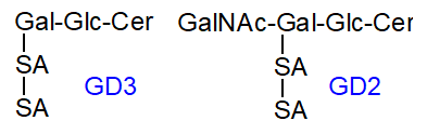
Figure \(\PageIndex{31}\): Structure GD2 and GD3 Specific gangliosides can have either positive or negative effects on the regulation of the malignant properties of cancer cells. Generally, disialyl glycosphingolipids or tandem-repeated sialic acid structures confer malignant properties in various cancer systems; they are not merely markers. For example, the disialo-gangliosides GD2 and GD3 are present in trace amounts only in normal tissues but are found at much higher concentrations in cancer cells, especially melanomas and neuroblastomas, with GD2 especially elevated in triple-negative breast cancer. These b-series gangliosides play a substantial part in the malignant properties of gliomas by mediating cell proliferation, migration, invasion, adhesion, and angiogenesis, and preventing immunosuppression. They are considered tumor-associated antigens, and the GD2 and GD3 synthases are important drug targets.
Ganglioside GM3(Neu5Gc), i.e., containing an abnormal sialic acid, is sometimes considered a tumor-specific antigen and a target for cancer immunotherapy. Aberrant sialylation occurs in many cancers, where neuraminidase levels are key factors in metastasis and cancer cell survival, and unusual gangliosides containing N-glycolyl sialic acid can accumulate in some cancers. N-Glycolyl-GM3, normally absent from human tissues, is present in all stage II breast cancers, and many other less common complex gangliosides accompany it. Similarly, the 5-N-deacetylated form of GM3 is expressed in metastatic melanomas but not in healthy tissue or in primary melanomas; it is considered a specific marker of metastatic disease and a potential therapeutic target. Increased synthesis of 9-O-acetyl-GD3, dependent on a sialyl-O-acetyltransferase - CAS1 Domain-Containing Protein 1, occurs in acute lymphoblastic leukemia and malignant melanomas, which appears to limit apoptosis. At the same time, O-acetylated GD2 (OAcGD2) is expressed in breast cancer and other tumors. A unique fucosyl-GM1, in which the terminal galactose is α-1,2-fucosylated at the non-reducing end, is found circulating in the serum of patients with many cancers, especially small-cell lung cancer, but rarely in normal conditions. It is also considered a potential indicator of cancer and a candidate for immunotherapy.
Clinical trials with an antibody to GD2 have been carried out successfully against the rare childhood cancer neuroblastoma, and the USDA has approved the use of this in combination with other drugs to treat this often lethal cancer. However, this antibody can have painful side effects due to an interaction with GD2 on neurons, and modified antibodies, which may be safer, are now being tested in multiple clinical trials. A phase I clinical trial with an antibody to GD3 has shown promising results in patients with malignant melanoma. Similarly, antibodies to OAcGD2 and fucosyl-GM1 have shown anti-tumor effects in vitro, and studies with human patients are underway.
Other diseases: Aberrant production of the ganglioside GM3 has been linked to pathophysiological changes associated with obesity, metabolic disorders, and type 2 diabetes mellitus through its effects on insulin receptors. It has a role in autoimmune disorders such as multiple sclerosis. In epilepsy, a deficiency in the enzyme ceramide synthase 1, which produces 18:0 ceramides, is believed to reduce ganglioside formation. Gangliosides influence allergic responses and autoimmune diseases because they are present in certain T cell subsets. As gangliosides are present on the surface of vascular, vascular-associated, and inflammatory cells, they may have a role in atherosclerosis and aging.
-
In contrast, monosialyl gangliosides, such as GM1, GM2, and GM3, may suppress the malignant properties of various cancer cells. The mechanism is believed to involve complex formation at the cell surface with membrane proteins, such as growth factor receptors and adhesion receptors, including those of the integrin family, leading to modified signals mediated by these receptors. Metastatic melanoma cells have higher GD3 levels than poorly metastatic cells or the normal counterpart, suggesting that GD3 may promote metastasis by suppressing the anti-tumor immune response.
Summary
(Summary written by Claude, Sonnet 4.6, Anthropic)
This chapter describes the biochemistry of sphingolipids — a structurally diverse family of bioactive and structural lipids defined by a long-chain sphingoid base backbone — tracing their biosynthesis from palmitoyl-CoA and serine through ceramide to complex sphingophospholipids, glycosphingolipids, and gangliosides, and connecting their metabolism to membrane organization, cellular signaling, neurological function, and inherited disease.
Sphingolipid biosynthesis begins with the PLP-dependent condensation of palmitoyl-CoA and L-serine, catalyzed by serine palmitoyltransferase (SPT) — a heterotrimeric integral membrane enzyme of the ER (SPTLC1 + SPTLC2 or SPTLC3, with regulatory ssSPTA/B small subunits) that is subject to negative feedback regulation by ORMDL proteins and serine availability. This is the committed, rate-limiting step of the entire pathway; genetic elimination of SPT subunits is embryonically lethal. The product, 3-keto-sphinganine, is rapidly reduced to sphinganine by an NADPH-dependent reductase. Sphinganine is then N-acylated by one of six ceramide synthases (CerS1–6) on the cytoplasmic face of the ER, each with distinct acyl-CoA chain-length specificity: CerS2 (C20–C26, abundant in brain, lung, liver), CerS1 (C18:0, brain/muscle), CerS3 (C26+, skin/testes), CerS5/6 (C16, lung/intestine). The Δ4-trans double bond of the sphingoid base is inserted afterward by dihydroceramide desaturase (DEGS1, ubiquitous; DEGS2, which can also hydroxylate to form phytoceramides in skin/intestine). The result is ceramide — the central hub of all sphingolipid metabolism. Ceramide is produced, catabolized, and reused through at least three interconnected pathways: de novo synthesis in the ER, the sphingomyelinase pathway (rapid hydrolysis of sphingomyelin in various membranes), and the salvage pathway (lysosomal ceramidase releases sphingosine for re-N-acylation by ceramide synthases, estimated to provide 50–90% of total sphingolipid biosynthesis).
The biological roles of ceramide and related metabolites follow an elegant regulatory logic. Ceramide and sphingosine are pro-apoptotic, promoting cell death, stress responses, growth arrest, and differentiation. In contrast, sphingosine-1-phosphate (S1P) and ceramide-1-phosphate promote cell survival, proliferation, and migration. The balance between these competing signals — the sphingolipid rheostat — is therefore a critical determinant of cell fate and requires exquisite spatial and temporal control in each cellular compartment. Many stimuli that trigger apoptosis (tumor necrosis factor-α, chemotherapeutic agents, radiation, interferon-γ, vitamin D₃) do so by activating sphingomyelinases at the plasma membrane, Golgi, or mitochondria to rapidly generate ceramide from the abundant sphingomyelin pool, providing a much faster signaling response than de novo biosynthesis could achieve.
Transport of ceramide from the ER to the Golgi for complex sphingolipid synthesis is mediated non-vesicularly by the CERT protein. CERT recognizes the ER via a short FFAT peptide motif and the trans-Golgi via an N-terminal PH domain that binds PI(4)P; its C-terminal START domain extracts ceramide from membranes with preference for d-erythro ceramides including dihydroceramide, holding them within a long amphiphilic cavity by hydrogen bonding with the sphingoid motif. This non-vesicular transfer maintains a steep ER-to-Golgi ceramide gradient that prevents toxic accumulation in the ER. A separate, ATP-independent vesicular mechanism delivers ceramide for glucosylceramide synthesis in the cis-Golgi. Thus, ceramide pools destined for sphingomyelin synthesis versus glycolipid synthesis are physically segregated.
Sphingomyelin (SM) is the most abundant sphingolipid in animal cells and is synthesized exclusively in animals (not plants or fungi). It is produced by sphingomyelin synthases SMS1 and SMS2, which transfer phosphocholine from phosphatidylcholine to ceramide, releasing DAG as a co-product — directly linking sphingolipid synthesis to the DAG/PKC signaling axis of glycerophospholipid metabolism. SMS1 operates on the luminal face of the trans-Golgi and is CERT-dependent; SMS2 is at the outer leaflet of the plasma membrane and regenerates sphingomyelin from locally produced ceramide. Both enzymes have phospholipase C activity in the absence of ceramide, providing additional regulatory coupling. Sphingomyelin is enriched in the outer leaflet of the plasma membrane and in myelin, where it constitutes up to 70% of some membrane lipids. Its catabolism is catalyzed by a family of sphingomyelinases at different pH optima and subcellular locations: lysosomal acid sphingomyelinase (SMPD1, requires saposin activator domain) is the housekeeping enzyme; secreted acid sphingomyelinase requires Zn²⁺ and operates at the plasma membrane; four neutral sphingomyelinases (NSM1–4) at the ER, Golgi, plasma membrane, and mitochondria generate signaling ceramide rapidly in response to inflammatory stimuli. Niemann-Pick disease types A and B result from acid sphingomyelinase deficiency with consequent accumulation of sphingomyelin and secondary cholesterol sequestration.
Glycosphingolipids are synthesized in the Golgi by sequential glycosylation of ceramide. Glucosylceramide synthase (GCS, a GT1-family enzyme) at the cytoplasmic face of the cis-Golgi adds UDP-glucose to ceramide; the product is then flipped to the luminal face by P4-ATPases (ATP10A/D) assisted by FAPP2 and GLTP for subsequent glycosylation. Glucosylceramide is the obligate precursor for lactosylceramide and all complex neutral oligoglycosphingolipids and gangliosides in animals. In the skin, it is essential for lamellar body formation and the epidermal water permeability barrier. Galactosylceramide synthase (restricted to oligodendrocytes, Schwann cells, kidney, and testes) adds UDP-galactose to ceramide in the ER lumen; the product is concentrated in myelin and is essential for myelin stability and oligodendrocyte differentiation. Sulfation of galactosylceramide produces sulfatide, another essential myelin component. Gaucher disease (the most common sphingolipidosis) results from GBA1 glucocerebrosidase deficiency, causing glucosylceramide accumulation in macrophage lysosomes to form foam "Gaucher cells," with splenomegaly, bone disease, and in severe forms, neurological involvement. Enzyme replacement therapy with recombinant glucocerebrosidase and oral glucosylceramide synthase inhibitors effectively treats non-neurological type 1 Gaucher disease.
Gangliosides — acidic oligoglycosphingolipids carrying one to five sialic acid (N-acetylneuraminic acid, Neu5Ac) residues — are the most complex sphingolipids and are especially abundant in the nervous system, where they comprise up to 12% of total brain lipid content, concentrated in the outer leaflet of neuronal plasma membranes. The four major brain gangliosides (GM1, GD1a, GD1b, GQ1b) account for ~95% of adult mammalian brain gangliosides; simpler forms (GM3, GD3) predominate during embryonic development, shifting to more complex forms as the brain matures. This developmental change is orchestrated by the regulated expression of specific sialyltransferases and glycosyltransferases positioned sequentially along the Golgi, with GM3 synthase in the cis-Golgi initiating the four biosynthetic series (0-, a-, b-, c-) from which all complex gangliosides derive. Gangliosides serve critical roles in neural development, synaptic transmission, myelin stabilization, and cell-cell recognition. They are concentrated in raft nanodomains along with sphingomyelin and cholesterol, and the unique hydrogen-bonding capacity of the "sphingoid motif" (the first five carbons of the sphingoid base) enables multiple noncovalent interactions with proteins, lipids, and carbohydrates within these domains. Catabolism occurs in lysosomes by sequential removal of monosaccharide and sialic acid residues by soluble exohydrolases and sialidases assisted by saposin activator proteins (A–D) and the GM2-activator protein, which perturb the membrane to allow enzyme access to these membrane-embedded substrates. Deficiencies in specific catabolic enzymes cause gangliosidoses — lysosomal storage diseases characterized by progressive neurodegeneration. The chapter's coverage of sphingolipid diversity, the sphingolipid rheostat, raft biology, and the consequences of metabolic dysfunction in storage diseases illustrates why this lipid class, despite its enigmatic past, remains a frontier of biomedical research with implications for neurology, cancer biology, inflammation, and metabolic disease.
This page titled 21.4: Biosynthesis of Membrane Sphingolipids was last modified on Wed, 16 Sep 2026 14:43:02 GMT and is shared under a not declared license and was authored, remixed, and/or curated by Henry Jakubowski and Patricia Flatt.
The book could not be loaded just now.