Opening the book…
Search Fundamentals of Biochemistry
by William (Bill) W. Christie and Henry Jakubowski.
This section is an abbreviated and modified version of material from the Lipid Web, an introduction to the chemistry and biochemistry of individual lipid classes, written by William Christie.
(Learning goals written by Claude, Sonnet 4.6, Anthropic)
Phosphatidic Acid and Lysophosphatidic Acid: Synthesis and Signaling
- Describe the four major biosynthetic routes to phosphatidic acid — sequential sn-glycerol-3-phosphate acylation (GPAT then AGPAT/LPAT), the DHAP pathway in peroxisomes (important for ether lipid synthesis), phospholipase D-mediated hydrolysis of phosphatidylcholine, and diacylglycerol kinase phosphorylation of DAG — explaining how each route supplies either the structural or signaling pool of this lipid and how lipins (phosphatidate phosphatases) regulate phosphatidic acid concentration to control the branch point between TAG storage and glycerophospholipid synthesis.
- Explain how lysophosphatidic acid (LPA) is generated extracellularly by autotaxin (a lysophospholipase D acting on plasma lysophosphatidylcholine) and intracellularly by phospholipase A2 acting on phosphatidic acid, and describe its roles as a growth-factor-like lipid mediator with effects on platelet activation, vascular development, and cancer cell proliferation and migration through specific GPCRs.
Biosynthesis of Glycerophospholipids: PC, PE, and PS
- Describe the CDP-choline (Kennedy) pathway for phosphatidylcholine synthesis — tracing phosphocholine → CDP-choline (catalyzed by the rate-limiting CTP:phosphocholine cytidylyltransferase/CCT, regulated by a membrane-sensing C-terminal domain) → PC via CEPT1/CPT1 in the ER/Golgi — and contrast this with the PE methyltransferase (PEMT) pathway (three sequential SAM-dependent methylations in liver) as a source of one-third of hepatic PC and an explanation for why elevated homocysteine is a cardiovascular risk factor.
- Describe the two pathways for phosphatidylethanolamine synthesis — the CDP-ethanolamine route (analogous to the Kennedy pathway for PC, proceeding through rate-limiting CTP:phosphoethanolamine cytidylyltransferase) in the ER/Golgi and the phosphatidylserine decarboxylase (PSD1) route in the inner mitochondrial membrane — and explain why the spatial separation of these pathways creates functionally distinct PE pools, with disruption of either pathway being embryonically lethal.
- Explain how phosphatidylserine is synthesized in animals exclusively by head-group base-exchange reactions at the mitochondria-associated membrane (MAM) — PS synthase I using PC and PS synthase II using PE as head-group donors — and describe the coupled metabolic cycle in which PS is transferred to the inner mitochondrial membrane, decarboxylated to PE by PSD1, and some PE returns to the ER to be re-converted to PS or methylated to PC, maintaining the tight co-regulation of PS and PE levels.
- Explain the Lands cycle of phospholipid remodeling — describing how phospholipase A2 cleaves the sn-2 fatty acid to generate a lysophospholipid intermediate, and how tissue- and chain-length-specific lysophospholipid acyltransferases (LPCAT1 for lung surfactant dipalmitoyl-PC, LPCAT2/3 for arachidonoyl enrichment in inflammatory cells, LPEAT for PE, MBOAT2/LPCAT4 for PS) reacylate to produce the final characteristic fatty acid composition — and explain why de novo synthesis determines gross composition while Lands cycle remodeling determines the precise molecular species distribution essential for membrane function and eicosanoid precursor availability.
Phosphatidylinositol, Polyphosphoinositides, and the Phosphatidylinositol Cycle
- Describe the biosynthesis of phosphatidylinositol via the CDP-diacylglycerol intermediate pathway (PA → CDP-DAG by CDP-DAG synthase CDS2, then PI synthase transfers myo-inositol to CDP-DAG in the ER), explaining how the distinctive 1-stearoyl-2-arachidonoyl molecular species composition is established by the substrate specificity of DGKε (which preferentially generates the 18:0/20:4 PA precursor) and LPIAT1/MBOAT7 (which remodels lysophosphatidylinositol to incorporate arachidonic acid at sn-2).
- Explain the phosphatidylinositol phosphate "lipid code" — describing how seven phosphoinositide isomers (PI(3)P, PI(4)P, PI(5)P, PI(3,4)P₂, PI(3,5)P₂, PI(4,5)P₂, and PI(3,4,5)P₃) are produced and maintained in specific membrane compartments by the concerted action of at least 20 kinases and 34 phosphatases, how PI(4,5)P₂ hydrolysis by phospholipase C generates IP₃ (triggering ER calcium release) and DAG (activating protein kinase C), and how the phosphatidylinositol cycle reconstitutes PI from these products through the sequential actions of DGKε, CDS2, and PI synthase at ER-plasma membrane contact sites at a cost of three ATP equivalents per turn.
Introduction
This section will explore the synthesis of membrane glycerophospholipids and their metabolic derivatives. We will start with the simplest one, phosphatidic acids, and end with phosphatidylinositol. To shorten the online text, we hide details about each lipid's properties in boxes with links for interested readers.
Phosphatidic Acids and Derivatives
Phosphatidic acid or 1,2-diacyl-sn-glycero-3-phosphate is a key intermediate in the biosynthesis of other glycerophospholipids and triacylglycerols. It is structurally one of the simplest phospholipids and was long thought to be important only as a precursor of other lipids, where it is indeed a key molecule. It is now known to have many other functions in animals, plants, and other organisms, including influencing membrane structure and dynamics and interacting with various proteins. As a lipid mediator, it modulates various signaling and cellular processes, including membrane tethering, conformational changes, the enzymatic activities of specific proteins, and vesicular trafficking. Moreover, its metabolite, lysophosphatidic acid, is recognized as a key signaling molecule with many biological effects, mediated through specific receptors.
Phosphatidic Acid – Occurrence and Biosynthesis
Phosphatidic acid is not an abundant lipid constituent of any living organism and is seldom present at concentrations greater than picomolar in cells. Still, it is extremely important as an intermediate in the biosynthesis of other glycerophospholipids and triacylglycerols and as a signaling molecule or a precursor of signaling molecules. Indeed, it is often overestimated in tissues, as it can arise from inadvertent enzymatic hydrolysis under inappropriate storage or extraction conditions during analysis. It is the simplest diacylglycerophospholipid and the only one with a phosphomonoester head group. The molecule is acidic and carries a negative charge, i.e., it is an anionic lipid. The structure of phosphatidic acid is shown in Figure \(\PageIndex{1}\).

At least four important biosynthetic pathways for phosphatidic acid occur in different organelles under various stimuli. The main pathway involves the sequential acylation of sn-glycerol-3-phosphate, derived from glucose catabolism, by fatty acid acyl-CoA derivatives. First, one acyltransferase catalyzes acylation at position sn-1 to form lysophosphatidic acid (1‑acyl-sn-glycerol-3-phosphate), and then a second specific acyltransferase catalyzes acylation at position sn-2 to yield phosphatidic acid. The synthesis of phosphatidic acid from glycerol-3-phosphate is shown in Figure \(\PageIndex{2}\).

In mammals, the glycerol-3-phosphate acyltransferase that catalyzes the first step exists in four isoforms, two in the mitochondrial outer membrane (designated GPAT1 and 2) and two in the endoplasmic reticulum (GPAT3 and 4); all are membrane-bound enzymes, which are believed to span the membranes. GPAT1 is highly expressed in the liver and adipose tissue and responds to changes in feeding status via sterol regulatory element-binding protein-1 (SREBP-1), a master transcriptional regulator of lipogenic enzymes. It directs fatty acyl-CoA esters toward glycerolipid synthesis rather than β-oxidation. GPAT3 is especially important for triacylglycerol storage in adipocytes, while GPAT4 is the main contributor to lysophosphatidic acid synthesis in the liver and brown adipose tissue.
For the second step in phosphatidic acid biosynthesis, five mammalian acyl-CoA:lysophosphatidic acid acyltransferases are known, of which three are in the endoplasmic reticulum (LPAAT or LPAT or AGPAT1, 2 and 3), with a further two (LPAT4 and 5) on the outer mitochondrial membrane. While LPAT1 and 2 have strict specificity for lysophosphatidic acid as an acyl acceptor, other isoforms can esterify other lysophospholipids. Human LPAT1 showed higher activity with 14:0-, 16:0-, and 18:2‑CoAs, while LPAT2 prefers 20:4-CoA and LPAT3 produces phosphatidic acid containing docosahexaenoic acid (22:6(n-3)). The latter is especially important in the retina and testes. LPAT4 and 5 prefer oleoyl-CoA and polyunsaturated acyl-CoAs as the acyl donor, suggesting a dual role in glycerolipid synthesis and remodeling. Endoplasmic reticulum activity predominates in adipose tissue, but mitochondrial forms are believed to account for half the activity in the liver. However, because phosphatidic acid traffics between the mitochondria and endoplasmic reticulum for remodeling or synthesis of other lipids, the relative contributions of the two can be difficult to assess.
In plants, the sn-glycerol-3-phosphate pathway exists in both plastids and the endoplasmic reticulum, with multiple isoforms of the two acyltransferases and differences in the acyl substrates. In brief, most plant lipid biosynthesis begins with fatty acid biosynthesis in the chloroplasts. In plastids, the acyltransferase ATS1 transfers 18:1 acyl groups from acyl-acyl carrier protein (acyl-ACP) to position sn-1 of glycerol 3-phosphate before ATS2 transfers a palmitoyl group from ACP to position sn-2, producing phosphatidic acid at the inner leaflet of the chloroplast inner envelope membrane (IEM). Fatty acids intended for the endoplasmic reticulum are released from ACP in the chloroplast stroma by IEM-associated thioesterases, exported, and then activated by outer envelope membrane acyl-CoA synthetases to produce species with C18 fatty acids in both positions. Thus, acyl-CoAs are used for phosphatidic acid biosynthesis in the endoplasmic reticulum, with marked differences in acyl-substrate specificity. Subsequently, plastids use phosphatidic acid to synthesize galactosyldiacylglycerols, while the endoplasmic reticulum uses it to synthesize triacylglycerols and phospholipids.
In bacteria, two enzyme families catalyze acylation at the sn-1 position of glycerol-3-phosphate. For example, one in Escherichia coli utilizes the acyl-acyl carrier protein (acyl-ACP) products of fatty acid synthesis as acyl donors, as well as acyl-CoA derived from exogenous fatty acids. In a second, broader group of bacteria, including cyanobacteria, enzymes (PlsX and PlsY) use the unique acyl donors, acyl phosphates derived partly from acyl-ACP, to acylate position sn-1. In this case, a further family of enzymes (PlsC) acylates position sn-2, using acyl-ACP as the acyl donor. However, some bacterial species may also use acyl-CoA.
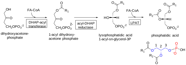
In animals, a second biosynthetic pathway utilizes dihydroxyacetone phosphate (DHAP) as the primary precursor for the peroxisomal enzyme DHAP acyltransferase, which produces acyl-DHAP. Acyl-DHAP reductase converts this intermediate to lysophosphatidic acid in an NADPH-dependent reaction. It is then acylated to form phosphatidic acid by the same LPAT as in the previous mechanism. This pathway is particularly important in ether lipid biosynthesis. The synthesis of phosphatidic acid from dihydroxyacetone phosphate is shown in Figure \(\PageIndex{3}\).
A third important route to phosphatidic acid is the hydrolysis of other phospholipids, especially phosphatidylcholine, by the enzyme phospholipase D (or by a family of related enzymes). The enzyme is readily available for study in plants, where the special functions of phosphatidic acid have long been known. Phospholipase D is now recognized in bacteria, yeasts, and most animal cells. In the latter, it exists in two main isoforms with differing specificities and cellular locations; PLD1 is found mainly in the Golgi-lysosome continuum, while PLD2 is present mainly in the plasma membrane. They are phosphoproteins whose activity is regulated by kinases and phosphatases and by binding to phosphatidylinositol-4,5-bisphosphate. In mitochondria, a distinctive enzyme of this type utilizes cardiolipin as a substrate. The mechanism uses water as the nucleophile to catalyze hydrolysis of phosphodiester bonds in phospholipids. Phospholipase D activity depends on and is regulated by neurotransmitters, hormones, small monomeric GTPases, and lipids. The hydrolysis of phosphatidylcholine to phosphatidic acids by phospholipase D is shown in Figure \(\PageIndex{4}\).

In addition to its function in generating phosphatidic acid mainly for signaling purposes but also for the maintenance of membrane composition, phospholipase D is involved in intracellular protein trafficking, cytoskeletal dynamics, cell migration, and cell proliferation, partly through protein-protein interactions; it is considered to be important in inflammation and cancer growth and metastasis as a downstream transcriptional target of proteins involved in the pathophysiology of these diseases. It also has an unusual activity as a guanine nucleotide exchange factor. A transphosphatidylation reaction with ethanol generates phosphatidylethanol, a useful biomarker for human ethanol consumption.
Under some conditions, diacylglycerol kinases can generate phosphatidic acid from 1,2-diacyl-sn-glycerols, for example, those produced from other phospholipids by phospholipase C. Such enzymes appear ubiquitous, although bacteria and yeast differ structurally from mammalian enzymes. Diacylglycerol kinases, of which at least ten isoforms (DGKα to DGKκ) exist with different subcellular locations and functions in animals, use ATP as the phosphate donor. While the epsilon isoform (DGKε) preferentially uses the 1-stearoyl-2-arachidonoyl species of diacyl-sn-glycerols to produce phosphatidic acid for phosphatidylinositol biosynthesis, other isoenzymes phosphorylate diverse diacylglycerol species. In addition to producing phosphatidic acid for phospholipid production or signaling, these enzymes may attenuate diacylglycerol signaling. For example, diacylglycerol kinases can contribute to cellular asymmetry and control cell polarity by regulating gradients in diacylglycerol and phosphatidic acid concentrations. Figure \(\PageIndex{5}\) shows the synthesis of phosphatidic acid via diacylglycerols and the reverse reaction.
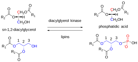
The reverse reaction, hydrolysis, is catalyzed by lipins. These enzymes regulate local phosphatidic acid concentrations and, therefore, its biological activity.
Another possible route to phosphatidic acid production for signaling is via acylation of lysophosphatidic acid, which can be produced independently for signaling, as discussed below. This pathway may be especially relevant in membranes, where the protein endophilin possesses LPAT activity and is believed to generate phosphatidic acid from lysophosphatidic acid, thereby altering membrane bilayer curvature.
Phosphatidic Acid - Role as a Lipid Precursor
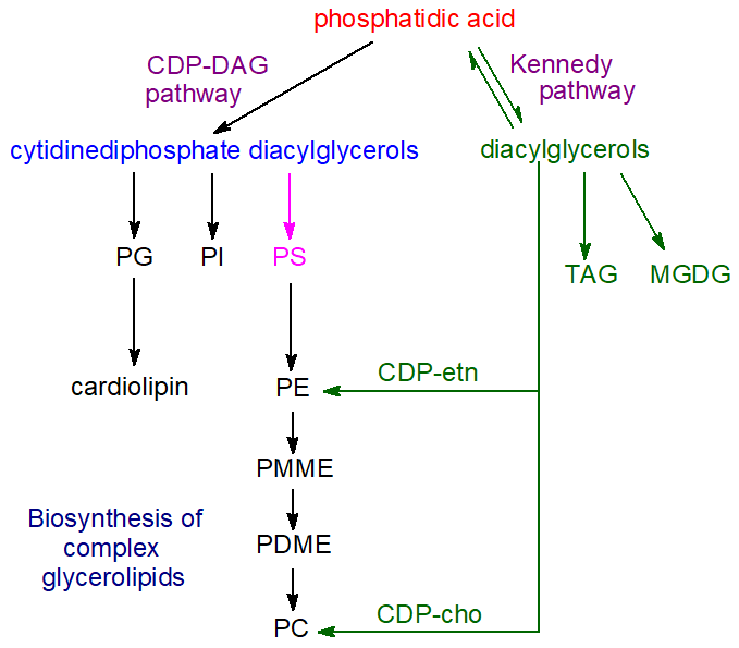
In summary, phosphatidic acid generated via 1-acyl-sn-glycerol-3-phosphate is the primary precursor of other glycerolipids, although other pathways may be more important for generating the lipid for signaling functions. Whether separate pools of this lipid exist for specific purposes is not certain, as its intracellular distribution changes dynamically under various cellular conditions. This may be due to inter-organelle transfer via vesicular transport or to lipid transfer proteins at membrane contact sites. Control of its concentration in membranes, especially in the endoplasmic reticulum, is therefore of great importance, and a transcriptional repressor, 'Opi1', which binds specifically to phosphatidic acid in membranes, is a key regulatory factor. However, many other phosphatidic acid-binding proteins have been identified that influence how phosphatidic acid is used, either as a biosynthetic precursor or for signaling. The mechanisms of phosphatidic acid homeostasis differ among animals, plants, yeasts, and bacteria, reflecting the differing functional requirements of these organisms. Figure \(\PageIndex{6}\) shows the pathways for biosynthesis of complex glycerolipids.
In addition to dietary, hormonal, and tissue-specific factors in animals, the extent to which fatty acids are channeled either into triacylglycerol synthesis for storage in lipid droplets and secretion in lipoproteins or into glycerophospholipids for membrane formation depends to a large extent upon the enzymes of the glycerol-3-phosphate pathway, their isoform expression, activities, and locations. On the other hand, phosphatidic acid is not only a biosynthetic precursor of other lipids but also a regulatory molecule in the transcriptional control of glycerolipid synthesis genes, and regulating its concentration in cells is similarly essential. For example, the local concentration of phosphatidic acid in the endoplasmic reticulum is an important factor in lipid droplet biogenesis.
The subsequent steps in the biosynthesis of triacylglycerols and the various glycerophospholipids using phosphatidic acid are described in separate documents on this website. Thus, hydrolysis of phosphatidic acid by phosphatidate phosphatase enzymes (including lipins 1, 2, and 3) is the source of most other glycerolipids, e.g., sn‑1,2‑diacylglycerols (DG), which are the precursors for the biosynthesis of triacylglycerols (TAG), phosphatidylcholine (PC), and phosphatidylethanolamine (PE) via the so-called Kennedy pathway (also of monogalactosyldiacylglycerols in plants). Via reaction with cytidine triphosphate, phosphatidic acid is the precursor of cytidine diphosphate diacylglycerol, which is the key intermediate in the synthesis of phosphatidylglycerol (PG), and thence of cardiolipin (CL), and of phosphatidylinositol (PI), and, in prokaryotes and yeast but not animals, phosphatidylserine (PS). Depending on the organism and other factors, phosphatidylserine can serve as a precursor for phosphatidylethanolamine, which can then give rise to phosphatidylcholine via mono- and dimethyl-phosphatidylethanolamine intermediates. Cytidine diphosphate diacylglycerol synthase is another enzyme that consumes phosphatidic acid, helping modulate phosphatidic acid levels in cells and regulate processes mediated by this lipid.
While the fatty acid composition of phosphatidic acid can resemble that of the eventual products, the latter are generally much altered by remodeling after synthesis via deacylation-reacylation reactions.
Click for more details.
- Link
-
Phosphatidic Acid - Biological Functions in Animals
In addition to its role as an intermediate in lipid biosynthesis, phosphatidic acid, especially that generated by the action of phospholipase D and by diacylglycerol kinases, may have signaling functions as a second messenger. However, it is not clear whether all the activities observed in vitro are also observed in vivo. Nonetheless, phosphatidic acid has been implicated in many aspects of animal cell biochemistry and physiology.
Some of the observed effects may be explained simply by the physical properties of phosphatidic acid, which tends to form a hexagonal II phase, especially in the presence of calcium ions. Thus, hydrolysis of phosphatidylcholine, a cylindrical, non-fusogenic lipid, converts it into cone-shaped phosphatidic acid, which promotes negative membrane curvature and membrane fusion. It differs from other anionic phospholipids in that its small anionic phosphomonoester head group lies very close to the hydrophobic interior of the lipid bilayer. In model systems, phosphatidic acid can induce membrane fusion, likely because it can form non-bilayer phases. For example, phosphatidic acid biosynthesis is believed to favor intraluminal budding of endosomal membranes, leading to exosome formation. In many cell types, vesicle trafficking, secretion, and endocytosis may require phosphatidic acid produced by phospholipase D.
Its overall negative charge is also relevant, and it is not always clear whether observed biological effects are specific to phosphatidic acid or to negatively charged phospholipids in general. In contrast to phosphoinositide-interacting proteins, which have defined structural folds, effector proteins that bind phosphatidic acid lack highly conserved binding motifs. However, positively charged lysine and arginine residues can bind phosphatidic acid with some specificity via hydrogen bonding to the phosphate group, distinguishing it from other phospholipids. An ‘electrostatic-hydrogen bond switch model’ has been proposed in which the head group of phosphatidic acid forms a hydrogen bond to amino acid residues, leading to deprotonation of the head group, increasing its negative charge from -1 to -2, and thus enabling stronger interactions with basic residues and tight docking with the membrane-interacting protein. In this way, phosphatidic acid can tether certain proteins to membranes, simultaneously inducing conformational changes, hindering ligand binding, and/or oligomerizing proteins to alter their catalytic activity, stability, and interactions with other molecules. It functions as a cellular pH sensor because protein binding depends on intracellular pH and the protonation state of its phosphate head group.
One key target of phosphatidic acid is mTOR, a serine/threonine protein kinase that, through a signaling cascade, regulates cell growth, proliferation, motility, and survival, as well as protein synthesis and transcription, by integrating nutrient and growth factor signals. This forms two distinct complexes of accessory proteins that regulate downstream targets. Of these, mTORC1 interacts directly with phosphatidic acid, and this interaction allosterically activates the enzyme complex to regulate protein synthesis, mitochondrial metabolism, and the transcription of enzymes involved in lipid synthesis. In contrast, phosphatidic acid appears to inhibit mTORC2 activity, for example, in relation to insulin signaling.
Phosphatidic acid is believed to regulate membrane trafficking events, and it is involved in activating the enzyme NADPH oxidase, which operates as part of the defense mechanism against infection and tissue damage during inflammation. It may increase or inhibit their activities by binding to targeted proteins, including protein kinases, protein phosphatases, and G-proteins. Effects on gene transcription have been observed linked to the inhibition of peroxisome proliferator-activated receptor (PPAR) activity. In yeast, phosphatidic acid in the endoplasmic reticulum binds directly to a specific transcriptional repressor to keep it inactive outside the nucleus; when the lipid precursor inositol is added, this phosphatidic acid is rapidly depleted, releasing the transcriptional factor so that it can be translocated to the nucleus, where it can repress target genes. Overall, this mechanism controls phospholipid synthesis.
In addition, phosphatidic acid regulates many aspects of phosphoinositide function. For example, murine phosphatidylinositol 4-phosphate 5-kinase, the main enzyme that generates the lipid second messenger phosphatidylinositol-4,5-bisphosphate, does not appear to function unless phosphatidic acid binds to it; this lipid, generated by phospholipase D, recruits the enzyme to the membrane and induces a conformational change that regulates its activity. It may promote phospholipase A2 activity, a key enzyme in producing eicosanoids from phosphoinositide precursors.
In relation to signaling activities, it should be noted that phosphatidic acid can be metabolized to sn-1,2-diacylglycerols or lysophosphatidic acid (see next section), both of which have distinctive signaling functions in their own right. Conversely, conversion back to phosphatidic acid can effectively deactivate both compounds.
Phospholipase D isoforms and phosphatidic acid have been implicated in a variety of pathologies, including neurodegenerative diseases, blood disorders, late-onset Alzheimer's disease, and cancer, prompting efforts to develop specific enzyme inhibitors for therapeutic use. Similarly, LPAT isoform expression can enhance the proliferation and chemoresistance of some cancer cells. Diacylglycerol kinase alpha (DGKα) is highly expressed in several refractory cancer cells, attenuating apoptosis and promoting proliferation. In addition, DGKα is highly abundant in T cells and induces a nonresponsive state, enabling advanced cancers to evade immune action. Inhibiting this enzyme is also a promising treatment strategy.
Phosphatidic Acid - Biological Functions in Plants
Phosphatidic acid is present at higher levels in plant roots than in leaves and is believed to play a role in root architecture. Similarly, its concentration is elevated in flowers and reproductive tissues, but its significance is unknown. In addition to its role as a central molecule in lipid biosynthesis, it facilitates lipid transport across plant membranes and serves as a key plant lipid second messenger, rapidly and transiently generated in response to many biotic and abiotic stresses. In contrast to animal metabolism, the diacylglycerol signaling pathway is believed to be relatively insignificant in plants.
The main source of phosphatidic acid for these purposes is phospholipase D (PLD) acting on membrane phospholipids, such as phosphatidylcholine and phosphatidylethanolamine. Plants contain numerous related enzymes: 12 in Arabidopsis and 17 in rice, compared with 2 in humans and 1 in yeast, and individual isozymes may elicit specific responses. Researchers group the isoforms into six classes based on gene architecture, sequence similarity, domain structure, and biochemical properties. These depend mainly on their lipid-binding domains, with some homologous to the human and yeast enzymes and most containing a characteristic ‘C2’ (calcium- and lipid-binding) domain. The most widespread of these is PLDα, which does not require binding to phosphatidylinositol 4,5-bisphosphate, unlike other PLD isoforms and the mammalian enzyme, but still requires millimolar levels of Ca2+. Studies with fluorescent biosensors suggest that phosphatidic acid accumulates in the subapical region of the cytosolic leaflet of the plasma membrane.
Phosphatidic acid can also be produced by the sequential action of phospholipase C and diacylglycerol kinase on membrane inositol phospholipids, with diacylglycerols as an intermediate (there are seven isoenzymes in A. thaliana). One difference from animal metabolism is that plants can synthesize diacylglycerol pyrophosphate from phosphatidic acid (see below).
Phosphatidic acid is required to bind and allosterically activate the monogalactosyldiacylglycerol synthase (MGDG1), located in the inner envelope membrane of the chloroplast. It may regulate thylakoid membrane biosynthesis. Phospholipase D activity and the phosphatidic acid it produces have long been recognized as important during germination and senescence, and they play an essential role in responses to stress damage and pathogen attack in both higher plants and green algae. High phosphatidic acid levels induced by phospholipase D activity during wounding or senescence can disrupt the membrane bilayer phase because this negatively charged phospholipid has a conical shape compared with the cylindrical shape of structural phospholipids. This change in ionization properties has crucial effects on lipid-protein interactions, as described by the electrostatic-hydrogen bond switch model above. Promoting negative curvature at the plasma membrane and binding to clathrin proteins are believed to facilitate endocytosis. Similar phenomena may explain why phosphatidic acid is important in responding to other forms of stress, including osmotic stress (salinity or drought), cold, and oxidation. Although much remains to be learned about the mechanism by which it exerts its effects, it is believed to promote the response to the plant hormone abscisic acid. In addition, phosphatidic acid may interact with salicylic acid to mediate defense responses.
In plants, phosphatidic acid is involved in many different cell responses induced by hormones, stress, and developmental processes. In cellular signaling, it often acts alongside phosphatidylinositol 4,5-bisphosphate by binding specific proteins rather than acting through a receptor. As in mammalian cells, targets of such signaling include protein kinases and phosphatases, as well as proteins involved in membrane trafficking and cytoskeletal organization. It can both activate and inhibit enzymes. If the target protein is soluble, binding to phosphatidic acid can sequester it into the membrane, affecting downstream targets. For example, it promotes pollen tube and root hair growth, reduces peroxide-induced cell death, and mediates signaling processes that lead to responses to ethylene and, again, to the hormone abscisic acid. Thus, in the 'model' plant Arabidopsis, phosphatidic acid interacts with a protein phosphatase to signal the closure of stomata promoted by abscisic acid; it also interacts with a further enzyme to mediate the inhibition of stomatal opening induced by abscisic acid. Together, these reactions constitute a signaling pathway that regulates plant water loss.
Notably, phosphatidic acid production can be initiated by opposing stress factors, such as cold and heat, as well as by antagonistic hormones, such as abscisic acid and salicylic acid. Phosphatidic acid molecules synthesized by the two main pathways may differ in composition and cellular distribution and may produce different responses, but this remains an open question. During low-temperature stress, diacylglycerol kinase generates phosphatidic acid. The cellular environment where the lipid is produced and the availability of target proteins or other molecules that can act synergistically likely control these differing activities. Researchers have manipulated genes encoding enzymes involved in phosphatidic acid metabolism to explore their potential for crop improvement, based on effects on plant growth, development, and stress responses.
As in animals, phosphatidic acid is catabolized, and its signaling functions are terminated by lipid phosphate phosphatases and phosphatidic acid hydrolases, as well as by acyl-hydrolases and lipoxygenases. These produce fatty acids and other small molecules that are subsequently absorbed and recycled.
Lysophosphatidic Acid
Figure \(\PageIndex{7}\) shows the structure of a lysophosphatidic acid (note the absence of an acyl group at C2).
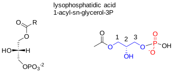
Lysophosphatidic acid (LPA), or 1-acyl-sn-glycerol-3-phosphate, differs structurally from phosphatidic acid by having only one mole of fatty acid per mole of lipid. As such, it is one of the simplest possible glycerophospholipids. It exists in many molecular species, i.e., esterified to fatty acids ranging from 16:0 to 22:6, and preliminary evidence suggests that saturated and polyunsaturated species may differ in their biological properties under some circumstances. As the sn-1-acylated form is more thermodynamically stable, facile isomerization ensures that it tends to predominate. It lacks one fatty acid compared to phosphatidic acid, making it much more hydrophilic. At the same time, the additional hydroxyl group strengthens hydrogen bonding within membranes, properties that may be important for its function in cells.
Although lysophosphatidic acid is present at very low levels only in animal tissues, it is biologically important and influences many biochemical processes. It is a biosynthetic precursor of phosphatidic acid, but it is also of particular interest as a lipid mediator with growth factor-like activities. For example, activated platelets rapidly produce and release it to influence target cells.
Biosynthesis: In the circulation, the most important source of lysophosphatidic acid is the activity of an enzyme with lysophospholipase D-like activity, known as ‘autotaxin ’, on lysophosphatidylcholine (200 μM in plasma) to yield LPA in an albumin-bound form, mainly, although it is relatively soluble in aqueous media because of its polarity and small size. This lipid is more abundant in serum (1 to 5 μM) than in plasma (100 nM) because its main precursor, lysophosphatidylcholine, is released from activated platelets during coagulation. Autotaxin belongs to the nucleotide pyrophosphatase-phosphodiesterase family. It is also present in cerebrospinal and seminal fluids, as well as in many other tissues, including cancer cell lines from which it was first isolated and characterized. Indeed, the name derives from the finding that it promoted chemotaxis in melanoma cells in an autocrine fashion. It binds to target cells via integrins and heparan sulfate proteoglycans, which may help deliver lysophosphatidic acid to its receptors. Genetic deletion of the enzyme in mice results in aberrant vascular and neuronal development and soon leads to embryonic death. However, the overexpression of autotaxin also causes physical defects and is eventually lethal to embryos.
Figure \(\PageIndex{8}\) shows the pathways for lysophosphatidic acid synthesis.

While autotaxin is the primary source of extracellular lysophosphatidic acid, it is now established that a wide variety of cell types produce it intracellularly through various mechanisms, often using phosphatidic acid derived from other phospholipids as the primary precursor via phospholipase D. For example, the hydrolysis of phosphatidic acid by a phospholipase A2 (PLA2) is the main mechanism in platelets. Other cellular enzymes include a phosphatidic acid-selective phospholipase A1 (PLA1), which produces sn-2-acyl-lysophosphatidic acid; a monoacylglycerol kinase (utilizing monoacylglycerols produced by lipid phosphate phosphatases); and glycerol-3-phosphate acyltransferase (the first step in phosphatidic acid biosynthesis). In particular, secretory PLA2-IIA (sPLA2-IIA) can induce the release of LPA from phosphatidic acid exposed on the surface of extracellular vesicles derived from platelets and Ca2+-loaded erythrocytes upon stimulation by pro-inflammatory cytokines.
Click the link below.
- Link
-
General function: Although lysophospholipids are relatively small molecules, they carry a high content of information through the nature of the phosphate head group, the positional distribution of the fatty acids on the glycerol moiety, the presence of ether or ester linkages to the glycerol backbone, and the chain-length and degree and position of saturation of the fatty acyl chains. Lysophosphatidic acid acts on nearly all cell types, often as a proliferative and pro-survival signal, inducing cellular invasion, migration, and differentiation while stimulating smooth muscle and fibroblast contraction, cytoskeletal rearrangement, cytokine/chemokine secretion, and numerous other effects. The 1-O-alkyl- and alkenyl-ether forms also display many of these activities, which can be derived from the platelet-activating factor. On the other hand, much of the lysophosphatidic acid produced intracellularly may be used to synthesize other phospholipids rather than for signaling purposes.
Receptors: The molecular information in the lysophosphatidic acid molecule confers selectivity in its functional interactions with cell receptors. Because most mammalian cells express lysophosphatidic acid receptors, this lipid may initiate signaling in the cells where it is produced and affect neighboring cells. Characterization of cloned lysophosphatidic acid receptors, combined with molecular genetic strategies, has allowed determination of signaling and biological effects dependent on receptor mechanisms. Researchers have identified at least six lysophosphatidic acid-specific G protein-coupled receptors in vertebrates, each found in particular organs and coupled to one or more of the four heterotrimeric Gα proteins, designated LPAR1 to LPAR6; LPAR1 is virtually ubiquitous in tissues. These vary appreciably in amino acid sequences but are classified into two subgroups, the EDG (LPAR1-3) and P2Y (LPAR4-6) families, with differing tissue distributions. Most cell types express these receptors in different combinations. They also interact with transient receptor potential cation channel V1 (TRPV1), peroxisome proliferator-activated receptor gamma (PPARγ), and other proteins. Plasma lysophosphatidic acid binds to its receptors while it is bound to albumin.
Experimental activation of LPAR receptors has shown that lysophosphatidic acid signaling through these receptors mediates a range of downstream signaling cascades. These include activation of adenylyl cyclase, cAMP production, intracellular Ca2+ and K+ production (by activating ion channels), protein kinases, phospholipase C, phosphatidylinositol 3-kinase, small GTPases (Ras, Rho, Rac), release of arachidonic acid, and much more. In this way, lysophosphatidic acid regulates cell survival, proliferation, cytoskeletal rearrangement, motility, cytokine secretion, cell differentiation, and many other vital cellular processes. Sometimes, lysophosphatidic acid appears to function in contradictory ways, with evidence that it promotes cell survival in some circumstances and programmed cell death in others.
Lysophosphatidic acid signaling has regulatory functions in the mammalian reproductive system, in both males and females, facilitating oocyte maturation and spermatogenesis through the action of receptors LPAR1 to LPAR3. During early gestation, it regulates vascular remodeling at the maternal-fetal interface. Lipids also support brain development by acting in neural progenitor cells, neurons, glia, and vascular remodeling. In the central nervous system, these receptors are thought to play a central role in triggering and maintaining neuropathic pain by mechanisms that may involve the demyelination of damaged nerves.
Lysophosphatidic acid has been detected in saliva at significant levels, and it has been suggested to be involved in wound healing in upper digestive organs, such as the mouth, pharynx, and esophagus. When applied topically to skin wounds, it has similar effects, probably by stimulating the proliferation of new cells to seal the wound. Receptor LPAR6, together with phospholipase A1, is required for hair follicle development, and it also regulates endothelial blood-brain barrier function. The proliferation and survival of stem cells and their progenitors are regulated by lysophosphatidic acid signaling. In contrast, in bone cells, lysophosphatidic acid acts via LPAR1 and is important for bone mineralization and repair.
Disease: Lysophosphatidic acid plays a particular role in various disease states and cancers, especially given reports of increased autotaxin expression and subsequent lysophosphatidic acid levels in several primary tumors. For example, a finding that lysophosphatidic acid is markedly elevated in the plasma and peritoneal fluid (ascites) of ovarian cancer patients compared to healthy controls may be especially significant. Also, elevated plasma levels were found in patients in the first stage of ovarian cancer, suggesting that it may represent a useful marker for the early detection of the disease. The secretory form of phospholipase A2 is believed to act preferentially on lipids from damaged membranes or microvesicles, such as those produced by malignant cells, thereby increasing this lipid's levels. Lysophosphatidic acid has been shown to stimulate the expression of genes encoding many different enzymes that promote the proliferation of ovarian and other cancer cells and may induce cell migration via receptors LPAR1 to LPAR3, and possibly LPAR6. At the same time, LPAR4 and LPAR5 have opposing effects. Autotaxin and LPARs have been implicated in resistance to chemotherapy and radiation treatment in cancer therapy.
As lysophosphatidic acid has growth-factor-like activities in many cell types, inducing cell proliferation and migration, altering cellular shape, and increasing endothelial permeability, it is perhaps not surprising that it is relevant to tumor biology. Treatment of various cancer cell types with lysophosphatidic acid increases the expression and release of interleukin-8 (IL-8), a potent angiogenic factor. Thus, it plays a critical role in cancer growth and spread by enhancing nutrient and oxygen availability. Evidence suggests that lysophosphatidic acid signaling is causally linked to hyperactive lipogenesis in cancer. For example, it activates the sterol regulatory element-binding protein (SREBP), together with the fatty acid synthase and AMP-activated protein kinase–ACC lipogenic cascades, leading to elevated de novo lipid synthesis. Autotaxin expression is increased in many cancer cell lines, and many lysophosphatidic acid surface receptors are aberrantly expressed in cancer cells. Cancer cells must evade the immune system during metastasis, and lysophosphatidic acid facilitates this evasion by inhibiting T cell activation. Therefore, lysophosphatidic acid metabolism is a pharmaceutical target in the search for new cancer therapeutics, aided by knowledge of the crystal structures of three receptors.
Lysophosphatidic acid signaling has been implicated in many aspects of chronic inflammation, which it promotes by affecting the endothelium in several ways, for example, by stimulating endothelial cell migration, secreting chemokines and cytokines, and regulating endothelial barrier integrity. Problems with lysophosphatidic acid signaling, together with changes in autotaxin expression, are believed to contribute to metabolic and inflammatory disorders such as obesity, insulin resistance, non-alcoholic fatty liver disease, rheumatoid arthritis, multiple sclerosis, and cardiovascular disease. Further, there is evidence that it contributes to neurological disorders, such as Alzheimer's disease and neuropathic pain, and asthma, fibrosis, and bone malfunction. Drugs that interact with lysophosphatidic acid receptors have been reported to attenuate symptoms of several diseases in animal models, and three have completed phase I and II clinical trials in human patients with idiopathic pulmonary fibrosis and systemic sclerosis. Drugs that target autotaxin production and lysophosphatidic acid catabolism are also in development, and the steroidal anti-inflammatory agent dexamethasone appears to be especially useful.
Under certain conditions, lysophosphatidic acid can become athero- and thrombogenic and might aggravate cardiovascular disease. As oxidized low-density lipoproteins promote lysophosphatidic acid production, their high levels in atherosclerotic plaques suggest they may serve as a biomarker for cardiovascular disease. Indeed, lysophosphatidic acid promotes pro-inflammatory events that lead to atheroma development and encourage disease progression. It may promote arterial thrombus formation by mediating platelet aggregation.
Related lipids: The sphingolipid analog, sphingosine-1-phosphate, shows a similar range of activities to lysophosphatidic acid, and the two lipids are often discussed in the same contexts, although they may have opposing effects. Acute leukemia cells produce methyl-lysophosphatidic acids (the polar head-group is methylated). Because these act as antigens that a specific group of human T cells reacts strongly to, they may be immunotherapy targets in hematological malignancies. Other lysophospholipids have distinctive biological functions.
Catabolism: A family of three lipid phosphate phosphatases (LPP1, 2, and 3) dephosphorylates lysophosphatidic acid to produce monoacylglycerols; they also dephosphorylate sphingosine-1-phosphate, phosphatidic acid, and ceramide 1-phosphate in a non-specific manner. These integral membrane proteins have active sites on the plasma membrane facing the extracellular environment, allowing them to access and hydrolyze extracellular lysophosphatidic acid and other phospholipids. Mice with a constitutive LPP3 deficiency are not viable, but this is not true for LPP1 and LPP2 knockout mice. Lysophosphatidic acid can be converted back to phosphatidic acid by a membrane-bound O-acyltransferase (MBOAT2) specific for lysophosphatidic acid (and lysophosphatidylethanolamine) with a preference for oleoyl-CoA as substrate.
Phosphatidylcholine and Related Lipids
Phosphatidylcholine - Structure and Occurrence
Phosphatidylcholine or 1,2-diacyl-sn-glycero-3-phosphocholine (once given the trivial name 'lecithin') is a neutral or zwitterionic phospholipid over a pH range from strongly acidic to strongly alkaline. It is usually the most abundant phospholipid in animals and plants, accounting for almost 50% of complex lipids. As such, it is a key building block of membrane bilayers. In particular, it makes up a very high proportion of the lipids of the outer leaflet of the plasma membrane in animals. Virtually all the phosphatidylcholine in human erythrocyte membranes is in the outer leaflet. In the plasma membranes of nucleated cells, 80-90% of this lipid is located on the outer leaflet. Phosphatidylcholine is also the principal phospholipid circulating in plasma, where it is an integral component of the lipoproteins, especially the HDL. On the other hand, it is less common in bacterial membranes, perhaps ~10% of species, but it is absent in the 'model' organisms Escherichia coli and Bacillus subtilis. In animal tissues, some of its membrane functions appear to be shared with the structurally related sphingolipid, sphingomyelin, although the latter has many unique properties.
Figure \(\PageIndex{9}\) shows the structure of phosphatidylcholine
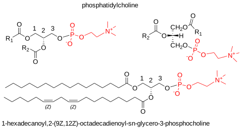
In animal tissues, phosphatidylcholine predominantly exists in the diacyl form. However, small proportions (compared with phosphatidylethanolamine and phosphatidylserine) of alkyl, acyl, and alkenylacyl forms may also be present. In general, animal phosphatidylcholine contains lower proportions of arachidonic and docosahexaenoic acids and higher proportions of C18 unsaturated fatty acids than the other zwitterionic phospholipid, phosphatidylethanolamine. Saturated fatty acids are most abundant in position sn-1, while polyunsaturated components are concentrated in position sn-2. Indeed, C20 and C22 polyenoic acids are exclusively in position sn-2, yet in the brain and retina, the unusual very-long-chain polyunsaturated fatty acids (C30 to C38) of the n-6 and n-3 families occur in position sn-1. Dietary factors influence fatty acid composition, but across animal species, we expect phosphatidylcholine in the same metabolically active tissue to have similar relative distributions of fatty acids between the two positions. Table \(\PageIndex{1}\) lists some representative data.
| Table \(\PageIndex{1}\). Positional distribution of fatty acids in the phosphatidylcholine of some animal tissues. | |||||||
| Position | Fatty acid | ||||||
|---|---|---|---|---|---|---|---|
| 16:0 | 16:1 | 18:0 | 18:1 | 18:2 | 20:4 | 22:6 | |
| Rat liver [1] | |||||||
| sn-1 | 23 | 1 | 65 | 7 | 1 | trace | |
| sn-2 | 6 | 1 | 4 | 13 | 23 | 39 | 7 |
| Rat heart [2] | |||||||
| sn-1 | 30 | 2 | 47 | 9 | 11 | - | - |
| sn-2 | 10 | 1 | 3 | 17 | 20 | 33 | 9 |
| Rat lung [3] | |||||||
| sn-1 | 72 | 4 | 15 | 7 | 3 | - | - |
| sn-2 | 54 | 7 | 2 | 12 | 11 | 10 | 1 |
| Human plasma [4] | |||||||
| sn-1 | 59 | 2 | 24 | 7 | 4 | trace | - |
| sn-2 | 3 | 1 | 1 | 26 | 32 | 18 | 5 |
| Human erythrocytes [4] | |||||||
| sn-1 | 66 | 1 | 22 | 7 | 2 | - | - |
| sn-2 | 5 | 1 | 1 | 35 | 30 | 16 | 4 |
| Bovine brain (gray matter) [5] | |||||||
| sn-1 | 38 | 5 | 32 | 21 | 1 | - | - |
| sn-2 | 33 | 4 | trace | 48 | 1 | 9 | 4 |
| Chicken egg [6] | |||||||
| sn-1 | 61 | 1 | 27 | 9 | 1 | - | - |
| sn-2 | 2 | 1 | trace | 52 | 33 | 7 | 4 |
| 1, Wood, R. and Harlow, R.D. Arch. Biochem. Biophys., 131, 495-501 (1969); DOI. 2, Kuksis, A. et al. J. Lipid Res., 10, 25-32 (1969); DOI. 3, Kuksis, A. et al. Can. J. Physiol. Pharm., 46, 511-524 (1968); DOI. 4, Marai, L. and Kuksis, A. J. Lipid Res., 10, 141-152 (1969); DOI. 5, Yabuuchi, H. and O'Brien, J.S. J. Lipid Res., 9, 65-67 (1968); DOI. 6, Kuksis, A. and Marai, L. Lipids, 2, 217-224 (1967); DOI. |
|||||||
Some exceptions exist: phosphatidylcholine in some tissues or organelles contains relatively high proportions of di-saturated molecular species. For example, lung phosphatidylcholine in most, if not all, animal species studied to date contains a high proportion (50% or more) of dipalmitoylphosphatidylcholine.
The positional distributions of fatty acids in phosphatidylcholine in representative plants and yeast are listed in Table \(\PageIndex{2}\). In the leaves of the model plant Arabidopsis thaliana, saturated fatty acids are concentrated at sn-1, but monoenoic fatty acids are distributed approximately equally between the two positions, and di- and triunsaturated fatty acids predominate at sn-2; the same is true for soybean lecithin. The pattern is somewhat similar in the yeast Lipomyces lipoferus, except that much of the 16:1 is in position sn-1.
| Table \(\PageIndex{2}\): Composition of fatty acids (mol%) in positions sn-1 and sn-2 in the phosphatidylcholine from plants and yeast. | ||||||
| Position | Fatty acid | |||||
|---|---|---|---|---|---|---|
| 16:0 | 16:1 | 18:0 | 18:1 | 18:2 | 18:3 | |
| Arabidopsis thaliana (leaves) [1] | ||||||
| sn-1 | 42 | 4 | 5 | 23 | 26 | |
| sn-2 | 1 | trace | 5 | 47 | 47 | |
| Soybean 'lecithin' [2] | ||||||
| sn-1 | 24 | 9 | 14 | 47 | 4 | |
| sn-2 | 5 | 1 | 13 | 75 | 6 | |
| Lipomyces lipoferus [3] | ||||||
| sn-1 | 24 | 18 | trace | 37 | 16 | 4 |
| sn-2 | 4 | 5 | trace | 39 | 31 | 19 |
| 1, Browse, J., Warwick, N., Somerville, C.R., and Slack, C.R. Biochem. J., 235, 25-31 (1986); DOI. 2, Blank, M.L., Nutter, L.J. and Privett, O.S. Lipids, 1, 132-135 (1966); DOI. 3, Haley, J.E. and Jack, R.C. Lipids, 9, 679-681 (1974); DOI. |
||||||
Phosphatidylcholine – Biosynthesis
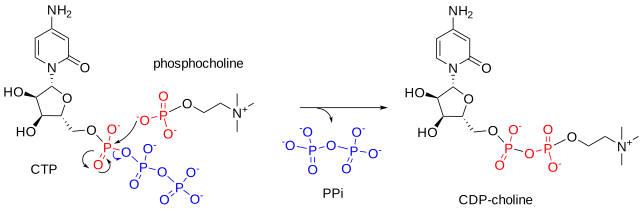
Several mechanisms support phosphatidylcholine biosynthesis in animals, plants, and microorganisms. Choline itself is not synthesized by animal cells. It is an essential nutrient for phospholipid synthesis and cholinergic neurotransmission (acetylcholine synthesis), and it provides methyl groups for numerous other metabolites. It must be obtained from dietary sources or by degradation of existing choline-containing lipids, for example, those produced by the second pathway described below. Once taken across membranes and into cells by specific transporters, choline is immediately phosphorylated by a choline kinase (1) in the cytoplasm of the cell to produce phosphocholine, which is reacted with cytidine triphosphate (CTP) by the enzyme CTP:phosphocholine cytidylyltransferase (CCT) (2) to form cytidine diphosphocholine (CDP-choline).
CTP + PC → CDP-choline + Pi
The latter enzyme exists in two isoforms, of which CCTα is the more important. It is a soluble protein found first in the nucleoplasm and then in the nucleoplasmic reticulum. This is considered the rate-limiting step in phosphatidylcholine biosynthesis, and the enzyme's activity is regulated by signals from a membrane sensor that reports on the relative abundance of the final product. However, choline kinase (ChoKα) also has regulatory functions.
Figure \(\PageIndex{10}\) shows an interactive iCn3D model of the mammalian (rat) CTP: Phosphocholine cytidylyltransferase catalytic domain (3HL4).

 Figure \(\PageIndex{10}\): Mammalian (rat) CTP-Phosphocholine cytidylyltransferase catalytic domain (3HL4). Click the image for a popup or use this external link: https://structure.ncbi.nlm.nih.gov/i...tBYRYk7vPKD7f6
The biologically active homodimer is shown. The B chain is colored by secondary structure, and the A chain is shown in gray. Key active-site residues (shown only in the A chain) are labeled and shown as CPK-colored sticks. A bound CDP-choline analog ((2-cytidylate-O'-phosphonyloxy)-ethyl-trimethylammonium) is shown in spacefill CPK colors.
This enzyme (CCT) catalyzes the key regulatory and rate-limiting step in PC synthesis. The C-terminal domain binds membrane lipids and regulates the enzyme. The iCn3D model above is for the catalytic domain with the regulatory domain deleted. Two nonconserved active site side chains, His 168 and Tyr 173, interact with and position the phosphocholine. Other active site residues include Arg-196 in L6, Lys-122 in L2, and Asp-94. Figure \(\PageIndex{11}\) shows a simplified mechanism for the reaction
In plants, nematodes, and certain parasites, phospho-base N-methyltransferases synthesize most phosphocholine by sequentially methylating phosphoethanolamine. However, only a few plant species methylate phosphatidylethanolamine this way. This is also the main route to free choline and betaine in plants.
The CDP-choline produced is acted upon by the membrane-bound enzyme CDP-choline:1,2-diacylglycerol choline/ethanolamine-phosphotransferase in the endoplasmic reticulum (CEPT1) and a related choline phosphotransferase 1 (CPT1) in the trans-Golgi, which catalyze the reaction with sn-1,2-diacylglycerols to form phosphatidylcholine. The first is responsible for most phosphatidylcholine biosynthesis, but it uses a somewhat different molecular species composition than the second, which prefers 1-alkyl precursors. This is the main pathway for phosphatidylcholine synthesis in animals and plants, and it is analogous to the major route to phosphatidylethanolamine; it is also found in a few bacterial species (e.g., Sinorhizobium meliloti). Phosphatidylcholine in mitochondria is obtained by transfer from the endoplasmic reticulum.
Figure \(\PageIndex{12}\) shows the main pathways for PC synthesis in plants and animals.

The discovery of this pathway's importance depended somewhat on serendipity. In experiments in Eugene Kennedy's laboratory, samples of adenosine triphosphate (ATP) contained a small amount of cytidine triphosphate (CTP) as an impurity. However, luck is of little value without receptive minds, and Kennedy and co-workers demonstrated that the impurity was an important metabolite essential for the formation of phosphatidylcholine.
The reaction above and the biosynthetic mechanism for phosphatidylethanolamine differ significantly from those for phosphatidylglycerol, phosphatidylinositol, and cardiolipin. Both use nucleotides, but with the latter, the nucleotide is covalently linked directly to the lipid intermediate, cytidine diphosphate diacylglycerol. However, a comparable pathway for phosphatidylcholine biosynthesis occurs in bacteria (see below).
The source of the sn-1,2-diacylglycerol precursor, which is also a key intermediate in forming phosphatidylethanolamine and phosphatidylserine, and triacylglycerols, is phosphatidic acid. In this instance, the important enzyme is phosphatidic acid phosphatase (also known as lipin, phosphatidate phosphatase, lipid phosphate phosphatase, or phosphatidate phosphohydrolase).
Figure \(\PageIndex{13}\) shows the biosynthesis of the diacyl precursor of PC
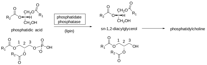
This enzyme is also important for producing diacylglycerols, which are essential intermediates in the biosynthesis of triacylglycerols and phosphatidylethanolamine. Yeasts contain two such enzymes: one Mg2+-dependent (PAP1) and the other Mg2+-independent (PAP2). Much of the phosphatidic acid phosphatase activity in mammals resides in three related cytoplasmic proteins, lipins-1, -2, and -3. Lipin-1 is found mainly in adipose tissue, while lipin-2 is present mainly in the liver. They are unique among glycerolipid biosynthetic enzymes in that they can move between cellular membranes rather than remain tethered. Of these, lipin-1 is the most important and exists in three isoforms, lipin-1α, lipin-1β, and lipin-1γ, with lipin-1α located mainly in the nucleus and lipin-1β in the cytoplasm. Lipin-1γ is present primarily in the brain.
The second pathway for phosphatidylcholine biosynthesis involves sequential methylation of phosphatidylethanolamine, with S-adenosylmethionine (SAM) as the methyl donor, mono- and dimethylphosphatidylethanolamine as intermediates, and phosphatidylethanolamine N‑methyltransferase as the catalyzing enzyme. A single enzyme (~20 kDa) in two isoforms catalyzes all three reactions in hepatocytes; the main form is located in the endoplasmic reticulum (ER), where it spans the membrane, while the second is found in the mitochondria-associated ER membrane. Yeast has at least two N-methyltransferases. This is a major pathway in the liver, generating one-third of the phosphatidylcholine in this organ but not in other animal tissues or, in general, in higher organisms. It may be the main route to phosphatidylcholine in those bacterial species that produce this lipid and in yeasts, but it appears to operate in only a few species of higher plants. This liver pathway is especially important when dietary choline is deficient.
Figure \(\PageIndex{14}\) shows PC synthesis via PE methylation.
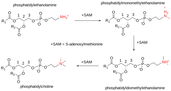
A by-product of phosphatidylcholine biosynthesis from phosphatidylethanolamine is the conversion of S‑adenosylmethionine to S‑adenosylhomocysteine, which the liver hydrolyzes to adenosine and homocysteine. An elevated level of the latter in plasma is a risk factor for cardiovascular disease and myocardial infarction.
Phosphatidylcholine biosynthesis via both pathways in the liver is necessary for normal secretion of plasma lipoproteins (VLDL and HDL) and is relevant to many human physiological conditions. These pathways for diacylphosphatidylcholine biosynthesis differ and are spatially separated from those producing alkyl-, acyl-, and alkenylacyl-phosphatidylcholines de novo. Phosphatidylcholine synthesis also does not occur uniformly throughout the endoplasmic reticulum. It occurs at membrane interfaces or where membranes meet other organelles, especially during dynamic membrane expansion.
The enzymes in the endoplasmic reticulum that synthesize all phospholipids are oriented, so their active sites face exclusively toward the cytosol. Problems would arise if the cytosolic leaflet expanded rapidly while the luminal leaflet did not, but a phospholipid transporter known as a scramblase enables rapid, bidirectional flip-flop of phospholipids between leaflets of the bilayer in an energy-independent manner. Compositional asymmetry first appears in the trans-Golgi and is completed before the plasma membrane forms, with phosphatidylcholine and sphingolipids present mainly in the exofacial (outer) leaflet. Phosphatidylethanolamine and phosphatidylserine are enriched in the cytosolic leaflet.
Dietary phosphatidylcholine is rapidly hydrolyzed in the proximal small intestine by pancreatic enzymes with lysophosphatidylcholine (and free fatty acids) formation. Further hydrolysis can occur in the jejuno-ileal brush-border membrane by membrane phospholipases, releasing glycerophosphocholine. Still, much of the lysophosphatidylcholine is reacylated by the lyso-PC-acyl-CoA-acyltransferase 3 for export in chylomicrons.
In plant cells, phosphatidylcholine biosynthesis occurs mainly in the endoplasmic reticulum, and it is a major component of most membranes other than the internal membranes of plastids; it is absent from the thylakoids and the inner envelope membrane but is the main glycerolipid of the outer monolayer of the outer envelope membrane. Further complications arise in plants, in that turnover or partial synthesis via lysophosphatidylcholine occurs in different organelles, using fatty acid pools or enzymes with differing specificities. In addition, fatty acids esterified to phosphatidylcholine serve as substrates for desaturases. As a result, an appreciable pool of diacylglycerols for the biosynthesis of triacylglycerols, galactosyldiacylglycerols, and other glycerolipids passes through phosphatidylcholine as an intermediate, so fatty acid compositions in different membranes change after the initial synthetic process. This mechanism differs from the remodeling of molecular species in animal tissues, which we discuss next. However, a comparable exchange of acyl groups does occur, in part catalyzed by acyltransferases (see the next section). Some phosphatidylcholine transfer per se from the endoplasmic reticulum to plastids may occur via contact points between the two membranes or may be facilitated by specific transport proteins.
While phosphatidylcholine is a major lipid in yeasts, recent work suggests that it is not essential if suitable alternative growth substrates are available, unlike in higher organisms, where perturbation of phosphatidylcholine synthesis can lead to growth inhibition or even cell death.
Remodeling of Phosphatidylcholine - the Lands' cycle
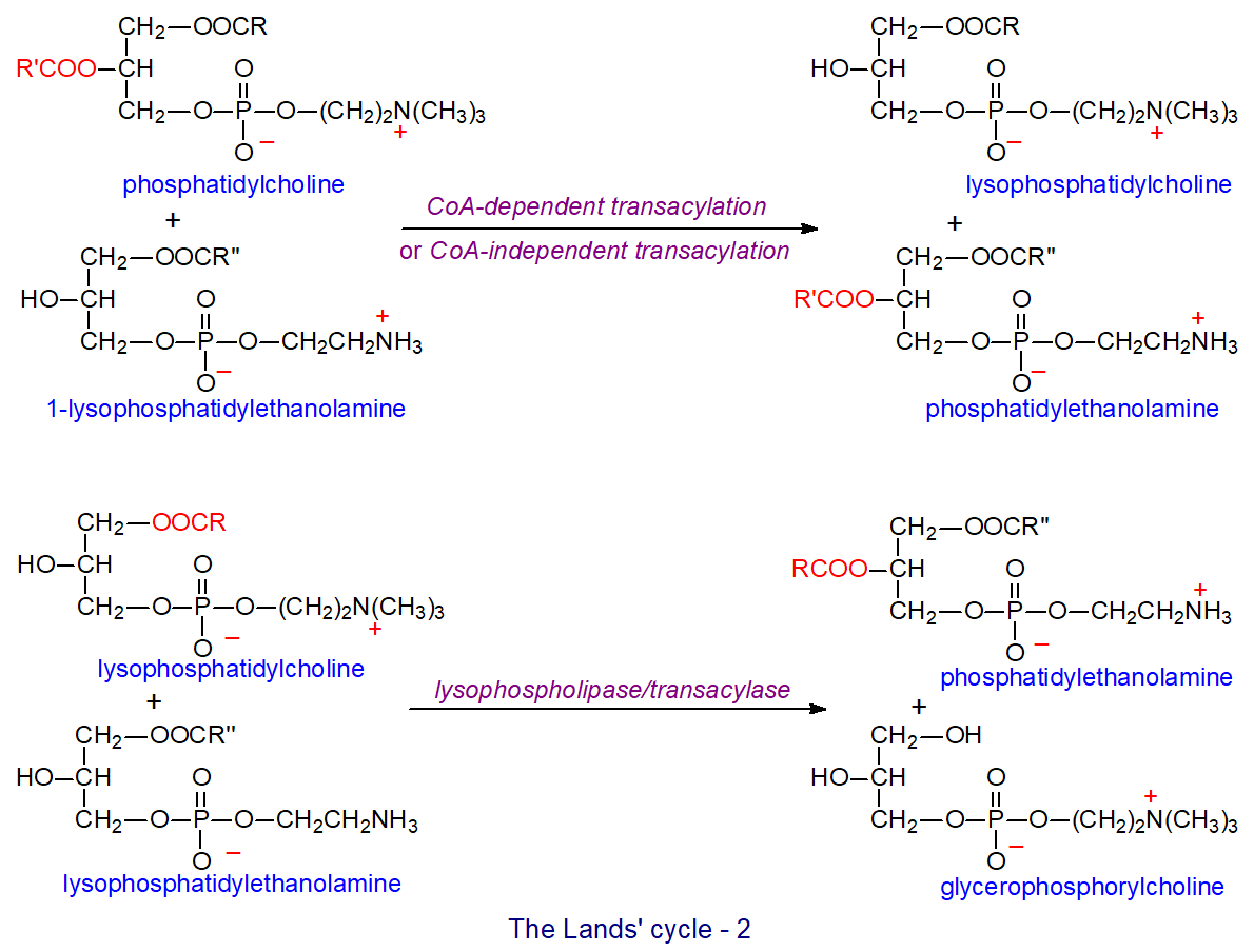
Whatever the mechanism of biosynthesis of phosphatidylcholine in animal tissues, it is apparent that the fatty acid compositions and positional distributions on the glycerol moiety are determined post-synthesis by extensive re-modeling involving orchestrated reactions of hydrolysis (phospholipase A2 mainly) to lysophosphatidylcholine, acyl-CoA synthesis and re-acylation by lysophospholipid acyltransferases or transacylases, a series of reactions that is sometimes termed the 'Lands' Cycle' after its discoverer W.E.M. (Bill) Lands. Similar processes occur with all glycerophospholipid classes.
The final lipid composition results from a mixture of de novo synthesis and remodeling. At least 15 groups of enzymes in the phospholipase A2 superfamily differ in calcium dependence, cellular location, and structure. All hydrolyze the sn-2 ester bond of phospholipids specifically, generating a fatty acid and a lysophospholipid, which have important functions beyond their role in the Lands cycle. A phospholipase A1 family of enzymes are esterases that can cleave the sn-1 ester bond but are less important in this context.
Figure \(\PageIndex{15}\) shows Land's cycle

Membrane-bound, coenzyme A-dependent lysophosphatidylcholine acyltransferases such as LPCAT3 (also designated ‘MBOAT5’) catalyze the re-acylation step; it is located chiefly in the endoplasmic reticulum, but also in mitochondria and the plasma membrane in organs such as the liver, adipose tissue, and pancreas. It maintains systemic lipid homeostasis by regulating lipid absorption and composition in the intestines, lipoprotein secretion, and de novo lipogenesis in the liver; it also incorporates linoleoyl and arachidonoyl chains specifically into lysophosphatidylcholine (as does the related enzyme LPCAT2). Inflammatory cells also express a CoA-independent acyltransferase that transfers arachidonic acid from phosphatidylcholine to ethanolamine-containing phospholipids. While LPCAT3 prefers 1-acyl lysophosphatidylcholine as an acyl acceptor, LPCAT2 utilizes 1-acyl and 1-alkyl precursors. LPCAT2 is highly expressed in inflammatory cells such as macrophages and neutrophils, which contain ether-phospholipids and contribute to the production of eicosanoid lipid mediators. The highly saturated molecular species of phosphatidylcholine found in lung surfactant are formed from species with a more conventional composition by remodeling by an acyltransferase with a high specificity for palmitoyl-CoA acid (LPCAT1). In other tissues, those species containing high proportions of polyunsaturated fatty acids depend more on de novo synthesis. These and further related enzymes are involved in remodeling all other phospholipids. Overexpression of the genes for these enzymes is associated with the progression of many different cancers and may be involved in other pathological conditions.
Phosphatidylcholine plays a central role in plant glycerolipid metabolism, and remodeling occurs for different reasons and by mechanisms that differ from those in animal cells, as briefly described above. For example, extensive remodeling involves fatty acid desaturation, the main entry point for acyl groups exported from the plastid into the endoplasmic reticulum. In addition, phosphatidylcholine remodeling provides fatty acids for triacylglycerol synthesis in developing seeds and diacylglycerols for synthesizing thylakoid lipids such as galactosyldiacylglycerols. In Arabidopsis, two lysophosphatidylcholine acyltransferases, LPCAT1 and LPCAT2, remodel phosphatidylcholine in developing seeds and leaves, preferring the sn-2 position and using fatty acids exported from the plastid. In some plant species, there is a strong preference for C18‑unsaturated acyl chains over 16:0. However, the lipases that generate lysophosphatidylcholine from phosphatidylcholine for this purpose are unknown. Some remodeling in plant membranes occurs in response to stress.
The yeast Saccharomyces cerevisiae can reacylate glycerophosphocholine, generated endogenously by the action of phospholipase B (an enzyme with both phospholipase A1 and A2 activities) on phosphatidylcholine, with acyl-CoA in the microsomal membranes using a glycerophosphocholine acyltransferase (Gpc1) to produce lysophosphatidylcholine, which can be converted back to phosphatidylcholine by the lysophospholipid acyltransferase (Ale1) with appreciable changes in the molecular species composition. This process is regulated in coordination with other major lipid pathways and affects yeast growth. The enzyme Gpc1 does not affect other phospholipids in yeast. A similar mechanism appears to operate in some plant species. Figure \(\PageIndex{16}\) shows the Land's cycle variants.
Catabolism
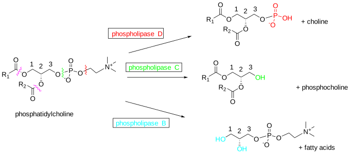
Phosphatidylcholine (and most other glycerophospholipids) in membranes can be metabolized by lipolytic enzymes, especially phospholipases, some of which are specific for particular lipid classes in humans. For example, in addition to the action of phospholipase A (discussed above), phospholipase C (six families in mammals differing in expression and subcellular distribution) yields diacylglycerols together with phosphocholine by hydrolyzing glycerophospholipids at the phosphodiester bond. This process is especially important in phosphoinositide metabolism. The sphingomyelin synthases also have phospholipase C activity (in the absence of ceramide). Phospholipase D generates phosphatidic acid and choline, while phospholipase B removes both fatty acids to yield glycerophosphocholine.
Figure \(\PageIndex{17}\) shows the activities of phospholipases on phosphatidylcholine.
In this way, catabolism recycles lipid components or confers signaling functions. At the same time, much of the choline is reused for phosphatidylcholine biosynthesis, often after being returned to the liver (the CDP-choline cycle). Some choline is oxidized in the kidney and liver to betaine, which serves as a methyl group donor for S-adenosylmethionine production, and some is lost through the excretion of phosphatidylcholine in bile. A proportion is used in nervous tissues to produce acetylcholine, a neurotransmitter important to learning, memory, and sleep. The liver takes up phosphatidylcholine from plasma high-density lipoproteins and, perhaps surprisingly, eventually converts a large proportion of it to triacylglycerols via diacylglycerol intermediates.
Click the link below.
- Link
-
Phosphatidylcholine – Biological Functions
Because of its generally cylindrical shape, phosphatidylcholine organizes spontaneously into bilayers, making it ideally suited to serve as the bulk structural element of biological membranes. As outlined above, it makes up a high proportion of the lipids in the outer leaflet of the plasma membrane. The unsaturated acyl chains are kinked, which confers membrane fluidity. Such properties are essential for balancing those lipids that do not form bilayers or that form specific microdomains, such as rafts. While phosphatidylcholine does not induce membrane curvature, which may be required for membrane transport and fusion processes, it can be metabolized to form lipids that do.
In contrast, dipalmitoyl phosphatidylcholine is the main surface-active component of human lung surfactant. However, in other animals, lung surfactant can be enriched in a combination of short-chain disaturated and monounsaturated species, mainly palmitoylmyristoyl- and palmitoylpalmitoleoyl-, in addition to the dipalmitoyl-lipid. This is believed to provide alveolar stability by decreasing the surface tension at the alveolar surface to a very low level during inspiration while preventing alveolar collapse at the end of expiration. Also, the internal lipids of the animal cell nucleus (after the external membrane has been removed) contain a high proportion of di-saturated phosphatidylcholine. This is synthesized entirely within the nucleus, unlike phosphatidylinositol, for example, and in contrast to other cellular lipids, its composition cannot be changed by extreme dietary manipulation; it has been suggested that it may have a role in stabilizing or regulating the structure of the chromatin, as well as being a source of diacylglycerols with a signaling function. A further unique molecular species, 1-oleoyl-2-palmitoyl-phosphatidylcholine, is located specifically at the protrusion tips of neuronal cells and appears essential for their function. 1-palmitoyl-2-arachidonoyl-phosphatidylcholine is important in regulating cell cycle progression and cell proliferation, independent of eicosanoid production.
Phosphatidylcholine binds noncovalently in the crystal structures of many membrane proteins, including cytochrome c oxidase and yeast cytochrome bc1. The ADP/ATP carrier protein has two phosphatidylcholine-binding sites, one on each side. In addition, 3‑hydroxybutyrate dehydrogenase must bind phosphatidylcholine to function optimally. Depending on the protein, the head group and the acyl chains may be involved in the interactions.
As noted above, phosphatidylcholine is the most abundant phospholipid component in plasma and all plasma lipoprotein classes. Although it is especially abundant in high-density lipoproteins (HDL), it strongly influences the levels of all circulating lipoproteins, especially very-low-density lipoproteins (VLDL), which are surrounded by a phospholipid monolayer. Indeed, phosphatidylcholine with polyunsaturated fatty acids in position sn-2 is essential for the assembly and secretion of VLDLs and chylomicrons in the liver and the intestines, and it must be synthesized de novo in the latter. Similarly, phosphatidylcholine synthesis is required to stabilize lipid droplet surfaces in tissues that store triacylglycerols.
Some phosphatidylcholine synthesized in the liver is secreted into bile by a specific flippase, along with bile acids, where it helps emulsify dietary triacylglycerols in the intestinal lumen to facilitate hydrolysis and uptake. Eventually, it is absorbed across the intestinal brush border membrane after hydrolysis to lysophosphatidylcholine, which may then initiate chylomicron formation in the endoplasmic reticulum of enterocytes by activating a protein kinase. In addition, enterocytes secrete phosphatidylcholine into the intestinal lumen, where it forms part of the hydrophobic mucus layer that protects the intestinal surface.
Phosphatidic acid, generated in plants by phospholipase D from phosphatidylcholine, plays key signaling roles. Similarly, phosphatidic acid generated from phosphatidylcholine in animals participates in phosphoinositide metabolism and signaling by activating phosphatidylinositol 4-phosphate 5-kinase, the main enzyme that generates the lipid second messenger phosphatidylinositol-4,5-bisphosphate. The plasmalogen form of phosphatidylcholine may also signal, as thrombin treatment of endothelial cells activates selective hydrolysis (phospholipase A2) of molecular species containing arachidonic acid at the sn-2 position, releasing this fatty acid for eicosanoid production. In contrast, the diacyl form of phosphatidylcholine may have a related function in signal transduction in other tissues. In addition, phosphatidylcholine may signal via diacylglycerol generation by phospholipase C, especially in the nucleus. Although the precursor pool is so large in many tissues that turnover is not easily measured, the presence of phospholipases C and D specific for phosphatidylcholine and activated by many agonists suggests this function, especially in the cell nucleus. Diacylglycerols formed this way would be much more saturated than those derived from phosphatidylinositol and would not be expected to be as active in some functions.
Phosphatidylcholine is the biosynthetic precursor of sphingomyelin and, as such, must influence the many metabolic pathways that constitute the sphingomyelin cycle. It is also a precursor of phosphatidic acid, lysophosphatidylcholine, platelet-activating factor, and phosphatidylserine, each with important signaling functions.
Because cancer cells and solid tumors have increased demand for membrane constituents, phosphatidylcholine synthesis is enhanced; the various biosynthetic and catabolic enzymes are potential targets for developing new therapeutic agents. Impaired phosphatidylcholine biosynthesis has been observed in many pathological conditions in the liver in humans, including the development of non-alcoholic fatty liver disease, liver failure, and impaired liver regeneration. Similarly, phosphatidylcholine deficiency or an imbalance in the phosphatidylcholine-to-phosphatidylethanolamine ratio negatively affects insulin sensitivity and glucose homeostasis in skeletal muscle.
Plants and bacteria: Beyond its structural role in plant membranes, phosphatidylcholine levels at the shoot apex correlate with flowering time, and this lipid is believed to bind to Flowering Locus T, a master regulator of flowering. Molecular species containing relatively low levels of α-linolenic acid are involved. Diacylglycerols formed by phospholipase C enzymes acting on phosphatidylcholine, rather than phosphatidylinositol, may be more important in plants, especially during phosphate deprivation, for generating precursors for galactolipid biosynthesis and perhaps for lipid remodeling more generally. In prokaryotes, phosphatidylcholine is essential for certain symbiotic and pathogenic microbe-host interactions. For example, in human pathogens such as Brucella abortus and Legionella pneumophila, this lipid is necessary for full virulence, and the same is true for plant pathogens such as Agrobacterium tumefaciens. Symbiotic bacteria, e.g., the rhizobial bacterium Bradyrhizobium japonicum, require it to establish efficient symbiosis and to form root nodules.
Figure \(\PageIndex{18}\) shows the structure of lysophosphatidylcholine
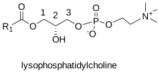
Figure \(\PageIndex{18}\): Lysophosphatidylcholine Lysophosphatidylcholine (LPC)
Lysophosphatidylcholine (LPC), with one mole of fatty acid per mole of lipid at the sn-1 position, occurs in trace amounts in most animal tissues. However, plasma concentrations are relatively high (150–500 µM). It is produced by hydrolysis of dietary and biliary phosphatidylcholine and is absorbed as such in the intestines, but it is re-esterified before export into the lymph. In addition, most tissues form it by hydrolyzing phosphatidylcholine using the phospholipase A2 superfamily, as part of the deacylation/reacylation cycle that controls overall molecular species composition. In many animal species, hepatocytes secrete much of the plasma LPC in a complex with albumin. Still, an appreciable amount forms in plasma through the action of the enzyme lecithin:cholesterol acyltransferase (LCAT), which the liver secretes. This catalyzes the transfer of fatty acids from the sn-2 position of phosphatidylcholine to free cholesterol in plasma, forming cholesterol esters and, of course, lysophosphatidylcholine, which consists of a mixture of molecular species with predominantly saturated and mono- and dienoic fatty acid constituents. Endothelial lipase action on phosphatidylcholine in HDL produces some LPC.
At high concentrations, lysophosphatidylcholine can disrupt membranes, while at low concentrations its biological effects may simply reflect its ability to readily diffuse into membranes, altering their curvature and indirectly affecting membrane protein properties. In plasma, albumin and lipoproteins bind it, reducing its effective concentration to a relatively safe level.
Lysophosphatidylcholine is considered to be an important factor in cardiovascular and neurodegenerative diseases. It is usually considered pro-inflammatory and is a pathological component of oxidized lipoproteins (LDL) in plasma and atherosclerotic lesions when generated by overexpression or enhanced activity of phospholipase A2. Its concentration is elevated in joint fluids from patients with rheumatoid arthritis. In addition, it is a major component of platelet-derived microvesicles and activates a specific platelet receptor, ultimately causing vascular inflammation and increasing atherosclerotic plaque instability. The intracellular acyltransferase LPCAT cannot remove lysophosphatidylcholine directly from plasma or lipoproteins, and no enzymes with lysophospholipase A1 activity appear to exist in the circulation. Lysophosphatidylcholine blocks the formation of early hemifusion intermediates required for cell-cell fusions. In insect bites, lysophosphatidylcholine attracts inflammatory cells to the site, enhances parasite invasion, and inhibits nitric oxide production, for example, in Chagas disease. Elevated levels of 26:0‑lysophosphatidylcholine in blood are reported to be characteristic of Zellweger spectrum disorders (resulting from a defect in peroxisome biogenesis).
Elevated levels of lysophosphatidylcholine have been identified in cervical cancer and may be diagnostic for the disease. In contrast, some cancers show reduced lysophosphatidylcholine levels, and it has protective effects in patients undergoing chemotherapy. Stearoyl-lysophosphatidylcholine plays an anti-inflammatory role in protecting against lethal sepsis in experimental animals through various mechanisms, including stimulating neutrophils to eliminate invading pathogens via a peroxide-dependent reaction. Similarly, reports indicate that lysophosphatidylcholine may have beneficial effects in rheumatoid arthritis and other diseases. However, some in vitro studies of lysophosphatidylcholine activity may be flawed because they used insufficient levels of carrier proteins. Another point to consider is that lysophosphatidylcholine is a precursor of the key lipid mediator lysophosphatidic acid via the enzyme autotaxin in plasma, and this may be the true source of some of the effects described for the former, especially on cell migration and survival.
There is evidence to suggest that lysophosphatidylcholine containing docosahexaenoic (DHA) and eicosapentaenoic (EPA) acids, presumably in position sn-2, in plasma targets more of these fatty acids into the brain, via a specific receptor/transporter at the blood-brain barrier known as the sodium-dependent LPC symporter 1 (MFSD2A), than occurs from the corresponding fatty acids in unesterified form. Hepatic lipase is especially important for generating these lipids. This finding is now being explored for potential therapeutic applications in neurological diseases, cognitive decline, and dementia. Similarly, at the maternal plasma/placental interface, phosphatidylcholine is taken up and hydrolyzed to sn‑2‑lysophosphatidylcholine, presumably by the endothelial lipase, to facilitate the transfer of polyunsaturated fatty acids across the basal membrane into the fetal circulation with the aid of the same LPC transporter.
Lysophosphatidylcholine has roles in cell signaling, and specific receptors (coupled to G proteins) have been identified, namely GPR119, GPR40, and GPR55. It activates phospholipase C, which releases diacylglycerol and inositol triphosphate, increasing intracellular Ca2+ and activating protein kinase C. Various cell systems have shown increased glucose-stimulated insulin secretion. Lysophosphatidylcholine also activates the mitogen-activated protein kinase in certain cell types, promoting demyelination in the nervous system. Lysophosphatidylcholine's interaction with TRPV4 ion channels in skin keratinocytes can cause persistent itching. Identifying a highly specific peroxisomal phospholipase A2γ that uniquely generates sn-2-arachidonoyl lysophosphatidylcholine suggests this may be relevant to eicosanoid generation and signaling. For example, 2-arachidonoyl-lysophosphatidylcholine is reportedly enriched in carotid atheroma plaque from patients with type 2 diabetes. In vascular endothelial cells, it induces the important pro-inflammatory mediator cyclooxygenase-2 (COX-2), a key enzyme in prostaglandin synthesis. However, it also benefits the innate immune system by activating macrophages and increasing their phagocytic activity in the presence of T lymphocytes.
Lysophospholipids in general, and lysophosphatidylcholine in particular, are important signaling molecules in mammalian cells. Their levels are tightly regulated, mainly by lysophospholipases A1 and A2 (LYPLA1 and LYPLA2), depending on the position of fatty acid esterification; these cytosolic serine hydrolases have esterase and thioesterase activity. The glycerophosphocholine produced can enter the Lands' cycle or be further degraded.
In plants, amylose-rich starch granules of cereal grains contain lysophosphatidylcholine as virtually the only lipid in the form of inclusion complexes or lining channels in the starch macromolecules.
Phosphatidylethanolamine and Related Lipids
Phosphatidylethanolamine – Structure and Occurrence
Phosphatidylethanolamine, or 1,2-diacyl-sn-glycero-3-phosphoethanolamine (once given the trivial name 'cephalin'), is usually the second most abundant phospholipid in animal and plant lipids, after phosphatidylcholine, and is frequently the main lipid component of microbial membranes. It can account for 20% of liver phospholipids and as much as 45% of those in the brain; mitochondria contain higher proportions than other organelles. As such, it is a key building block of membrane bilayers, and it is present exclusively in the inner leaflet of the plasma membrane in animal cells, for example. It is a neutral or zwitterionic phospholipid (at least in the pH range 2 to 7), with the structure shown (a specific molecular species is illustrated as an example).
Figure \(\PageIndex{19}\) shows the structure of phosphatidylethanolamine.
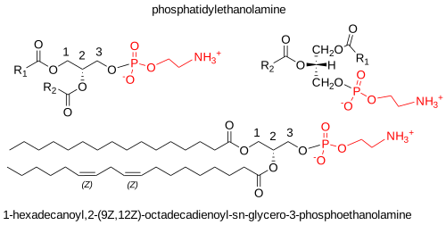
In animal tissues, phosphatidylethanolamine exists in diacyl, alkyl, alkenyl, and acyl forms. In some cell types (especially inflammatory cells, neurons, and tumor cells), up to 70% of phosphatidylethanolamine may have an ether linkage. Still, in the liver, the plasmalogen form of phosphatidylethanolamine, i.e., with an O‑alk-1’-enyl linkage, accounts for only 0.8% of total phospholipids. Generally, PE has a much higher proportion of ether linkages than PC. If physiological conditions inhibit plasmalogen biosynthesis, the diacyl form replaces it, thereby maintaining overall phospholipid content.
Animal phosphatidylethanolamine tends to contain higher proportions of arachidonic and docosahexaenoic acids than the other zwitterionic phospholipid, phosphatidylcholine. These polyunsaturated components are concentrated in position sn-2, with saturated fatty acids most abundant in position sn-1, as illustrated for rat liver and chicken egg in Table \(\PageIndex{3}\). In most other species, phosphatidylethanolamine in the same metabolically active tissues would be expected to show similar features.
| Table \(\PageIndex{3}\): Positional distribution of fatty acids in phosphatidylethanolamine in animal tissues. | |||||||
| Position | Fatty acid | ||||||
|---|---|---|---|---|---|---|---|
| 14:0 | 16:0 | 18:0 | 18:1 | 18:2 | 20:4 | 22:6 | |
| Rat liver [1] | |||||||
| sn-1 | 25 | 65 | 8 | ||||
| sn-2 | 2 | 11 | 8 | 8 | 10 | 46 | 13 |
| Chicken egg [2] | |||||||
| sn-1 | 32 | 59 | 7 | 1 | |||
| sn-2 | 1 | 1 | 25 | 22 | 29 | 12 | |
| 1, Wood, R. and Harlow, R.D., Arch. Biochem. Biophys., 131, 495-501 (1969); 2, Holub, B.J. and Kuksis, A. Lipids, 4, 466-472 (1969); |
|||||||
The O-alkyl and O-alkenyl chains at the sn-1 position of the analogous ether lipids generally consist of 16:0, 18:0 or 18:1 chains. In contrast, arachidonic and docosahexaenoic acids are the most abundant components at the sn-2 position.
The positional distributions of fatty acids in phosphatidylethanolamine from the model plant Arabidopsis thaliana leaves are listed in Table \(\PageIndex{4}\). Here, saturated fatty acids are also concentrated at position sn-1, and di- and tri-unsaturated fatty acids predominate at position sn-2. The pattern differs somewhat in the yeast Lipomyces lipoferus, where the compositions of the two positions are relatively similar.
| Table \(\PageIndex{4}\): Composition of fatty acids (mol%) in positions sn-1 and sn-2 in the phosphatidylethanolamine from leaves of Arabidopsis thaliana and from Lipomyces lipoferus. | ||||||
| Position | Fatty acid | |||||
|---|---|---|---|---|---|---|
| 16:0 | 16:1 | 18:0 | 18:1 | 18:2 | 18:3 | |
| A. thaliana [1] | ||||||
| sn-1 | 58 | trace | 4 | 5 | 15 | 18 |
| sn-2 | trace | trace | trace | 2 | 60 | 38 |
| L. lipoferus [2] | ||||||
| sn-1 | 29 | 18 | 4 | 28 | 13 | 6 |
| sn-2 | 23 | 15 | 3 | 34 | 17 | 6 |
| 1, Browse, J., Warwick, N., Somerville, C.R., and Slack, C.R. Biochem. J., 235, 25-31 (1986); DOI. 2, Haley, J.E. and Jack, R.C. Lipids, 9, 679-681 (1974); DOI . |
||||||
Phosphatidylethanolamine – Biosynthesis
The two main pathways employed by mammalian cells for the biosynthesis of phosphatidylethanolamine are the CDP-ethanolamine pathway, i.e., one of the general routes to phospholipid biosynthesis de novo in plants and animals, and the phosphatidylserine decarboxylase pathway, which occurs in two spatially separated organelles - the endoplasmic reticulum and mitochondria, respectively. Plants produce ethanolamine by decarboxylating serine, whereas in animals most comes from dietary sources and requires facilitated transport into cells. A small amount of ethanolamine phosphate comes from the catabolism of sphingosine-1-phosphate, which is essential for the survival of the protozoan Trypanosoma brucei. The initial steps in phosphatidylethanolamine biosynthesis occur in the cytosol, where two specific ethanolamine kinases phosphorylate ethanolamine to produce ethanolamine phosphate; ethanolamine phosphate phosphorylase catalyzes the reverse reaction and may regulate the pathway in some tissues. The second step is rate-limiting: the product reacts with cytidine triphosphate (CTP) to form cytidine diphosphoethanolamine, catalyzed by CTP:phosphoethanolamine cytidylyltransferase.
In the final step, a membrane-bound enzyme, CDP-ethanolamine:diacylglycerol ethanolaminephosphotransferase, catalyzes the reaction of cytidine diphosphoethanolamine with diacylglycerol to form phosphatidylethanolamine. Two such enzymes exist: ethanolamine phosphotransferase 1 (EPT1) in the Golgi and choline/EPT1 (CEPT1) in the endoplasmic reticulum, but EPT1 is more important for the biosynthesis of the plasmalogen form, 1-alkenyl-2-acyl-glycerophosphoethanolamine, especially molecular species containing polyunsaturated fatty acids. At the same time, CEPT1 produced species with shorter-chain fatty acids. The diacylglycerol precursor is formed from phosphatidic acid by the enzyme phosphatidic acid phosphohydrolase.
Figure \(\PageIndex{20}\) shows the synthesis of phosphatidylethanolamine in the ER/Golgi.
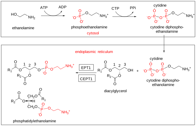
The second major pathway is the mitochondrial conversion of phosphatidylserine (decarboxylation) to phosphatidylethanolamine. The conservation of this pathway from bacteria to humans suggests that it has been preserved to optimize mitochondrial performance. Mitochondria are not connected with the rest of the cell's membrane network by classical vesicular routes but must receive and export small molecules through nonvesicular transport at zones of proximity with other organelles at membrane contact sites, such as a specific domain of the endoplasmic reticulum termed the mitochondria-associated membrane (MAM). Lipid transport of phosphatidylserine is then enabled by tethers that bridge two membranes, lipid transfer proteins, and recruitment proteins. In this process, the lipid must traverse two aqueous compartments, the cytosol and the mitochondrial intermembrane space, to reach the inner mitochondrial membrane.
Phosphatidylserine decarboxylase is located on the external surface of the mitochondrial inner membrane, and most mitochondrial phosphatidylethanolamine is derived from this pathway. While various isoforms of phosphatidylserine decarboxylase exist in prokaryotes, yeasts, and mammals, the main forms designated 'PSD1' are found only in mitochondria and are structurally related. An isoform designated 'PSD2' is located in the endosomal membranes of yeasts, and the phosphatidylethanolamine formed is the preferred substrate for phosphatidylcholine biosynthesis. Cellular concentrations of phosphatidylethanolamine and phosphatidylserine are closely related and tightly regulated.
Figure \(\PageIndex{21}\) shows the synthesis of phosphatidylethanolamine in mitochondria.

In prokaryotic cells, such as E. coli, where phosphatidylethanolamine is the most abundant membrane phospholipid, it is derived entirely from phosphatidylserine decarboxylation. In this instance, the enzyme undergoes autocleavage for activation and utilizes a pyruvoyl moiety to form a Schiff base intermediate with phosphatidylserine, facilitating decarboxylation. This pathway is also important in mammalian cells and yeasts. However, the relative contributions of the two main pathways of phosphatidylethanolamine synthesis in mammalian cells appear to vary with cell type. In cultured cells, more than 80% of phosphatidylethanolamine is reported to be derived from the phosphatidylserine decarboxylase pathway. In hamster hearts and rat hepatocytes, only ~5% of phosphatidylethanolamine is synthesized via this route, and most is made via the CDP-ethanolamine pathway. In yeast, PSD1 generates 70% of phosphatidylethanolamine. The spatially distinct pools within the cell are functionally distinct, but both are essential to life. For example, disruption of the phosphatidylserine decarboxylase gene causes misshapen mitochondria and has lethal consequences in embryonic mice, although phosphatidylethanolamine synthesis continues for a time in other cellular regions. Similarly, eliminating the endoplasmic reticulum route is embryonically lethal.
Three additional minor biosynthetic pathways are known. Phosphatidylethanolamine can form by enzymatic exchange of ethanolamine with phosphatidylserine or by re-acylation of lysophosphatidylethanolamine. The second is associated with the mitochondria-associated membrane, where phosphatidylserine synthase II is located. Finally, the bacterial plant pathogen Xanthomonas campestris can synthesize phosphatidylethanolamine by condensing cytidine diphosphate diacylglycerol with ethanolamine.
Ether lipids: These pathways for diacyl-phosphatidylethanolamine biosynthesis are distinct and spatially separated from those producing alkyl-, acyl-, and alkenyl-acyl-phosphatidylethanolamines, suggesting functional differences. In the protozoan T. brucei, for example, diacyl and ether phosphatidylethanolamine pools have separate functions and cannot substitute for each other.
Lands cycle: The various mechanisms produce different pools of phosphatidylethanolamine species, often in different cellular compartments with distinctive compositions. Studies with mammalian cell types in vitro suggest that the CDP-ethanolamine pathway preferentially produces molecular species with mono- or di-unsaturated fatty acids at the sn-2 position. At the same time, the phosphatidylserine decarboxylation reaction mainly generates species with polyunsaturated fatty acids at the sn-2 position. However, as with other phospholipids, animal tissues attain their final fatty acid composition through a remodeling process known as the Lands cycle. The first step is hydrolysis by a phospholipase A2 to lysophosphatidylethanolamine, followed by reacylation using various acyl-CoA:lysophospholipid acyltransferases. The enzymes LPCAT1, 2, and 3, involved in phosphatidylcholine biosynthesis, are also active on phosphatidylethanolamine, while LPEAT1 mainly uses lysophosphatidylethanolamine and is specific for oleoyl-CoA. Some of these isoforms appear to be confined to particular tissues. A CoA-independent acyltransferase in macrophages also transfers arachidonic acid from phosphatidylcholine to ethanolamine-containing phospholipids.
Click the link below for more information.
- Link
-
Phosphatidylethanolamine – Biological Function
Physical properties: Although PE has sometimes been equated with PC in biological systems, their chemistry and physical properties differ significantly, and they serve different functions in biochemical processes. Both are key components of membrane bilayers, and their ratio may be important for many cellular functions. However, PE has a smaller head group, which gives the lipid a cone shape. On its own, it forms inverted hexagonal phases rather than bilayers. In a bilayer, it is believed to exert lateral pressure that modulates membrane curvature and stabilizes membrane proteins in their optimal conformations. It can hydrogen bond to adjacent lipids and proteins via its polar head group. For example, phosphate can be stabilized at the binding site by interactions with lysine and arginine side chains or with hydroxyl groups on tyrosine side chains. It can also interact strongly with the phospholipid acyl chains.
In contrast to phosphatidylcholine, phosphatidylethanolamine is concentrated with phosphatidylserine in the inner leaflet of the plasma membrane. On the other hand, it is present in both the inner and outer membranes of mitochondria, but especially the former, where it is present at much higher levels than in other organelles and facilitates membrane fusion and protein movement across membranes in addition to many other essential mitochondrial functions, including oxidative phosphorylation via the electron transport chain. It is an important functional component of membrane contact sites between the endoplasmic reticulum and mitochondria. Studies suggest that eukaryotic cells, especially those of insects, perform a function similar to cholesterol in membranes, increasing the bilayer's rigidity to maintain membrane fluidity. In bacterial membranes, phosphatidylethanolamine appears to primarily dilute the high negative charge density of anionic phospholipids.
Protein Interactions: Membrane proteins make up 30% of the genome and carry out innumerable biochemical functions, including transport, energy production, biosynthesis, signaling, and communication. Within a membrane, most integral proteins consist of hydrophobic α-helical transmembrane domains that zigzag across it and are connected by hydrophilic loops. Of those parts of the proteins outside the bilayer, positively charged residues are much more abundant on the cytoplasmic side of membrane proteins than on the trans side (the positive-inside rule). Phosphatidylethanolamine is believed to play a key role by inhibiting the localization of negative amino acids on the cytoplasmic side, supporting the positive-inside rule, and by having an appropriate charge density to balance that of the membrane surface and the protein. However, under certain conditions, it can also permit negatively charged residues on the cytosolic surface, thereby supporting protein function.
Phosphatidylethanolamine is vital for mitochondrial function, as shown by defects in oxidative phosphorylation in the absence of PSD1 and by its role in regulating mitochondrial dynamics and protein biogenesis in the outer mitochondrial membrane. Synthesis of phosphatidylethanolamine in the inner mitochondrial membrane is critical for cytochrome bc1 complex (III) function, which has a conserved lipid-binding site in a specific subunit.
Phosphatidylethanolamine binds non-covalently to a superfamily of cytosolic proteins with multiple functions termed 'phosphatidylethanolamine-binding proteins'. While four members have been identified in mammals (PEBP1-4), more than 400 family members conserved during evolution are known from bacteria to higher eukaryotes. These have many functions, including lipid binding, neuronal development, inhibition of serine proteases, and regulation of several signaling pathways; in plants, they control shoot growth and flowering. PEBP4 is particularly interesting because it is highly expressed across many cancers and can increase their resistance to therapy. In animal tissues, phosphatidylethanolamine is especially important in the sarcolemmal membranes of the heart during ischemia; it is involved in the secretion of nascent very-low-density lipoproteins from the liver, and it plays roles in membrane fusion and fission. It has a functional role in the Ca2+-ATPase: one molecule of phosphatidylethanolamine is bound in a cavity between two transmembrane helices, acting as a wedge to keep them apart. This is displaced when Ca2+ is bound to the enzyme. Many other important proteins bind non-covalently to phosphatidylethanolamine similarly, including rhodopsin and aquaporins.
The content of phosphatidylethanolamine in newly secreted VLDL particles and apoB-containing particles isolated from the Golgi lumen is much higher than in circulating VLDLs, suggesting that this lipid is involved in VLDL assembly and/or secretion. However, it is rapidly and efficiently removed from circulating VLDL. In cells, lipid droplets are believed to promote the coalescence of smaller droplets into larger ones.
Although the mechanism has yet to be fully elucidated, effects on protein conformation are believed to underlie the finding that phosphatidylethanolamine is the primary factor in the brain required for mammalian prion propagation and infectivity. Host defense peptides are antimicrobial agents produced by both prokaryotic and eukaryotic organisms, and many have a high affinity for phosphatidylethanolamine as a lipid receptor, thereby modulating their activity. For example, the peptide antibiotics cinnamycin and duramycins from Streptomyces have a hydrophobic pocket that fits around phosphatidylethanolamine, stabilizing binding via an ionic interaction between the lipid's ethanolamine group and the peptide's carboxylate moiety; this complex is active against other Gram-positive organisms, such as Bacillus species.
Much of the evidence for PE's unique properties comes from studies of the bacterium E. coli, in which PE is a major membrane lipid. Gram-negative bacteria have two membrane bilayers in the cell wall. As much as 90% of the phospholipids in the inner leaflet of the outer membrane are phosphatidylethanolamine, with a high proportion in the cytoplasmic leaflet of the inner membrane also. In particular, PE plays a specific role in supporting active lactose transport via lactose permease, and other transport systems may require it or be stimulated by it. Evidence suggests that phosphatidylethanolamine acts as a 'chaperone' during the assembly of this and other membrane proteins, guiding their folding pathways and aiding the transition from the cytoplasmic to the membrane environment.
Autophagy and ferroptosis: A covalent conjugate of phosphatidylethanolamine with a protein designated 'Atg8' is formed by the action of cysteine protease ATG4 (belonging to the caspase family) and various other proteins, and is involved in the process of autophagy (controlled degradation of cellular components) in yeast by promoting the formation of membrane vesicles containing the components to be degraded (phosphatidylinositol 3-phosphate is also essential to this process). Similarly, oxidatively modified phosphatidylethanolamine is an important factor in ferroptosis, a form of apoptosis in which disturbances to iron metabolism lead to an accumulation of hydroperoxides.
Precursor of other lipids: Phosphatidylethanolamine is a precursor for the synthesis of N-acyl-phosphatidylethanolamine (see below) and thence of anandamide (N‑arachidonoylethanolamine), and it is the donor of ethanolamine phosphate during the synthesis of the glycosylphosphatidylinositol anchors that attach many signaling proteins to the surface of the plasma membrane. In bacteria, it functions similarly in the biosynthesis of lipid A and other lipopolysaccharides. It is also the substrate for the hepatic enzyme phosphatidylethanolamine N-methyltransferase, which accounts for about a third of hepatic phosphatidylcholine production.
Miscellaneous other functions: Phosphatidylethanolamine is the precursor of an ethanolamine phosphoglycerol moiety bound to two conserved glutamate residues in eukaryotic elongation factor 1A, an essential component in protein synthesis. This unique modification appears to be important for plant resistance to pathogen attack. Francisella tularensis, the cause of tularemia, suppresses host inflammation and the immune response when infecting mouse cells. The effect is due to a distinctive phosphatidylethanolamine species containing 10:0 and 24:0 fatty acids, and the synthetic lipid produces the same effects in vitro in human cells infected with the dengue fever virus. This lipid is hoped to be a potent anti-inflammatory therapeutic agent.
Plants: In the seeds of higher plants, a deficiency of phosphorylethanolamine cytidylyltransferase, a rate-limiting enzyme in phosphatidylethanolamine biosynthesis, profoundly affects embryo viability and maturation.
Lysophosphatidylethanolamine
Figure \(\PageIndex{22}\) shows the the structure of lysophosphatidyethanolamine

Figure \(\PageIndex{22}\): Lysophosphatidyethanolamine Lysophosphatidylethanolamine (LPE), with one mole of fatty acid per mole of lipid, is found in trace amounts only in animal tissues other than plasma (10 to 50µM, or ~1% of total serum phospholipids). The enzyme phospholipase A2 forms it by hydrolyzing phosphatidylethanolamine as part of a de-acylation/re-acylation cycle that controls its overall molecular species composition, as discussed above. A membrane-bound O-acyltransferase (MBOAT2) specific for LPE (and lysophosphatidic acid) has been characterized by a preference for oleoyl-CoA as a substrate. Reports describe LPE involvement in cellular functions, including the differentiation and migration of certain neuronal cells and various cancer cells. For example, oleoyl-LPE in the brain stimulates neurite outgrowth and protects against glutamate toxicity.
In plants, lysophosphatidylethanolamine specifically inhibits phospholipase D, a key enzyme in degrading membrane phospholipids during the early stages of plant senescence. Through this action, it retards the senescence of leaves, flowers, and post-harvest fruits. Indeed, it has many horticultural applications when applied externally, e.g., stimulating ripening and extending the shelf life of fruit, delaying senescence, and increasing the vase life of cut flowers. In bacteria, lysophosphatidylethanolamine displays chaperone-like properties, promoting the functional folding of citrate synthase and other enzymes. Some biological properties have been reported in animal tissues in vitro, but researchers have not yet identified a specific receptor.
Lysophospholipids, especially lysophosphatidylethanolamines, are produced in bacterial envelope membranes by many endogenous and exogenous factors. Flippases must transport them back into the bacterial cell, where the peripheral enzyme acyl-ACP synthetase/LPL acyltransferase converts them back to the diacyl forms. Lysophosphatidylethanolamines produced by certain bacteria act synergistically with the sulfonolipid rosette-inducing factors (RIFs) to maximize the latter's activity and induce choanoflagellates to move from a unicellular to a multicellular state.
N-Acyl Phosphatidylethanolamine
In N-acyl phosphatidylethanolamine, a fatty acid acylates the free amino group of phosphatidylethanolamine. This lipid has been detected in rather small amounts in several animal tissues (~0.01%), but especially in the brain, nervous tissues, and the epidermis, when the N-acyl chain is often palmitic or stearic acid (human plasma: N16:0-PE (40%), N18:1-PE (23.3%), N18:0-PE (19%), N18:2-PE (16.6%) and N20:4-PE (1.4%)). Under conditions of degenerative stress, it can accumulate in significant amounts, for example, as a result of ischemic injury, infarction, or cancer. It is present in plasma after rats are fed a high-fat diet, and it can then cross into the brain, where it accumulates in the hypothalamus.
Figure \(\PageIndex{23}\) shows the structures of N-Acyl Phosphatidylethanolamine.
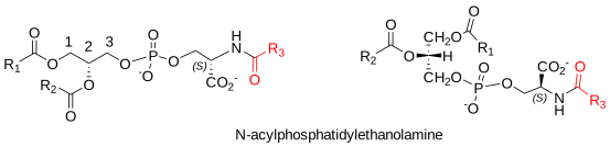
Figure \(\PageIndex{23}\): N-Acyl Phosphatidylethanolamine In animals, N-acyl phosphatidylethanolamine is particularly important as a precursor of anandamide and other biologically important ethanolamides (e.g., N-oleoylethanolamide) in the brain and other tissues, especially the intestines. In brief, it is formed biosynthetically by the action of a transferase (cytosolic phospholipase A2ε), exchanging a fatty acid from the sn-1 position of a phospholipid (probably phosphatidylcholine) to the primary amine group of phosphatidylethanolamine (without a hydrolytic step). Both diacyl- and alkenylacyl-phosphatidylethanolamine can serve as acceptors. In addition, some transfer can also occur from phosphatidylethanolamine per se by an intramolecular reaction. However, some N-acyl phosphatidylethanolamine can form artefactually due to faulty extraction procedures during analysis.
In plants, N-acyl phosphatidylethanolamine is a common constituent of cereal grains (e.g., wheat, barley, and oats) and of some other seeds (1.9% of the phospholipids of cotton seeds, but 10-12% of oats). In other plant tissues, it is detected most often under conditions of physiological stress. In contrast to animals, synthesis involves the direct acylation of phosphatidylethanolamine with a free fatty acid, catalyzed by a membrane-bound transferase using a reverse serine-hydrolase mechanism. The activation of N-acyl phosphatidylethanolamine metabolism in plants, with the release of N-acylethanolamines and the formation of phosphatidic acid, appears to be associated with cellular stress. However, research is still in the early stages. However, both N-acyl lipid classes have been implicated in physiological processes such as elongation of main and lateral roots, regulation of seed germination, seedling growth, and defense against pathogens.
N-Acyl phosphatidylethanolamine occurs in many microbial species, while N-acetyl phosphatidylethanolamine was detected in the filamentous fungus Absidia corymbifera, where it comprised 6% of total membrane lipids. It was accompanied by an even more unusual lipid, 1,2‑diacyl-sn-glycero-3-phospho(N-ethoxycarbonyl)-ethanolamine.
-
In contrast, it inhibits the folding of some multi-helical proteins. Without this lipid, the transport membranes may not have the correct tertiary structure and will not function correctly. Whether the lipid is required once the protein is correctly assembled is not fully understood in all cases, but it may be needed to orient enzymes correctly in the inner membrane. Some studies suggest that this organism can survive without phosphatidylethanolamine, but life processes are inhibited.
Phosphatidylserine and Related Lipids
Phosphatidylserine, or 1,2-diacyl-sn-glycero-3-phospho-L-serine, is an important anionic phospholipid that provides essential physical properties to membranes in eukaryotes and prokaryotes. It also has many biological functions in cells, including effects on blood coagulation and apoptosis, and it is the biosynthetic precursor of phosphatidylethanolamine in prokaryotes and eukaryotic mitochondria. Its metabolite, lysophosphatidylserine, has signaling functions and operates through specific receptors. There is also increasing interest in a structurally related lipid, phosphatidylthreonine, and other phospholipids linked to amino acids.
Phosphatidylserine - Structure and Occurrence
Although phosphatidylserine is widely distributed among animals, plants, and microorganisms, it usually accounts for less than 10% of total phospholipids, with the highest concentration in myelin from brain tissue. For example, the brain and liver of mice contain 14% and 3% phosphatidylserine, respectively. However, it may comprise 10 to 20 mol% of total phospholipids in the plasma membrane, where, under normal conditions, it is concentrated in the inner leaflet and in the endoplasmic reticulum. In the yeast Saccharomyces cerevisiae, it is a minor component of most cellular organelles other than the plasma membrane, where it can, surprisingly, account for more than 30% of total lipids. It is a minor membrane constituent in most bacteria, although it is important as an intermediate in phosphatidylethanolamine biosynthesis. The 1‑octadecanoyl-2-docosahexaenoyl molecular species, especially important in brain tissue, is illustrated here.
Figure \(\PageIndex{24}\) shows the structure of phosphatidylserine
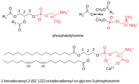
Phosphatidylserine is an acidic (anionic) phospholipid with three ionizable groups, i.e., the phosphate moiety, the amino group, and the carboxyl function. As with other acidic lipids, it occurs naturally as a salt. However, it strongly tends to chelate calcium via the charged oxygen atoms of both the carboxyl and phosphate moieties, thereby altering the conformation of the polar head group. This interaction may be highly relevant to phosphatidylserine's biological function, especially during bone formation.
In animal cells, the fatty acid composition of phosphatidylserine varies by tissue. Still, it does not appear to resemble the precursor phospholipids, either because specific molecular species are selectively used for biosynthesis or because lipid remodeling via deacylation-reacylation reactions with lysophosphatidylserine as an intermediate occurs (see below). In human plasma, 1-stearoyl-2-oleoyl and 1-stearoyl-2-arachidonoyl species predominate, but in the brain (especially grey matter), retina, and many other tissues, 1-stearoyl-2-docosahexaenoyl species are especially abundant and appear essential for normal nervous system function. Indeed, the n-3-to-n-6 fatty acid ratio in brain phosphatidylserine is much higher than in most other lipids. The positional distribution of fatty acids in phosphatidylserine from rat liver and bovine brain is listed in Table \(\PageIndex{5}\). As with most phospholipids, saturated fatty acids are concentrated at sn-1 and polyunsaturated fatty acids at sn-2.
| Table \(\PageIndex{5}\): Positional distribution of fatty acids in phosphatidylserine from rat liver and bovine brain | ||||||
| Position | Fatty acid | |||||
|---|---|---|---|---|---|---|
| 16:0 | 18:0 | 18:1 | 18:2 | 20:4 | 22:6 | |
| Rat liver [1] | ||||||
| sn-1 | 5 | 93 | 1 | |||
| sn-2 | 6 | 29 | 8 | 4 | 32 | 19 |
| Bovine brain [2] | ||||||
| sn-1 | 3 | 81 | 13 | |||
| sn-2 | 2 | 1 | 25 | trace | 1 | 60 |
| 1. Wood, R. and Harlow, R.D. Arch. Biochem. Biophys., 135, 272-281 (1969); DOI. 2. Yabuuchi, H. and O'Brien, J.S. J. Lipid Res., 9, 65-67 (1968); DOI. |
||||||
In the leaves of Arabidopsis thaliana, used as a 'model' plant in many studies, the fatty acid composition of phosphatidylserine resembles that of phosphatidylethanolamine. An intriguing report states that the acyl chain lengths of phosphatidylserine increase with age and stress, and that 22:0 and 24:0 fatty acids have been reported to occur in this lipid in the plasma membrane of some plant species.
Compared to PE, PS with ether-linked moieties (alkyl and alkenyl) is uncommon in animal tissues. However, it is reported to be relatively abundant in the human retina and macrophages (first found in rat lung). In general, phosphatidylserine is most concentrated in plasma membranes and endosomes but is very low in mitochondria. Because it lies entirely on the inner monolayer of the plasma membrane (and other cellular membranes) and is the most abundant anionic phospholipid, it may contribute to interfacial effects through nonspecific electrostatic interactions. This normal distribution is disturbed during platelet activation and cellular apoptosis.
N-Acylphosphatidylserine is reportedly present in the frontal cortex of patients with schizophrenia as a minor component of the lipids of sheep erythrocytes, bovine brain, and the central nervous system of freshwater fish and Bryozoans, amongst others. The N-arachidonoyl form may be the precursor of the endocannabinoid N-arachidonoylserine.
Biosynthesis of Phosphatidylserine
L-serine is a non-essential amino acid that most organisms actively synthesize. In animals, nearly all cell types produce it, though in the brain astrocytes synthesize it, not neurons. Neurons must be supplied with this amino acid for the biosynthesis of phosphatidylserine (and of sphingoid bases).
In animal tissues, phosphatidylserine is synthesized solely by calcium-dependent base-exchange reactions in which the polar head-group of an existing phospholipid is exchanged for L-serine. Two routes involve distinct enzymes (PS synthases I and II) that share 30% homology and several membrane-spanning domains and can use different substrates. Both enzymes synthesize phosphatidylserine on the cytosolic face of the endoplasmic reticulum (ER), mainly in a specific domain termed the mitochondria-associated membrane ('MAM'), because it is transiently tethered to the mitochondrial outer membrane, presumably by a protein bridge. A complex of integrated proteins ('ERMES') has been characterized in yeast with a similar function. The reaction involves exchanging L-serine with either phosphatidylcholine or phosphatidylethanolamine, catalyzed by PS synthase I (long thought to accept only phosphatidylcholine as a substrate).
In contrast, PS synthase II catalyzes a similar exchange with diacyl-phosphatidylethanolamine and its plasmalogen form. Both enzymes are subject to feedback regulation by their product phosphatidylserine, thereby maintaining the correct amounts of this lipid. Figure \(\PageIndex{25}\) shows the synthesis and metabolism of phosphatidylserine in animals.
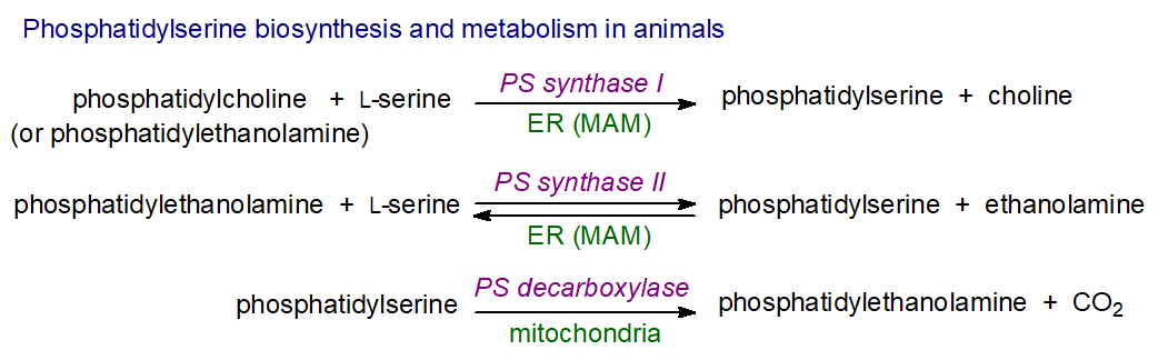
Phosphatidylserine synthase I is expressed in all mouse tissues, especially the kidney, liver, and brain, while phosphatidylserine synthase II is most active in the brain and testis and much less so in other tissues. The latter enzyme has a high specificity for molecular species containing docosahexaenoic acid. It is unknown why such a complex series of coupled reactions is necessary or why two enzymes are required. Still, one virtue is that the free ethanolamine and choline formed are rapidly re-utilized for phospholipid synthesis. Thus, phosphatidylserine and phosphatidylethanolamine are produced without reducing phosphatidylcholine levels. Eliminating both enzymes is embryonically lethal in knockout mice. Still, each can be knocked out separately, and the mice survive, even though they have substantially reduced levels of phosphatidylserine and phosphatidylethanolamine.
As with other phospholipids, animal tissues attain their final fatty acid composition through remodeling, known as the Lands cycle. The first step is hydrolysis by a phospholipase A2 to lysophosphatidylserine, followed by reacylation by various acyl-CoA:lysophospholipid acyltransferases. One membrane-bound O-acyltransferase (LPCAT4 or MBOAT2) prefers oleoyl-CoA, while a second (LPCAT3 or MBOAT5) incorporates linoleoyl and arachidonoyl chains (and also uses lysophosphatidylcholine).
Following synthesis, phosphatidylserine molecules can diffuse laterally in a concentration-dependent manner to different membrane regions to fulfill their physiological functions. In humans, cytosolic transport proteins transfer phosphatidylserine and other acidic phospholipids between membranes, and this can also occur via vesicular transport.
Some of the newly synthesized phosphatidylserine is transferred to the plasma membrane. At the same time, a proportion is transported to the mitochondria, probably again via transient membrane contact (MAM), where a specific decarboxylase in the inner mitochondrial membrane decarboxylates it to produce phosphatidylethanolamine. In yeast, molecular species containing two monoenoic fatty acids are preferred for transport and metabolism; this process also occurs at the Golgi/endosome membranes. All the phosphatidylethanolamine in mitochondria is formed this way. Still, some can return to the endoplasmic reticulum, where it may be converted back to phosphatidylserine by the action of the PS synthases. The CDP-ethanolamine pathway does not fully complement the mitochondrial production of phosphatidylethanolamine from phosphatidylserine, as mice lacking the enzyme do not survive for long. Cellular concentrations of these two lipids are intimately related and tightly regulated. Figure \(\PageIndex{26}\) shows the mitochondrial metabolism of phosphatidylserine
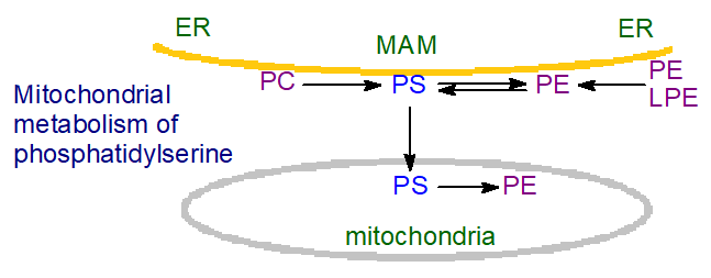
Thus, much of the phosphatidylserine is decarboxylated to phosphatidylethanolamine, which may be the major route to the latter in bacteria. As phosphatidylcholine in yeast is produced via methylation of phosphatidylethanolamine, phosphatidylserine is the primary precursor for this phospholipid in these organisms.
Bacteria and plants: In bacteria, other prokaryotic organisms and yeast, phosphatidylserine is synthesized by a mechanism comparable to that of most other phospholipids, i.e., by reaction of L-serine with CDP-diacylglycerol, and depends on Mg2+ or Mn2+. Phosphatidylserine synthases belong to two different families: type I (non-integral membrane form) in the phospholipase D-like family, as in E. coli, and type II (integral membrane form) in the CDP-alcohol phosphotransferase family, as in Bacillus sp. and the yeast S. cerevisiae. However, the latter shows no homology with the bacterial enzymes.
In many plants, including the model plant Arabidopsis, much phosphatidylserine is produced by a calcium-dependent base-exchange reaction in which the head group of an existing phospholipid is exchanged for L-serine in the luminal leaflet of the endoplasmic reticulum (i.e., mechanistically similar to PS synthase I). Flippases transfer it to the cytoplasmic membrane leaflet, then to post-Golgi compartments, before it eventually accumulates at the plasma membrane. However, some vesicular transport may occur, or direct transfer may occur at membrane contact sites. A CDP-diacylglycerol (prokaryotic-like) biosynthetic pathway exists in some species, e.g., wheat.
Let's explore the mechanism of the Methanocaldococcus jannaschii phosphatidylserine synthase (MjPSS). The organism is a hyperthermophilic methanogen. Figure \(\PageIndex{27}\) shows substrate binding by MjPSS.

The large binding pocket for CDP-DAG in MjPSS extends from the hydrophobic membrane core to the active site near the cytoplasmic surface in the center of the dimer. In the closed structures (left), both CDP-DAG alkyl chains adopt similar conformations within the binding pocket. In contrast, helices 7 and 8 positions in the open structures (right) allow one alkyl chain to reach the membrane via a different path. Serine molecules are only found in open structures (right). In one closed conformation, a citrate is near the substrate-binding site, whereas in the other closed structures, this position is empty. A chain of three chloride ions extends parallel to the dimer interface from the active site to the cytoplasmic interface with the N-terminal helix hH from the other protomer.
Figure \(\PageIndex{28}\) shows the reaction cycle of MjPSS during the synthesis of PS from CDP-DAG and serine in the presence of Mg2+ and Ca2+.

The CDPDAG binding site in MjPSS forms and is stabilized by the divalent cations Mg2+ and Ca2+ (a). In the absence of CDP-DAG, Ca2+ most likely is coordinated by water molecules, as shown in a, or by residues in nearby loops that would be flexible in the absence of CDP-DAG. CDP-DAG binding (b) is driven by coordination of Ca2+ by the negatively charged phosphates. Serine binds to the binding pocket after CDP-DAG (c). For nucleophilic attack, the serine molecule positions its hydroxyl group near the β-phosphate of CDP-DAG. The serine molecule is activated by deprotonation, attacks the β-phosphate, and forms the penta-coordinated transition state (d). A water molecule in the interaction network of Asp66, Arg101, and nearby chloride ions likely removes the serine proton. Hydrolysis of the CDP-DAG/serine complex at the transition state forms the MjPSS complex with the products PS and CMP (e). The next cycle starts after product release and CDP-DAG binding. Structural data are available for the state with bound CDP-DAG (b), CDP-DAG and serine (c), and for the transition state of the CDP-DAG/serine complex (d).
Figure \(\PageIndex{29}\) shows an interactive iCn3D model of the Methanocaldococcus jannaschii phosphatidylserine synthase (PSS) in the open state with bound CDP-DAG and serine (7B1L). The enzyme is also named CDP-diacylglycerol--serine O-phosphatidyltransferase.

Figure \(\PageIndex{29}\): Bacterial phosphatidylserine synthase (PSS) in the open state with bound CDP-DAG and serine (7B1L) (Note the actual PDB file title is for the closed state.) Click the image for a popup or use this external link: https://structure.ncbi.nlm.nih.gov/i...B3Rn3gVN4c4ge6
The A chain of the homodimer is shown in gray and the B in plum. The active site residues in the A chain in the above mechanism are shown in stick representation, with CPK colors and labels. Hover over the large ligand (58A) in the gray subunit. Two free serines are shown near it, but only one is probably the substrate.
Click the link below.
- Link
-
Phosphatidylserine – Biological Function
Membrane location: Phosphatidylserine modulates membrane charge locally, enabling recruitment of soluble cations and proteins and contributing to the organization of processes within cell membranes. Its membrane distribution is tightly controlled because it facilitates signaling across cellular compartments. Thus, it transitions from the luminal leaflet of the endoplasmic reticulum to the cytosolic leaflet of the trans-Golgi network, likely via flippase and scramblase activity in the Golgi, and is highly enriched on the inner leaflet of the plasma membrane. Transport to the plasma membrane against a concentration gradient is aided in part by proteins designated 'ORP5' and 'ORP8' in humans (Osh 6 and Osh7 in yeast) with a 'PH' binding domain for phosphatidylinositol 4,5-bisphosphate and an 'ORD' domain for phosphatidylserine. At a membrane contact site between the endoplasmic reticulum and plasma membrane, phosphatidylserine is exchanged for phosphatidylinositol 4-phosphate. Such transfer requires energy input, which can be supplied by ATP or phosphoinositides. Although it does not take part in membrane raft formation, phosphatidylserine is present in caveolae, where it is believed to interact with caveolin-1. It is also present in appreciable amounts in the endosomal compartment.
The asymmetric structure of the plasma membrane, with high concentrations of anionic lipids such as phosphatidylserine in the cytosolic leaflet and zwitterionic lipids in the extracellular leaflet, creates two surfaces with greatly different electrostatic potentials that influence protein association with the membrane surface and the activities of integral membrane proteins. This distribution is maintained and can be altered, after specific activation, by various flippases (transfer back into the cytoplasmic leaflet), floppases (transfer out of the cytoplasmic leaflet), and scramblases (bidirectional transfer), including ATP-dependent translocases selective for phosphatidylserine. Phosphatidylserine is highly enriched in the cytosolic leaflet of recycling endosomal membranes, which replenish the lipids and proteins of the plasma membrane, and it is essential for their function.
Enzyme activation: Besides its role as a membrane component and a precursor for other phospholipids, phosphatidylserine is an essential cofactor that binds to and activates many proteins, especially those with signaling activities. The lipid's negative charge facilitates protein binding through electrostatic interactions or Ca2+ bridges. For example, appreciable amounts of phosphatidylserine on the cytosolic leaflet of endosomes and lysosomes enable these compartments to bind proteins with specific phosphatidylserine-binding domains, including several important signaling and fusogenic effectors. The cytoskeletal protein spectrin binds phosphatidylserine this way, and enzymes such as neutral sphingomyelinase and the Na+/K+-ATPase also require it, with the 18:0/18:1 molecular species especially important. It is believed that the fatty acyl components of this species in the inner leaflet of the plasma membrane (and potentially other intracellular membranes) may interact (interdigitation or "handshake") with the very-long chains of sphingolipids in the outer leaflet in raft microdomains, resulting in a high local concentration of the anionic phospholipid and an accumulation of negative surface charge to which specific poly-cationic proteins in the membranes can bind. This may then enable signal transfer across the membrane to the cytosol.
Similarly, phosphatidylserine participates directly in key brain signaling pathways by binding to cytosolic proteins involved in neuronal signaling, thereby activating them. At least three major pathways are affected, including phosphatidylinositol 3-kinase and protein kinase C. For example, most enzymes of the protein kinase C family contain a 'C2' calcium-dependent cysteine-rich region that recognizes phosphatidylserine and, in coordination with the 'C1' domain that binds to diacylglycerols, is essential for activating and localizing them to the plasma membrane of appropriately stimulated cells. Phosphatidylserine is not involved in cell signaling through metabolite formation, as with phosphatidylinositol.
Blood coagulation: Phosphatidylserine is an important component of the blood coagulation process in platelets, where it is transported from the inner to the outer surface of the plasma membrane upon platelet activation. Here, exposed phosphatidylserine enhances prothrombin activation to thrombin (the key molecule in the blood clotting cascade) by triggering a cascade of reactions and providing a negatively charged platform that enables calcium ions to bridge γ-carboxyglutamic acid-containing domains on coagulation factors. Platelets can also release membrane vesicles with surface-exposed phosphatidylserine, promoting coagulation. Apolipoprotein A-1 in high-density lipoproteins plays a regulatory role. It neutralizes these procoagulant properties by orienting the phospholipids in surface areas too small to accommodate the prothrombinase complex. Blood coagulation is beneficial when it prevents blood loss from the circulatory system. However, it is detrimental when it causes thrombosis, and phosphatidylserine is essential for regulating the process.
Apoptosis: In addition, in response to specific calcium-dependent stimuli, phosphatidylserine plays an important role in regulating apoptosis, or programmed cell death, the natural process by which aged or damaged cells are removed from tissues before they can exert harmful effects. When cells are damaged, scramblase activation disrupts the normal distribution of this lipid by moving it from the inner leaflet of the plasma membrane bilayer to the outer leaflet. This occurs because aminophospholipid translocases, which return the lipid to the inner leaflet, are inhibited. In erythrocytes, phosphatidylserine is located in the inner leaflet of the membrane bilayer under low Ca2+ conditions, when membrane cholesterol suppresses the phospholipid scramblase. It is exposed to the outer leaflet under elevated Ca2+ concentrations, which activate the scramblase. After this asymmetry collapses and phosphatidylserine is transferred to the outer leaflet of an effete cell, it is believed to be recognized by a cohort of receptors, either directly or indirectly, through bridging ligands on the surface of macrophages and related scavenger cells. These activate a family of cysteine-dependent aspartate-specific proteases, the caspases, and other enzymes to facilitate the engulfment of the apoptotic cells and their potentially toxic or immunogenic contents in a non-inflammatory manner. Notably, the transition from a pro-inflammatory to an anti-inflammatory state is defined by macrophage phagocytosis of neutrophils via this phosphatidylserine-dependent process.
During apoptosis, reactive oxygen species are generated, mainly hydrogen peroxide, which, together with the enzyme cytochrome c, brings about rapid oxidation of the fatty acids in phosphatidylserine before this lipid is externalized. Indeed, molecular species of phosphatidylserine with an oxidatively truncated sn-2 acyl group that incorporates terminal γ-hydroxy (or oxo)-α,β-unsaturated acyl moieties are especially potent signals for scavenger receptors in macrophages and a prerequisite for engulfment of apoptotic cells.
This has been described as "a dominant and evolutionarily conserved immunosuppressive signal that promotes tolerance and prevents local and systemic immune activation" or, more succinctly, as an "eat-me signal" (externalized phosphatidylinositol 3,4,5-trisphosphate (PI(3,4,5)P) may have a similar function). Phosphatidylserine binding to specific proteins, such as apolipoprotein H (β2-glycoprotein 1), enhances their recognition and clearance. This process is essential for lung and brain development and is also relevant to clinical situations in which apoptosis plays an important role, such as cancer, chronic autoimmunity, and infections. For example, phosphatidylserine is a necessary component of the TAM family of receptor tyrosine kinases, and the receptor-ligand complex is particularly important in cancer cells, where phosphatidylserine-TAM signaling regulates many aspects of inflammation and immune resolution and is a target for therapeutic intervention. Phosphatidylserine exposure is increased substantially on the surface of tumor cells or tumor cell-derived microvesicles, which have innate immunosuppressive properties and facilitate tumor growth and metastasis. Targeting phosphatidylserine is a promising strategy in cancer immunotherapy. In atherosclerosis, phosphatidylserine is believed to have anti-inflammatory and protective effects as a component of high-density lipoproteins, probably via an apoptotic mechanism. In contrast, because this mechanism is important for erythrocyte turnover, it is relevant to thrombus formation and blood clot stabilization. Numerous viruses and bacteria have hijacked the innate immunosuppressive effect of externalized phosphatidylserine to facilitate infection.
A similar apoptotic mechanism operates in retinal pigment epithelial cells to remove the large amounts of photoreceptor cell debris generated continuously. In addition, an analogous mechanism translocates appreciable amounts of phosphatidylserine to the surface of T lymphocytes that express low levels of the transmembrane enzyme tyrosine phosphatase. This redistribution signals and modulates the activity of several membrane proteins. The anticoagulant protein annexin V binds phosphatidylserine with high specificity and is used as a probe to detect apoptotic cells. Notably, phosphatidylserine is a major component of microvesicle membranes in animal cells, and its translocation to the outer leaflet upon cellular activation is essential for biogenesis. In addition, exposure of phosphatidylserine on the cell surface has been reported as a factor in non-apoptotic forms of regulated inflammatory cell death, such as necroptosis.
Role in infections: Unfortunately, viruses such as Ebola and HIV can hijack this apoptosis machinery by incorporating phosphatidylserine into their viral envelopes to convince cells to engulf them; the viral glycoprotein/cellular receptor complex may then facilitate entry into other cells. Similarly, parasites ingested this way, including Leishmania and Trypanosoma species, use host phosphatidylserine to establish infections and facilitate disease progression, as they do not elicit proinflammatory cytokine production. This mechanism has been termed 'apoptotic mimicry' and is critical for parasite survival within the macrophage.
Other activities: Phosphatidylserine is required for the transmembrane movement of excess cholesterol, derived initially from the lysosomal degradation of low-density lipoproteins, from the plasma membrane to the endoplasmic reticulum, thereby maintaining membrane integrity and ensuring cell survival. It is, therefore, an important element in cholesterol homeostasis. The mechanism is believed to involve proteins known as GRAMD1s embedded in the endoplasmic reticulum membrane at sites in contact with the plasma membrane. These have two functional domains: the StART-like domain, which binds cholesterol, and the GRAM domain, which binds anionic lipids such as phosphatidylserine, linking the two membranes and enabling cholesterol transfer.
A further unusual function of phosphatidylserine is that it is a key component of lipid-calcium-phosphate complexes that act as nucleation centers for hydroxyapatite formation and initiate mineral deposition during bone formation. Phosphatidylserine and inorganic phosphate must be present before calcium ions are introduced, when phosphatidylserine's high affinity for calcium ions becomes important. Nucleation is facilitated by the protein annexin V. Similarly, during bone repair and maintenance, the fusion of osteoclasts requires the non-apoptotic exposure of phosphatidylserine at the surface of fusion-committed cells with the aid of a transmembrane protein (DC-STAMP) expressed in dendrocytes. This activity is relevant to cardiovascular disease, particularly the phenomenon of "hardening of the arteries," in which atherosclerotic plaques can mineralize via hydroxyapatite deposition.
Among many other functions of phosphatidylserine, it is believed to be an essential surface membrane component for fusion between cell types other than osteoclasts, including fiber formation in muscle cells and the fusion of macrophages into inflammatory giant cells and myoblasts into myotubes. Such cell fusions require non-apoptotic exposure of phosphatidylserine on the surfaces of fusing cells, where it interacts with phosphatidylserine-recognizing proteins to regulate the time and place of cell fusion. Phosphatidylserine provides stable membrane domains in spermatozoa that are essential for fertilization, and it is also an essential component of plasma membrane microdomains known as caveolae, where it is required for their formation and stability, possibly through specific binding to caveolin proteins.
The high concentrations of docosahexaenoic acid (DHA) in the brain and retinal phosphatidylserine are certainly important for the development and function of these tissues. Accumulation of phosphatidylserine in neuronal membranes is promoted by DHA. This is important for maintaining neuronal survival. Phosphatidylserine may also be a reservoir of DHA for protectin formation in neuronal tissue. On the other hand, the Food and Drug Administration in the USA considers that there is little scientific evidence to support claims that dietary supplements of phosphatidylserine reduce the risk of dementia or cognitive dysfunction in the elderly, and other nutritional claims appear to be dubious also. Antibodies to phosphatidylserine are formed in some disease states, including thrombosis and recurrent spontaneous pregnancy loss. A mutation in the phosphatidylserine synthase gene causes the rare genetic disease Lenz-Majewski syndrome I, which greatly increases the enzyme's activity while preventing feedback inhibition. Abnormal phosphatidylserine metabolism has been implicated in other diseases.
In yeasts such as Candida albicans, phosphatidylserine and the enzyme phosphatidylserine decarboxylase, which generates phosphatidylethanolamine, are both essential for virulence toward a host.
Lysophosphatidylserine
Figure \(\PageIndex{30}\) shows the structure of lysophosphatidylserine
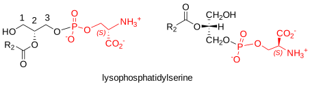
Figure \(\PageIndex{30}\): lysophosphatidylserine Lysophosphatidylserine, i.e., with a fatty acid in only one position, mediates many biological processes, especially in the immune system of animal tissues. It occurs in the thymus, peripheral lymphoid tissues, central nervous system, and colon, but is barely detectable in plasma. Phospholipases primarily deacylate the diacyl lipid. For example, a secreted isoform that is phosphatidylserine-specific (PLA1A) removes the sn-1 acyl group of phosphatidylserine to generate sn‑2‑lysophosphatidylserine containing unsaturated fatty acids, and is greatly upregulated by various inflammatory stimuli. This extracellular enzyme utilizes phosphatidylserine exposed on the cell membrane as a substrate, although other phospholipases may operate intracellularly and produce sn‑1‑lysophosphatidylserine. In addition, platelets in some species (not significantly in humans) secrete a phospholipase A2 group IIA (ABHD16A), which generates saturated sn‑1‑lysophosphatidylserine (and other lysophospholipids).
Lysophosphatidylserine has been detected after injury to animal tissues (tumor growth, graft rejection, burns). It may have a similar function to lysophosphatidic acid in cell signaling, for example, in regulating calcium flux and stimulating immune cells through G protein-coupled receptors, of which three (GPR34, P2Y10, and GPR174, LPS1-3) have been detected in mice and humans. For example, GPR174 mediates lysophosphatidylserine-induced suppression of T-cell proliferation in vitro. When cells are damaged, lysophosphatidylserine can be generated by a reaction dependent on NADPH oxidase activation. It can diffuse and transmit the information to other cells, especially mast cells, and it is produced to enhance the clearance of activated and dying neutrophils. It thus helps resolve inflammation. One specific molecular species, i.e., 1‑(11Z‑eicosenoyl)-glycero-3-phosphoserine, is reported to be a true agonist of the Toll-like receptor 2/6 heterodimer, which is important for the immune response to pathogens; both its polar head group and the length of its acyl chain are required for this activity. In contrast, sn-2-lysophosphatidylserine has proinflammatory effects, as it augments mast cell degranulation and mast cell-dependent anaphylactic shock; most other lysophospholipids lack this activity.
Deregulated lysophosphatidylserine metabolism has been linked to certain cancers, cardiometabolic disorders, night blindness, and the human genetic neurological disorder PHARC. High serum levels of PLA1A are associated with such autoimmune disorders as Graves' disease and systemic lupus erythematosus, and there is increased expression of the enzyme in metastatic melanomas. It is necessary for hepatitis C virus assembly, and it can play a role in the innate immune response. In schistosomiasis, lysophosphatidylserine from the parasite is believed to be a key activator molecule in the host.
Negatively charged lysophosphatidylserine species tend to organize in non-bilayer structures and are believed to facilitate the folding of certain membrane proteins in situ better than bilayer-forming lipids.
Phosphatidylinositol and Related Phosphoinositides
Although it had long been recognized that phosphatidylinositol or 1,2-diacyl-sn-glycero-3-phospho-(1'-myo-inositol) was a key membrane constituent, it was initially something of a surprise when the manifold biological activities of this lipid, and then of the derived phosphatidylinositol phosphates and their hydrolysis products, were discovered in animals, plants, and microorganisms. Many years after the initial discoveries in the 1950s, these lipids remain a major focus for research efforts worldwide, with considerable relevance to human health. Phosphatidylinositol and its various metabolites and relevant enzymes can be located and function within different membrane regions in cells, and they form part of what have been termed phosphoinositide and phosphatidylinositol cycles, their versatility stemming from the inositol head group, a six-carbon hexahydroxy ring, which can be reversibly phosphorylated on the 3, 4, and 5 positions. Beyond their structural role in membranes, these lipids are intimately involved in innumerable aspects of membrane trafficking and signaling in eukaryotic cells. These functions are essential to cell growth and metabolism. Only a brief overview of such a highly complex topic is possible here. Glycosyl-phosphatidylinositol (GPI) is a related lipid that anchors proteins.
Phosphatidylinositol
Structure and Occurrence: Phosphatidylinositol is an important lipid, both as a membrane constituent and as a participant in essential metabolic processes in all plants and animals, directly and via several metabolites. It is an acidic (anionic) phospholipid with a phosphatidic acid backbone, linked via the phosphate group to inositol (hexahydroxycyclohexane). In most organisms, the stereochemical form is myo-D-inositol (with one axial hydroxyl at position 2 and the remainder equatorial, i.e., a chair-like structure). However, other forms (scyllo- and chiro-) have occasionally been found in plants. The 1‑stearoyl,2-arachidonoyl molecular species, which is biologically important in animals, is illustrated.
Figure \(\PageIndex{31}\) shows the structure of phosphatidylinositol.
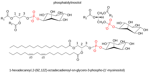
Phosphatidylinositol is especially abundant in brain tissue, where it can account for 10% of phospholipids. It is present in all tissues, cell types, and membranes at relatively low levels compared to many other phospholipids. In rat liver, it amounts to 1.7 micromoles/g, i.e., less than phosphatidylcholine, phosphatidylethanolamine, and phosphatidylserine. Under normal conditions, it is present entirely in the inner leaflet of the erythrocyte membrane and in the plasma membrane of nucleated cells. Phosphatidylinositol per se is rarely found in prokaryotes other than the Actinomycetales, although the thermophilic α-proteobacterium Rhodothermus marinus contains dialkylether glycerophosphoinositides.
The fatty acid composition of phosphatidylinositol is rather distinctive, as shown in Table \(\PageIndex{6}\). Thus, in almost all animal tissues, the characteristic feature is a high content of stearic and arachidonic acids. All stearic acid is linked to position sn-1, and all arachidonic acid to position sn-2. As much as 78% of the total lipid may consist of the single molecular species sn-1-stearoyl-sn-2-arachidonoyl-glycerophosphorylinositol (see Table \(\PageIndex{7}\) below). Although 1-alkyl- and alkenyl- forms of phosphatidylinositol are known, they are much less abundant than the diacyl form. In plant phosphatidylinositol (e.g., Arabidopsis thaliana), palmitic acid is the main saturated fatty acid at sn-1, while linoleic and linolenic acids are the main unsaturated components at sn-2. Similarly, in yeast, palmitic acid is predominantly at sn-1, with oleic and palmitoleic acids at sn-2; in Amoebozoa, a C16 alkyl group is at sn-1 and cis-vaccenic acid at sn-2.
| Table \(\PageIndex{6}\): Fatty acid composition of phosphatidylinositol (wt % of the total) in animal and plant tissues. | ||||||||||
| Tissue | Fatty acids | |||||||||
|---|---|---|---|---|---|---|---|---|---|---|
| 16:0 | 18:0 | 18:1 | 18:2 | 18:3 | 20:3 | 20:4 | 22:3 | 22:5 | 22:6 | |
| Bovine brain [1] | 8 | 38 | 10 | 1 | - | 5 | 34 | 2 | tr. | 1 |
| Bovine liver [2] | 5 | 32 | 12 | 6 | 1 | 7 | 23 | 4 | 3 | 5 |
| Rat liver [3] | 5 | 49 | 2 | 2 | 4 | 35 | 1 | |||
| A. thaliana [4] | 48 | 3 | 2 | 24 | 24 | |||||
| [1] = Holub, B.J. et al.. J. Lipid Res.., 11, 558-564 (1970); DOI. [2] = Thompson, W. and MacDonald, G., J. Biol. Chem., 250, 6779-6785 (1975); DOI. [3] = Wood, R. and Harlow, R.D. Arch. Biochem. Biophys., 135, 272-281 (1969); DOI. [4] = Browse, J. et al. Biochem. J., 235, 25-31 (1986); DOI. | ||||||||||
Biosynthesis: The basic mechanism for the biosynthesis of phosphatidylinositol and phosphatidylglycerol is sometimes termed a branch point in phospholipid synthesis, as phosphatidylcholine and phosphatidylethanolamine are produced by a somewhat different route.
Phosphatidylinositol is found in all eukaryotes, which can generally synthesize inositol de novo from glucose-6-phosphate. As with phosphatidylglycerol (and hence cardiolipin), phosphatidylinositol is formed biosynthetically from phosphatidic acid via the intermediate cytidine diphosphate diacylglycerol, which is produced by the action of a CDP-diacylglycerol synthase believed to be the rate-limiting enzyme in phosphatidylinositol biosynthesis. Then, the enzyme CDP-diacylglycerol inositol phosphatidyltransferase ('phosphatidylinositol synthase' or 'PIS') catalyzes a reaction with myo-inositol to produce phosphatidylinositol.
Figure \(\PageIndex{32}\) shows the synthesis of phosphatidylinositol in eukaryotes.
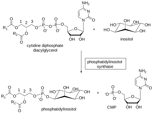
In mammals, PIS is located in the endoplasmic reticulum, in part in a subcompartment associated with mitochondria (mitochondria-associated membranes, MAM) and in mitochondria per se. Indeed, PIS is present in a mobile ER-derived subcompartment that transiently contacts other organelles, including the plasma membrane, and facilitates the distribution of phosphatidylinositol to other subcellular compartments. The other product is cytidine monophosphate (CMP). Because PIS catalyzes the reverse reaction, the phosphatidylinositol synthesis rate depends on the relative concentrations of the precursors and product, and the latter must be transported away from the site of synthesis for the reaction to continue. Vesicular transport delivers much of the phosphatidylinositol to other membranes. A family of soluble phosphatidylinositol transfer proteins (PITPα, PITPβ, and PITPNC1) provides phosphatidylinositol from the ER to kinases for phosphorylation (see below).
Molecular species specificity: The phosphatidylinositol synthase per se does not exhibit the fatty acyl specificity observed in the final product, but earlier in the biosynthetic process, 1-stearoyl-2-arachidonoyl species of diacyl-sn-glycerols are converted preferentially into phosphatidic acid by the epsilon isoform of diacylglycerol kinase (DGKε), anchored to the membrane via its N-terminal hydrophobic helix segment; ATP is the phosphate donor. In addition, one CDP-diacylglycerol synthase (CDS2) has similar specificity and generates the immediate precursor CDP-diacylglycerol from phosphatidic acid. Some specificity may be introduced via lysophosphatidylinositol, formed as a by-product of eicosanoid formation (see below) or as an intermediate as part of the normal cycle of deacylation-acylation of phosphatidylinositol in tissues in which the fatty acid composition is remodeled to give the final distinctive composition. A membrane-bound O-acyltransferase (MBOAT7 or LPIAT1) specific for position sn-2 of lysophosphatidylinositol, with a marked preference for arachidonoyl-CoA, is ubiquitously expressed in animal tissues and may regulate free arachidonic acid and eicosanoid levels.
In macrophages subjected to inflammatory stimuli, remodeling reactions produce phosphatidylinositol containing two molecules of arachidonate, and evidence suggests it is a novel bioactive phospholipid that regulates innate immune responses in these cells. Further specificity may be introduced by lysocardiolipin acyltransferase (LYCAT; also known as LCLAT1 or ALCAT1), which exhibits a preference for lysophosphatidylinositol and lysophosphatidylglycerol over other phospholipids in vitro, and incorporates 18:0 rather than shorter chain fatty acids into position sn-1 of phosphatidylinositol and other phosphoinositides, especially phosphatidylinositol-4,5-bisphosphate and phosphatidylinositol-3-phosphate; this enzyme may be located adjacent to the phosphatidylinositol synthase in the endoplasmic reticulum. Some of the phosphatidylinositol in membranes is derived from recycling polyphosphoinositides via the phosphatidylinositol cycle, which could influence the molecular species composition (see below).
The highly specific distribution of fatty acids on the glycerol moiety of phosphatidylinositol is disrupted in some cancer cells, especially those with a mutation in the p53 gene, one of the most frequently mutated genes in cancer.
Plants and bacteria: In contrast to animals, plants have two phosphatidylinositol synthase isoforms, PIS1 and PIS2, which display specificities for particular species of the CDP-diacylglycerol substrate. PIS1 preferentially generates phosphatidylinositol with saturated or monounsaturated fatty acids, while PIS2 generates polyunsaturated species; the two forms may have different functions. In protozoan parasites, such as Trypanosoma brucei, phosphatidylinositol synthase's active site may be located in the endoplasmic reticulum and the Golgi lumen. Evidence suggests two distinct product pools in this organism: a bulk membrane form derived from inositol imported from the environment, and a second used to synthesize GPI anchors, which uses myo-inositol synthesized de novo. In yeast, some biosynthesis may occur on the cytosolic side of the plasma membrane.
The enzyme is a transmembrane protein and uses CDP-diacylglycerol as a donor and either inositol (eukaryotes) or inositol phosphate (prokaryotes) as the acceptor alcohol. The structure of a similar enzyme, phosphatidylinositol-phosphate synthase from Renibacterium salmoninarum, is shown in Figure \(\PageIndex{33}\).

Figure \(\PageIndex{33}\): Structure and reaction of phosphatidylinositol-phosphate synthase from Renibacterium salmoninarum. Clarke, O., Tomasek, D., Jorge, C. et al. Structural basis for phosphatidylinositol-phosphate biosynthesis. Nat Commun 6, 8505 (2015). https://doi.org/10.1038/ncomms9505. Creative Commons Attribution 4.0 International License. http://creativecommons.org/licenses/by/4.0/
Panel A shows the reaction for PIP synthases, which transfer a diacylglycerol-substituted phosphate group (purple/red) from the CDP-DAG donor to the inositol phosphate acceptor (green), generating PIP and CMP. Panel B shows the structure of the RsPIPS-Δ6N homodimer in ribbon representation viewed from two orthogonal orientations (in the plane of the membrane on the left; towards the cytosol down the dimer axis on the right). One protomer is colored grey, and the helices of the other are depicted in spectral coloring, from blue (JM1) to red (TM6). The Af2299 extramembrane domain used to facilitate crystallization is not shown here.
Figure \(\PageIndex{34}\) shows a large cavity that contains the active site of RsPIPS.
Panel (a) shows the structure of RsPIPS-Δ6N in ribbon representation, with one protomer colored grey and the other colored by the Kyte–Doolittle hydrophobicity scale, from −4.5 (most polar, light blue) to 4.5 (most hydrophobic, orange). Two orthogonal representations are shown; on the left is a view in the plane of the membrane, and on the right is a view from the cytosol along the dimer axis. A transparent purple surface delineates the borders of the interfacial cavity, which contains three subregions: 1, the inositol phosphate acceptor-binding pocket; 2, the nucleotide-binding pocket between TM2 and TM3; and 3, a hydrophobic groove between TM2 and JM1. (b) Detail of the active site viewed in the plane of the membrane, with side chains that contact the bound Mg2+ and SO42- ions labeled and depicted in stick representation.
A nucleotide-binding site formed from transmembrane segments 1, 2, and 3 contains 8 conserved residues (D1xxD2G1xxAR…G2xxxD3xxxD4). The first three aspartic acid side chains coordinate a metal ion, while the fourth likely acts as a general base in catalysis.
Figure \(\PageIndex{35}\) shows an interactive iCn3D model of the phosphatidylinositolphosphate (PIP) synthase with bound CDP-DAG from Renibacterium salmoninarum (5D92).

Figure \(\PageIndex{35}\): Phosphatidylinositolphosphate (PIP) synthase with bound CDP-DAG from Renibacterium salmoninarum (5D92).. Click the image for a popup or use this external link: https://structure.ncbi.nlm.nih.gov/i...i9nhSw9U32Man8
The two identical subunits of the homodimer are shown in gray and plum. The two CDP-DAGs are shown in spacefill and CPK colors.
Function: In addition to functioning as negatively charged building blocks of membranes, the inositol phospholipids (including the phosphatidylinositol phosphates or 'polyphosphoinositides' discussed below) have crucial roles in the interfacial binding of proteins and the regulation of protein activity at the cell interface. Because phosphoinositides are polyanionic, they can engage in strong, nonspecific electrostatic interactions with proteins. However, they bind especially efficiently to so-called ‘PH’ domains of cellular proteins. For example, at least three phosphatidylinositol molecules are present in the crystal structure of human erythrocyte glycophorin, and they are believed to influence binding to other proteins via their head groups. The lipid is a structural component of the yeast cytochrome bc1 complex.
In animal tissues, phosphatidylinositol is the primary source of arachidonic acid required for eicosanoid biosynthesis, including prostaglandins, via phospholipase A2, which releases fatty acids from the sn-2 position. The reverse reaction also occurs.
Figure \(\PageIndex{36}\)s shows the generation of arachidonic acid and eicosanoids from PI using phospholipase A2

Similarly, phosphatidylinositol and its phosphates are the main sources of diacylglycerol, a signaling molecule in animal and plant cells, via a family of highly specific enzymes collectively known as phospholipase C. In brief, diacylglycerols regulate the activity of a group of at least a dozen related enzymes known as protein kinase C, which control many key cellular functions, including differentiation, proliferation, metabolism, and apoptosis. Indeed, the biological actions of the various components released have been the subject of intensive study over many years. 2‑Arachidonoylglycerol, an endogenous cannabinoid receptor ligand, may also be a product of phosphatidylinositol catabolism.
Phosphatidylinositol Phosphates (Polyphosphoinositides) in Animals
Structure and Occurrence: Pioneering work by Mable and Lowell Hokin in the 1950s showed that phosphatidylinositol is converted to polyphosphoinositides, which have important signaling and other functional activities, including cell communication via signal transduction, cell survival and proliferation, membrane trafficking, and modulation of gene expression. Many substrate-selective kinases phosphorylate phosphatidylinositol at positions 3, 4, and 5 of the inositol ring, with distinct phosphatases and phospholipases maintaining the balance. Seven different isomers are known (mono-, bis-, and tris-phosphorylated), produced in a tightly coordinated manner, and each has characteristic biological activities. Each turns over much more rapidly than the parent phosphatidylinositol molecule. In addition, the molecular species of each isomer differ in the nature of their fatty acyl groups. Although phosphatidylinositol 4-phosphate and phosphatidylinositol 4,5-bisphosphate were long thought to be the most significant in quantitative and possibly biological terms, phosphatidylinositol 3-phosphate and its metabolites are now recognized as biologically important.
Figure \(\PageIndex{37}\) shows the structures of phosphatidylinositol phosphates

These lipids are usually present at low levels in tissues, typically about 0.5 to 1% of the total lipids in the inner leaflet of the plasma membrane, so they are unlikely to have an appreciable structural role. On the other hand, static measurements of rapidly turning-over lipids do not meaningfully assess their cellular functions. The positional distributions of fatty acids in the phosphatidylinositol, phosphatidylinositol 4-phosphate, and phosphatidylinositol 4,5-bisphosphate of ox brain are listed in Table \(\PageIndex{7}\). In each, saturated fatty acids are concentrated at sn-1 and polyunsaturated fatty acids, especially arachidonate, at sn-2; differences among the three lipids are minimal.
| Table \(\PageIndex{7}\): Distribution of fatty acids (mol% of the total) in positions sn‑1 and sn‑2 in phosphatidylinositol (PI) and the phosphatidylinositol mono- and diphosphates of ox brain. | ||||||
| Fatty acids | PI | PI monophosphate | PI diphosphate | |||
|---|---|---|---|---|---|---|
| sn-1 | sn-2 | sn-1 | sn-2 | sn-1 | sn-2 | |
| 16:0 | 15 | 9 | 7 | |||
| 18:0 | 74 | 69 | 69 | |||
| 18:1 | 10 | 10 | 20 | 13 | 21 | 10 |
| 18:2 | 1 | 2 | trace | 1 | 1 | 1 |
| 20:3(n-9) | 5 | 10 | 10 | |||
| 20:3(n-6) | 5 | 11 | 12 | |||
| 20:4(n-6) | 67 | 49 | 52 | |||
| 22:3 | 7 | 10 | 7 | |||
| 22:6(n-3) | trace | trace | trace | |||
| Data from Holub, B.J. et al., J. Lipid Res., 11, 558-564 (1970); DOI. Molecular species data, see Traynor-Kaplan, A. et al., Biochim. Biophys. Acta, 1862, 513-522 (2017); DOI. |
||||||
Biosynthesis: Phosphatidylinositol per se is the ultimate precursor of all phosphoinositides; their head groups have different charges and structures that directly affect membrane properties and, via metabolic interactions, can act as chemical switches. Membranes maintain individual phosphoinositides at steady-state levels through a continuous, sequential series of phosphorylation and dephosphorylation reactions catalyzed by specific kinases, phosphatases, and phospholipase C enzymes, which are regulated and/or relocated via cell-surface receptors for extracellular ligands. While this has been termed a ‘futile cycle’, which can consume a significant proportion of cellular ATP production, it is only part of a wider pattern of reactions - the phosphatidylinositol cycle (see below). Controlled synthesis of these phosphoinositides occurs in distinct intracellular compartments, supporting distinct, independently regulated functions and spatially distinct target enzymes or receptors. In mammals, researchers have identified 18 phosphoinositide interconversion reactions to date, mediated by at least 20 phosphoinositide kinases and 34 phosphoinositide phosphatases spanning 8 and 10 classes, respectively; some remain uncharacterized. Most of these enzymes are conserved across all eukaryotes, and each has distinct functions and specificities that related isoforms cannot replace.
Generally, most monophosphorylations occur in endomembranes, such as the endosomes and the Golgi network. In contrast, the second and third phosphorylations occur primarily at the plasma membrane, as reflected in each membrane's lipid composition. While these enzymes are believed to act independently and sequentially to produce a specific product, some may participate in protein complexes to coordinate their activities. Specific transporters, especially the 'Nir2' protein, facilitate phosphoinositide exchange between membranes. Phosphatidylcholine metabolism is also linked, as phospholipase D can hydrolyze it to phosphatidic acid, an important activator of key kinases. Figure \(\PageIndex{38}\) overviews polyphosphoinositide metabolism in animal tissues.
Thus, as an example, phosphatidylinositol 4-phosphate (PI(4)P) is produced by the action of a phosphatidylinositol 4-kinase (PI4K) in the Golgi, and is in turn phosphorylated by a phosphatidylinositol phosphate 5-kinase (PIPK I) to form phosphatidylinositol 4,5-bisphosphate (PI(4,5)P) at the plasma membrane. However, a specific 4-kinase (PIPK II) can also form it by phosphorylating phosphatidylinositol 5-phosphate. Four PI4K isoforms in two structural families operate in different subcellular membrane compartments to produce phosphatidylinositol 4-phosphate for specific signaling functions. Some selectivity in forming molecular species or remodeling may further enrich arachidonic acid content.
Researchers later discovered that a 3-kinase (PI3K III, or the VPS 34 complex) also phosphorylates phosphatidylinositol to produce phosphatidylinositol 3-phosphate (PI(3)P) in early endosomes. Three phosphatidylinositol 3-kinase families (eight isoforms) have been described, each with distinct substrate specificities. A second phosphoinositide signaling pathway involves activation of two of these 3‑kinases, stimulated by growth factors and hormones, which phosphorylate phosphatidylinositol 4,5-bisphosphate (by PI3K I - four isoforms) and phosphatidylinositol 4‑phosphate (by PI3K II - three isoforms) to produce phosphatidylinositol 3,4,5-trisphosphate (PI(3,4,5)P) and phosphatidylinositol 3,4‑bisphosphate (PI(3,4)P), respectively. Although phosphatidylinositol 3-phosphate and other 3‑phosphorylated metabolites account for only about 0.5% of total phosphoinositides in resting mammalian cells, they are now recognized as profoundly important for cellular metabolism.
In addition to kinase activity, specific phosphoinositide phosphatases regulate these metabolite levels; these enzymes are highly conserved in eukaryotes and dephosphorylate phosphoinositides at the 3, 4, and 5 positions of the inositol ring. For example, so-called ‘SHIP’ phosphatases convert phosphatidylinositol 4,5‑bisphosphate back to phosphatidylinositol 4-phosphate by hydrolysis of the 5-phosphate group. 3‑Phosphorylated phosphoinositides are only degraded by phosphatases, especially those of the PTEN family, and not by phospholipase C (see below).
Cell organelles have membranes with distinct functions and molecular compositions. Yet, all phosphatidylinositol precursors are primarily formed in the endoplasmic reticulum, and membrane lipids must be transported between sites via specific trafficking processes/proteins. Effector proteins are selectively recruited to particular membranes by binding a single phosphoinositide, then interact with phosphoinositide-binding proteins and enzymes that channel phosphoinositide production toward the required biological outcomes and regulate signaling. For example, much of the phosphatidylinositol 4‑phosphate and phosphatidylinositol 4,5-bisphosphate involved in signaling is believed to be formed at contact sites between the endoplasmic reticulum and plasma membrane.
A concept has emerged in which each phosphoinositide has its own role – the ‘lipid code’ hypothesis, in which defined lipids act as labels for each cellular membrane to organize cells into dynamic and responsive membrane-bound compartments and maintain the orderly flow required for the complexities of membrane trafficking and spatio-temporal signaling reactions. Thus, phosphatidylinositol 4-phosphate, phosphatidylinositol 4,5‑bisphosphate, phosphatidylinositol 3-phosphate, and phosphatidylinositol 3,5‑bisphosphate are found mainly on the Golgi, plasma membrane, early endosomes, and late endocytic organelles, respectively, where they are sometimes regarded as landmarks for these compartments. For example, phosphatidylinositol 4,5‑bisphosphate is present throughout the plasma membrane and is considered a general marker. In contrast, phosphatidylinositol 3,4,5-triphosphate is a characteristic component of the basolateral region of this membrane in a polarized cell but is absent from the apical part. However, this map of phosphoinositides to specific organelles is derived from their steady-state distributions. Still, the highly dynamic generation and consumption of different phosphoinositides in response to stimuli across various subcellular compartments in living cells, driven by kinases and phosphatases, together with lipase reactions, may lead to transient pools of distinct molecular forms. The precursors must be continuously replenished by new synthesis.
Click the link below.
- Link
-
Function: The distinctive subcellular localization of different phosphoinositide species and the rapid, reversible nature of their phosphorylation give them a central role in cell signaling cascades and intracellular membrane trafficking. The precise locations of particular phosphoinositides give each organelle a specific identity and, in some cases, each face of an organelle, such as the cis and trans faces of the Golgi apparatus, enabling directional transport of cellular constituents between organelles or membranes. Phosphoinositides can signal directly by binding to specific cytosolic domains of membrane proteins via their polar head groups, triggering downstream signaling cascades, often in conjunction with an acidic phospholipid, such as phosphatidylserine or phosphatidic acid, at an adjacent binding site. The term 'lipidon' has been coined to describe the unique collection of co-located lipids that distinguish biological membrane nano-environments and provide the context for PI recognition in vivo. In this way, they can regulate the function of innumerable proteins integral to membranes, for example, by relocating a protein from one area of the cell to another (e.g., from the cytosol to the inner leaflet of the plasma membrane) or by attracting cytoskeletal and signaling components to the membrane. Proteins that bind phosphoinositides in this way include phospholipases, protein kinases, regulators of membrane trafficking, and cytoskeletal, scaffold, and ion channel proteins. Dysregulation of phosphoinositide metabolism and signaling contributes to many diseases, including cancer.
Binding usually involves electrostatic interactions with the negative charges of the phosphate groups on the inositol ring, with characteristic clusters of basic amino acid residues in proteins to recruit them to intracellular membranes, often leading to specific folding and increased activity of unstructured peptides. Researchers have identified at least 70 distinct types of phosphoinositide-binding sites in proteins. In particular, the pleckstrin homology (PH) domain, a ~100-amino-acid binding region, is the most abundant lipid-binding domain, with more than 225 examples identified; it can show strong specificity for particular polyphosphoinositides and often binds simultaneously with other proteins. While nonspecific electrostatic interactions initially drive the interaction, specific binding follows and increases membrane residence time. The phox homology (PX) domain family, with 49 members in humans, is unique in that it can recognize all seven phosphoinositide forms, whereas proteins with a FYVE domain, which is enriched in cysteine and stabilized by two zinc atoms, bind specifically to phosphatidylinositol 3-phosphate (PI(3)P). The protein kinase C family has C1 or C2 domains that recognize phosphatidylinositol 4,5-bisphosphate and phosphatidylinositol 3,4,5-trisphosphate specifically (and sometimes other lipids). The distinctive phosphoinositide composition of membranes in different organelles strengthens and specifies these interactions through cooperative binding with other membrane proteins.
Phosphatidylinositol 3-phosphate and the other phosphatidylinositol monophosphates are present in cells at low levels, although their levels do not appear to fluctuate greatly. PI(3)P has been implicated in membrane trafficking through interactions with specific endosomal proteins. In particular, it plays a pivotal role in initiating autophagy, i.e., the controlled internal degradation and turnover of cellular constituents. At the same time, PI(3,5)P2 is important for autophagosome–lysosome fusion and the subsequent acidification of this organelle. After sorting lysosomal contents, components of the internalized cargo are recycled to the plasma membrane; a specific phosphatase dephosphorylates PI(3)P to phosphatidylinositol, which is then phosphorylated to PI(4)P. Thus, internalization, sorting, and trafficking of membrane proteins depend on the interconversion of phosphoinositide species through coordinated phosphorylation-dephosphorylation reactions.
PI(3)P generally controls cellular processes by recruiting effector proteins through low- to moderate-affinity interactions with specific PI(3) P-binding domains. A protein designated Akt (protein kinase B) is recognized as a direct effector of the PI3K signaling cascade, with receptor tyrosine kinases as the main upstream activators, for example, but it is now known that every phosphatidylinositol phosphate has a specific set of effector proteins that are recruited to target membranes or are allosterically regulated by the specific receptors; each function may require a different effector. PI(3)P also regulates the final stage of cell division (cytokinesis), and the lipid accumulates at sites of cell division. Because class I PI3K isoforms, in particular, have been implicated in the etiology and maintenance of various diseases and metabolic disorders, including cancer, inflammation, and autoimmunity, drug companies are actively pursuing inhibitors. In particular, they mediate insulin-independent glucose transport and many of insulin's physiological actions. In lung cancer, RAS proteins, key signaling switches that control proliferation, differentiation, and survival in eukaryotic cells, regulate the activity of type I phosphatidylinositol 3-kinase (PI3K); this is essential for tumor initiation and maintenance.
Phosphatidylinositol 4-phosphate is a precursor for 4,5-bisphosphate, but it also binds to a protein on the cell's cytoskeleton and has its own characteristic functions. It is the most widely distributed phosphoinositide and, in addition to the Golgi and the plasma membrane, is present in late endosomes, lysosomes, secretory vesicles, and autophagosomes. As a part of protein-lipid complexes, it is believed to have a role in essential nuclear processes. In yeast, it functions in anterograde transport from the trans-Golgi and retrograde transport from the Golgi to the endoplasmic reticulum; it is also necessary for the formation of secretory vesicles in the Golgi that are targeted to the plasma membrane. Some of that in the plasma membrane is exchanged for phosphatidylserine by the action of specific transport proteins at junctions with the endoplasmic reticulum.
In addition, PI(4)P is essential for the structure and function of the late endosomes, where it is required for the recruitment of specific proteins that control cargo exit (following hydrolysis of PI(3)P). Some of these participate in vesicle formation, while others like the oxysterol binding protein (OSBP) are involved in lipid transfer. After initiation of the process by PI(3)P, PI(4)P, PI(4,5)P2, and their binding proteins are modulators of autophagy at most stages of the process. PI(4)P has been called the 'fuel' that drives cholesterol transport, as its hydrolysis provides the energy that enables the establishment of active sterol concentration gradients across membrane-bound compartments with the aid of OSBP, which is a key regulator of cholesterol, oxysterol, and PI(4)P concentrations in membranes. In the plasma membrane, PI(4)P supports ion channel function and anchors proteins with polybasic domains. However, it is not utilized to synthesize PI(4,5)P2 in this membrane. In contrast, PI(4)P derived from PI(4,5)P2 in the membrane of primary cilia in the retina is important for vision. PI(4)P plays an important role in the progression of many diseases, including virus replication, cancer, and various inflammatory diseases, and inhibitors of PI4-kinase are under investigation for their therapeutic potential.
While the biological properties of phosphatidylinositol 5-phosphate have taken longer to unravel because of the difficulties of separating this isomer, it is now apparent that it is involved in osmoregulation in plants and animals. It also has signaling functions; although it is the least abundant phosphatidylinositol monophosphate, it is involved in signaling in the nucleus and the cytoplasm, modulating cellular responses to various stresses, hormones, and growth factors. In endosomes, it regulates protein sorting.
Although phosphatidylinositol 4,5-bisphosphate (PI(4,5)P2) is found primarily in the inner leaflet of the plasma membrane, where it may define membrane identity in eukaryotic cells, it is also present in endosomes, the endoplasmic reticulum, and the nucleus. It is an essential precursor of lipid second messengers, such as diacylglycerols, with vital signaling functions mediated by plasma membrane G-protein coupled receptors, receptor tyrosine kinases, and immune receptors. Because of its large head group and multivalent negative charge, PI(4,5)P2 has been described as an "electrostatic beacon" interacting with membrane proteins, other lipids, and cellular cations in various ways. As a result, despite its relatively low concentration, it is a key regulator of innumerable events at the plasma membrane, including cell adhesion and motility, vesicle endocytosis and exocytosis, and ion channel function, especially for potassium, calcium, and sodium. For example, ion channels appear to be obligatory factors, increasing their activity by activating key proteins, while phospholipase C hydrolysis reduces this activity.
PI(4,5)P2 interacts with cationic residues on a large array of proteins, in concert with cholesterol, to form localized membrane domains distinct from sphingolipid-enriched rafts. Indeed, it is present at much higher concentrations than other phosphoinositide species in cells, although most of it is, in effect, sequestered by binding proteins. Phosphatidylinositol 4,5-bisphosphate and its diacylglycerol metabolites are also important for membrane vesicle formation. For example, the clathrin-coated vesicle pathway is a major cellular pathway for internalizing cell-surface proteins such as transferrin. PI(4,5)P2 is essential to this process because it binds to membrane machinery, increasing the number of clathrin-coated pits and facilitating protein internalization. It also plays a related role in caveolae, where it is concentrated at the rim.
Through its attachment to the apical plasma membrane, phosphatidylinositol 4,5-bisphosphate is intimately involved in actin cytoskeleton development, thereby controlling cell shape, motility, and many other processes. In particular, it binds with high specificity to effectors such as vinculin, a membrane-cytoskeletal protein that links integrin adhesion molecules to the actin cytoskeleton. Dysregulation of this function has been implicated in tumor cell migration and metastasis. In yeast, stearic acid at position sn-1 appears essential for this function. In the cell nucleus, this lipid is believed to be involved in maintaining chromatin, the complex combination of DNA, RNA, and protein that makes up chromosomes in a transcriptionally active conformation, and is a precursor for further signaling molecules. It plays a role in gene transcription and RNA processing, especially in modulating RNA polymerase activity and in other nuclear processes.
Lipids are essential components of the immune response of animal tissues to toxic bacterial lipopolysaccharides, as they bind to specific proteins. It is also involved in the pathophysiology of HIV via an interaction with the Tat protein secreted by infected cells.
PI(4,5)P2 is the primary precursor of the endocannabinoid 2-arachidonoylglycerol in neurons. It is also an essential cofactor for phospholipase D and affects cellular phosphatidic acid production through its specific signaling functions. By binding specifically to ceramide kinase, the enzyme that synthesizes ceramide-1-phosphate, it influences sphingolipid metabolism. Like ceramide-1-phosphate, it binds to and activates the Ca2+-dependent phospholipase A2, which generates the arachidonate for eicosanoid production. One molecule of phosphatidylinositol 4,5‑bisphosphate binds to each subunit of the mammalian GIRK2 potassium channel, enabling a conformational change that supports the protein's transport function.
Perhaps the best-characterized phosphoinositide signaling function results from the hydrolysis of phosphatidylinositol phosphates by phospholipase C isoforms, producing sn-1,2-diacylglycerol and inositol 3,4,5-trisphosphate (see below), which act as second messengers. Only those polyunsaturated diacylglycerol species derived from PI(4,5)P2 can bind and activate protein kinase C (α, ε, δ) isoforms in vitro and in vivo. This lipid is important because it binds strongly to these enzymes via a basic patch distal to a Ca2+ binding site, thereby targeting them selectively to the plasma membrane. Aberrant expression of phospholipase Cγ2 may contribute to neurodegenerative diseases. Via PI3 kinase, PI(4,5)P2 is a precursor of PI(3,4,5)P3, which has distinctive signaling properties.
Phosphatidylinositol 3,4-bisphosphate can be produced by two routes and regulates a variety of cellular processes relevant to health and disease, including B cell activation and autoantibody production, insulin sensitivity, neuronal dynamics, endocytosis, and cell migration. It binds selectively to many proteins and acts as a secondary messenger by recruiting the protein kinase Akt (protein kinase B). It may influence the cell cycle, cell survival, angiogenesis, and glucose metabolism. During endocytosis in the endolysosomal system, it is produced from PI(4,5)P2 and controls the maturation of endocytic-coated pits. Its synthesis and turnover are spatially segregated within the endocytic pathway. In epithelial cells, it is located on the apical membrane (i.e., facing the lumen), rather than the basolateral membrane, and is believed to determine apical membrane identity and function.
Phosphatidylinositol 3,5-bisphosphate is present at low levels only in cells (0.04-0.1% of the total phosphatidylinositides) unless stimulated by growth factors. Still, it is important for membrane and protein trafficking, especially in late endosomes in eukaryotes and in yeast vacuoles. For example, converting PI(3)P to PI(3,5)P2 promotes endosomal maturation and degradative sorting. It mediates signaling in response to stress and hormonal cues and controls ion transport in membranes. Genetic studies confirm that it is essential for healthy embryonic development, especially in the nervous system.
Phosphatidylinositol 3,4,5-trisphosphate (PI(3,4,5)P3) is almost undetectable in quiescent cells. Yet its intracellular level rises rapidly through synthesis at the plasma membrane in response to agonists such as extracellular growth factors and hormonal stimuli. By recruiting proteins with pleckstrin homology (PH) domains to the plasma membrane, it has been implicated in various cellular functions, including growth, cell survival, proliferation, cytoskeletal rearrangement, intracellular vesicle trafficking, and cell metabolism. In particular, it is an important component of a signaling pathway in the cell nucleus. In epithelial cells, it is located on the basolateral membrane, i.e., facing adjacent cells, where it may determine the identity and function of this membrane. In contrast to phosphatidylinositol 3-phosphate, it opposes autophagy by binding to and activating Akt's PH domain, thereby inducing cell proliferation. During feeding, various physiological responses trigger insulin secretion, which phosphorylates phosphatidylinositol 4,5-bisphosphate to phosphatidylinositol 3,4,5-trisphosphate and initiates a signaling cascade that suppresses autophagy. When impaired, this pathway contributes to insulin resistance in metabolic diseases, including obesity and diabetes. It has been implicated in tumor cell migration and metastasis. PI(3,4,5)P3 is also present in the nucleus and nucleoli, where it is believed to function in RNA processing/splicing, cytokinesis, protein folding, and DNA repair. In contrast, like phosphatidylserine, it is reportedly transferred to the outer leaflet of the plasma membrane in aged or damaged cells as an 'eat‑me' signal for phagocytes and apoptosis.
The human immune system uses neutrophils, highly mobile cells, to eliminate pathogens from infected tissue. The first step is to track and then pursue molecular signals, such as cytokines, emitted by pathogens. Two phospholipids act in sequence to orient neutrophils in the correct direction. The first is phosphatidylinositol 3,4,5-trisphosphate, which binds to the protein DOCK2 and enables its translocation to the plasma membrane. Then, phosphatidic acid, generated by phospholipase D acting on phosphatidylcholine, takes over and directs DOCK2 to the leading edge of the plasma membrane. This causes actin polymerization within the cell and, in effect, reshapes the neutrophil, pointing it toward the source of the pathogens' signals. In contrast, Mycobacterium tuberculosis can subvert phosphoinositide signaling to arrest phagosome maturation by dephosphorylating phosphatidylinositol 3-phosphate.
Water-Soluble Inositol Phosphates
As mentioned briefly above, hydrolysis of phosphatidylinositol phosphates by calcium-dependent phospholipase C (or 'phosphoinositidase C') leads to the generation of sn‑1,2‑diacylglycerols, which act as second messengers in animal cells and are of enormous metabolic importance. Many enzymes of this type exist, but phosphoinositide-specific phospholipase C activity is an essential step in inositol signaling pathways. The enzyme exists in six families, each with at least 13 isoenzymes, all of which share conserved regions, such as the pleckstrin homology (PH) binding domain. Each has a distinctive role and can have a characteristic cell distribution linked to a specific function. Signaling molecules such as G-protein-coupled receptors, receptor tyrosine kinases, Ras-like GTPases, and calcium ions stimulate these enzymes, linking phosphatidylinositol phosphate hydrolysis to a wide range of other cellular signals. Because phospholipase C is a soluble cytosolic protein, translocation to the plasma membrane is crucial for signal transduction. Regulation of these isoenzymes, and PLCγ1 in particular, is vital for health, as they can activate or inhibit important pathophysiological processes, especially cancer.
Some phosphatidic acid is synthesized from diacylglycerols produced within the plasma membrane by diacylglycerol kinases. This acid is transported back to the endoplasmic reticulum and can ultimately be reutilized for phosphatidylinositol biosynthesis.
The other products of the phospholipase C reaction that are especially relevant because of their many essential functions are water-soluble inositol phosphates. Up to 60 different compounds of this type are possible, and at least 37 have been found in nature, all of which are biologically important. However, polyphosphoinositides with a phosphate in position 3 are not substrates for phospholipase C.
Figure \(\PageIndex{39}\) shows the generation of inositol phosphates by phospholipase C.

For example, in response to various physiological stimuli in animals, phospholipase C hydrolyzes phosphatidylinositol 4,5-bisphosphate in the plasma membrane, releasing inositol 1,4,5-trisphosphate. This important cellular messenger diffuses into the cytosol and stimulates calcium release from an ATP-loaded store in the endoplasmic reticulum via ligand-gated calcium channels (the diacylglycerols remain in the membrane to recruit and activate members of the protein kinase C family). The increase in calcium concentration, together with the altered phosphorylation status, activates or deactivates many different protein targets, enabling cells to respond appropriately to the extracellular stimulus. To enable rapid replenishment of the phosphatidylinositol 4,5‑bisphosphate used in this way, a cycle of reactions - the phosphatidylinositol cycle - must occur (see below). In contrast, a recent publication suggests that phosphatidylinositol 4-phosphate in the plasma membrane may be a more important source of diacylglycerol following stimulation of G protein–coupled receptors.
The various inositol phosphates appear to control cellular events in specific ways, especially by organizing key signaling pathways, rearranging the actin cytoskeleton, or regulating intracellular vesicle trafficking. They have been implicated in gene transcription, RNA editing, nuclear export, and protein phosphorylation. These remarkable compounds can be rapidly synthesized and degraded in discrete membrane domains or even subnuclear structures, making them ideal regulators of dynamic cellular mechanisms. Structural studies of inositol polyphosphate-binding proteins suggest that inositides may, at least in part, modify protein function as structural cofactors, ensuring proteins adopt optimal conformations. In addition, phosphoinositides and inositol polyphosphates are key components of the cell's nucleus, where they have many essential functions, including DNA repair, transcription regulation, and RNA dynamics. They may serve as activity switches for the nuclear complexes responsible for these processes, with the phosphorylation state of the inositol ring being of primary importance. As different isomers appear to have specific functions at each gene expression level, extracellular events must coordinate the production of these compounds in a highly synchronous manner.
From plants to mammals, kinases add an extra regulatory tier by generating energetic diphosphate (pyrophosphate)-containing molecules from inositol phosphates. Conversely, polyphosphate phosphohydrolase enzymes can dephosphorylate them, regenerating the original inositol phosphates. These inositol pyrophosphates and the enzymes involved in their metabolism also regulate cellular processes by modulating protein activity through various mechanisms.
Phospholipase C isoenzymes regulate the concentration of phosphatidylinositol 4,5-bisphosphate and related lipids and, hence, their activities, in addition to generating new biologically active metabolites.
Phosphatidylinositides in Plants
In plants, as in animals, phosphatidylinositol and polyphosphoinositides have essential biological functions, exerting regulatory effects by acting as ligands that bind protein targets via specific lipid-binding domains and thereby altering protein localization and enzymatic activity. However, polyphosphoinositide metabolism appears to have evolved differently after the divergence of the animal and plant kingdoms. As a result, the details of each process differ substantially, not least because phosphoinositides are localized differently in plants and animals. Phosphatidylinositol per se is the precursor of the phosphorylated forms and determines their fatty acid compositions. It also inhibits programmed cell death by serving as the biosynthetic precursor of the sphingolipid ceramide phosphoinositol and by reducing ceramide levels.
As in animals, the various phosphoinositides (five in total) are produced and interconverted rapidly by a series of kinases and phosphatases (in many isoforms) in different cellular membranes in response to environmental or developmental cues. For example, the endoplasmic reticulum generates phosphatidylinositol. At the same time, PI 4-kinases and their products are located in the trans-Golgi network and nucleus, and PI4P 5-kinases and their products are present at the plasma membrane. During polyphosphoinositide biosynthesis, the first phosphorylation occurs at the hydroxyl group at position 3 or 4 of the inositol ring, catalyzed by the appropriate kinases, while the second phosphorylation then occurs at position 5; phosphatase action on PI 3,5‑bisphosphate produces PI 5-phosphate. Most other metabolites are produced via phosphatidylinositol 3-phosphate, and reports that some phosphatidylinositol 3,4,5-trisphosphate may be produced from phosphatidylinositol 4,5‑bisphosphate require confirmation. In contrast to mammalian phosphatidylinositol 3-kinases, which accept phosphatidylinositol and its monophosphates as substrates, the plant enzyme acts only on the former.
Figure \(\PageIndex{40}\) shows polyphosphoinositide metabolism in plants
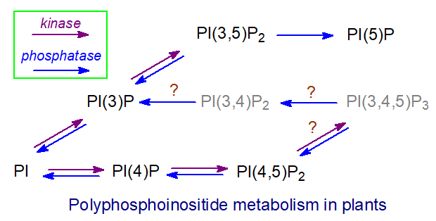
The reverse reaction in plants is carried out by phosphoinositide phosphatases, which fall into three main families: the phosphatase/tensin (PTEN) family, 5-phosphatases (5-PTases), and phosphatases containing Suppressor of Actin (SAC) domains, each with distinct subcellular locations, substrate specificities, and regulatory mechanisms.
Although plants have what might be considered normal levels of phosphatidylinositol 4-phosphate, the concentrations of phosphatidylinositol 4,5‑bisphosphate and other phosphoinositides are extremely low (10 to 20-fold lower than in mammalian cells), yet they still have vital functions. Cell types differ, but in Arabidopsis epidermal root cells, PI(4,5)P2 is most abundant in the plasma membrane (apex region) and nucleus. At the same time, PI4P slowly distributes between the plasma membrane and Golgi, with the highest concentration in the former. Multivesicular bodies/late endosomes accumulate PI3P and PI(3,5)P2; the tonoplast and autophagosomes contain PI3P. How the various metabolites are transported across membranes remains to be determined. Non-vesicular transport is believed to occur at membrane contact sites, and vesicular transport probably occurs as well.
Highly polarized distributions of phosphoinositides are found within membranes, generally oriented toward the cytosolic leaflet, and they are believed to be organized in nanoclusters with other lipids and proteins. For example, phosphatidylinositol-4-phosphate is an important constituent of the plasma membrane in plant cells, where it controls the electrostatic state and is involved in cell division. It may control the location and function of many membrane proteins, including those required for development, reproduction, immunity, nutrition, and signaling. PI(4)P is the only phosphoinositide present at the cell plate, i.e., the membrane separating two daughter cells during cell division. In addition, PI(4)P may interact with salicylic acid in the plant immune response, and it is produced during salt stress. However, researchers are now discovering specific functions for each plant phosphoinositide; these lipids are rapidly produced in response to osmotic and heat stress, and continuous turnover is essential for cell growth and development. For example, they strongly affect the growth of many cell types and guard cell function. In the nucleus, proteins have been identified that bind phosphoinositides via their acyl chains, leaving the head group exposed for enzymatic modification and signal transduction.
Phosphoinositides are especially important in microdomains at the tips of growing tissues such as the shoot apical meristem, pollen tubes, and root hairs, where phosphatidylinositol 4,5-bisphosphate functions in stem cell maintenance and organogenesis. In the plasma membrane, it is enriched in the detergent-resistant component, which is commonly equated with rafts. Although its concentration is low, PI(4,5)P2 has been shown to signal by binding many target proteins with characteristic binding domains. For example, with phosphatidic acid, PI(4,5)P2 regulates the activity of many actin-binding proteins, which in turn control actin cytoskeleton activity. This plays a key role in plant growth, subcellular organelle movement, cell division and differentiation, and plant defense. In addition, this lipid regulates ion channels, ATPases, and phospholipase C-mediated lipid degradation, as well as the production of additional second messengers. It is an important factor in both clathrin-mediated endocytosis and exocytosis. The specificity of these interactions may depend on the fatty acid composition of the lipid and on the activity of phosphatidylinositol 4-phosphate 5-kinase.
As in animals, phosphoinositides have a role in endosomal sorting through the central vacuole, a plant-specific organelle with both lytic and storage functions. Phosphatidylinositol 3,5-bisphosphate is the least abundant phosphoinositide, but it is a crucial lipid for membrane trafficking. The PI to PI(3)P to PI(3,5)P2 cascade, whose second step requires the kinase FAB1, is required for endosomal sorting events leading to membrane protein degradation or retrieval, vacuolar morphogenesis, and autophagy. PI(3,5)P2 is involved in stomatal closure and root hair growth, and it is also induced by salt stress.
Several phospholipase C enzymes specific for polyphosphoinositides have been isolated from higher plants; Ca2+ activates them, and, unlike their mammalian counterparts, G proteins do not regulate them. It is uncertain whether phosphatidylinositol is a substrate for these enzymes in vivo. Less is known about the metabolism of the water-soluble inositol phosphates produced compared to animals, and plants appear to lack a receptor for inositol 1,4,5-trisphosphate (IP3). However, it is the most abundant metabolite of this type and has been reported to induce calcium ion release, triggering stomatal closure. However, increasing evidence supports lipid signaling mediated by phospholipase C in plant abiotic stress tolerance and development. There is a general, if contested, belief that inositol hexakisphosphate (phytic acid or IP6), produced at least in part by sequential phosphorylation of inositol 1,4,5-trisphosphate, is a more important cellular messenger in plants and mobilizes an endomembrane store of calcium ions. Inositol-1,2,4,5,6-pentakisphosphate (IP5) is a structural co-factor of the jasmonic acid receptor coronatine insensitive 1, linking phosphoinositide signaling with phytohormone-controlled pathways.
In contrast to animals, diacylglycerols, the other product of phospholipase C hydrolysis of phosphoinositides, are rapidly converted to phosphatidic acid by diacylglycerol kinases and have not been considered important in signal transduction in plants. Plants lack protein kinase C, but they do have proteins with related properties that appear to be influenced by diacylglycerols. Via phospholipase D, inositol phospholipids provide phosphatidic acid, which has well-characterized signaling functions in plants, especially in defense.
Lyso-Phosphoinositides
Figure \(\PageIndex{41}\) shows the structure of lysophosphatidylinositol
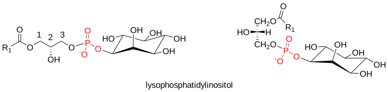
Lysophosphatidylinositols: Lysophosphatidylinositols (LPI), i.e., with a single fatty acid only linked to the glycerol moiety, are formed as intermediates in the remodeling of the fatty acid compositions of the lipids by the action of phospholipase A1 or phospholipase A2 (e.g., cPLA2α), and when arachidonic acid is released for eicosanoid biosynthesis (see above). In ovarian cancer, LPI is elevated appreciably to around 15µM in ascites, and it is also present at high levels in obese subjects.
It has become apparent relatively recently that, like other lysophospholipids, phosphatidylinositol and polyphospho-analogs may have messenger functions. For example, it has long been known to stimulate insulin release from pancreatic cells, suggesting a role in glucose homeostasis. sn-2-Arachidonoyl-lysophosphatidylinositol, in particular, is an endogenous ligand for the G protein-coupled receptor GPR55 and can thereby induce rapid phosphorylation of certain enzymes, including a protein kinase, which promotes cancer cell proliferation, migration, and metastasis. Indeed, lysophosphatidylinositol is a biomarker for poor prognosis in cancer patients, and its concentration is significantly elevated in highly proliferative cancer cells in vitro. GPR55 is expressed in many brain regions, the intestines, endocrine pancreas, and islets (where it may stimulate insulin release). It has been implicated in macrophage activation and inflammation. Beyond its role in cancer, lysophosphatidylinositol has been implicated in many metabolic diseases. It is reported to be a precursor of the endocannabinoid 2‑arachidonoylglycerol via human glycerophosphodiesterase 3, a lysophospholipase C. This enzyme suppresses the lysophosphatidylinositol receptor, thereby acting as a switch between GPR55 and endocannabinoid (CB2) signaling.
Glycerophosphoinositol: Sequential removal of both fatty acids from phosphatidylinositol by a specific phospholipase A2 (PLA2IVα) with both phospholipase A2 and lysophospholipase activities releases water-soluble glycerophosphoinositol. A glycerophosphodiester phosphodiesterase can hydrolyze this to inositol 1-phosphate; however, glycerophosphoinositol per se has distinctive biological activities and functions, as do related compounds derived from phosphatidylinositol phosphates. In particular, glycerophosphoinositol has anti-inflammatory activity, as it inhibits the inflammatory and thrombotic responses induced by bacterial lipopolysaccharides (endotoxins).
Figure \(\PageIndex{42}\) shows the structure of glycerophosphoinositol

The Phosphatidylinositol Cycle
Phosphatidylinositol can be considered the center of a cycle of reactions and intermediates involved in innumerable aspects of cellular signaling in animals (a similar cycle can be described for plants). We discuss these in detail above, but it is useful to note how each component fits into a larger pattern. In brief, as illustrated, the various synthetic and hydrolytic reactions involved in phosphoinositide metabolism can be considered to constitute a phosphatidylinositol cycle with enzymes located both in the endoplasmic reticulum and plasma membrane, so lipids have to be transferred across the cytosol in both directions between the two to complete the cycle, probably via adjacent membrane structures and facilitated by proteins of the phosphatidylinositol transfer protein membrane-associated family (PITPNM or nir2), which may channel phosphoinositide production to specific biological outcomes. Phospholipase C and phosphatidylinositol-4-phosphate 5-kinase (PI4P 5K) are located in the plasma membrane, while the cytidine diphosphate-diacylglycerol synthase (CDS2) and phosphatidylinositol synthase are in the endoplasmic reticulum. The epsilon isoform of diacylglycerol kinase (DGKε) is located at contact sites between the endoplasmic reticulum and plasma membrane. Still, nine additional isoforms with distinct cellular and subcellular localizations may be involved in the cycle. Each turn of the cycle consumes a great deal of energy and requires three moles of ATP, as well as cytidine triphosphate and inositol. If endogenous pyrophosphatases hydrolyze pyrophosphate to inorganic phosphate, the cycle can proceed in only one direction.
Figure \(\PageIndex{43}\) shows the phosphatidylinositol cycle.
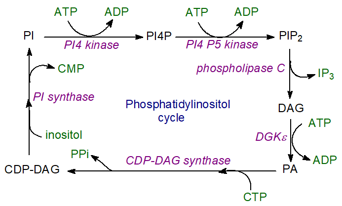
Factors such as membrane curvature must be taken into account, and the diagram is necessarily a considerable oversimplification. In addition to participating in this cycle, many lipid intermediates can serve as precursors for other lipids; for example, diacylglycerols can be precursors for triacylglycerols, while phosphatidic acid is a precursor for phosphatidylcholine and phosphatidylethanolamine. Each lipid intermediate undergoes acyl-chain remodeling via the Lands cycle, and the released polyunsaturated fatty acids can be used for eicosanoid production. A further byproduct of the cycle is inositol triphosphate, which regulates intracellular calcium levels.
It has been suggested that the unique molecular species composition of phosphoinositides (18:0-20:4) could influence their selective recycling back into phosphatidylinositol, as many of the enzymes involved prefer this substrate. A further proposal is that the phosphatidylinositol cycle could enrich this species through multiple rounds of cycling.
Summary
(Summary written by Claude, Sonnet 4.6, Anthropic)
This chapter describes the biosynthesis, remodeling, and signaling roles of the major membrane glycerophospholipids — phosphatidic acid, lysophosphatidic acid, phosphatidylcholine (PC), phosphatidylethanolamine (PE), phosphatidylserine (PS), and phosphatidylinositol (PI) and its polyphosphorylated derivatives — tracing the flow of carbon from glycerol-3-phosphate through the branched network of glycerolipid synthesis to specific membrane and signaling functions.
Phosphatidic acid (PA) is the central committed intermediate in glycerophospholipid synthesis and a signaling molecule in its own right. It is produced by four routes: the primary route involves sequential acylation of sn-glycerol-3-phosphate by GPAT (sn-1) and AGPAT/LPAT (sn-2), with multiple isoforms of each enzyme in the mitochondria and ER that differ in tissue distribution, acyl-CoA specificity, and regulatory responses to feeding state and insulin signaling; the DHAP pathway in peroxisomes produces LPA via reduction of acyl-DHAP by NADPH, channeling substrates preferentially into ether lipids; phospholipase D (PLD1 in the Golgi-lysosome continuum, PLD2 at the plasma membrane) hydrolyzes PC to release PA and choline, generating PA for signaling in membrane trafficking, cytoskeletal dynamics, cell migration, and cancer; and diacylglycerol kinases (ten isoforms, DGKα–κ) phosphorylate DAG to PA, with the ε-isoform specifically generating the 1-stearoyl-2-arachidonoyl PA used for PI synthesis. The interconversion between PA and DAG, catalyzed by lipins (Mg²⁺-dependent phosphatidate phosphatases with a HAD-family DxDxT motif), is a critical regulatory branch point: PA channels carbon to PI and cardiolipin through the CDP-DAG branch, while DAG channels carbon to PC, PE, and TAG through the Kennedy branch. Lipin activity is regulated by phosphorylation, membrane association, and dual nuclear functions as a transcriptional coactivator that regulates lipogenic gene expression. Phosphatidic acid itself plays regulatory roles as a membrane curvature sensor, protein recruiter, and transcriptional regulator (by binding the Opi1 repressor in yeast), and its local concentration in the ER is a key determinant of lipid droplet biogenesis.
Lysophosphatidic acid (LPA) is a potent bioactive lipid mediator despite its simple structure (a single acyl chain on glycerophosphate). The primary extracellular source is autotaxin — a nucleotide pyrophosphatase-phosphodiesterase that cleaves lysophosphatidylcholine (abundant in plasma) to LPA — with LPA then acting through specific GPCRs on target cells to regulate platelet activation, vascular and neural development, and cancer cell proliferation and migration. Genetic deletion of autotaxin in mice causes embryonic lethality from vascular and neuronal defects, while overexpression is also lethal, demonstrating tight requirement for precise LPA levels. Intracellularly, LPA is produced from PA by phospholipase A1 or A2 in platelets and other cells.
Phosphatidylcholine, the most abundant phospholipid in animal membranes (often ~50% of total phospholipids, concentrated in the outer leaflet of the plasma membrane), is synthesized primarily by the CDP-choline (Kennedy) pathway in the ER: choline is phosphorylated by choline kinase; the rate-limiting step is catalyzed by CTP:phosphocholine cytidylyltransferase (CCT), which has a regulatory C-terminal domain that senses membrane composition and translocates from nucleoplasm to ER membrane when PC is insufficient; CDP-choline is combined with DAG by CEPT1 in the ER or CPT1 in the trans-Golgi. Because animal cells cannot synthesize choline de novo, dietary supply and recycling from phospholipid catabolism are essential. A second pathway — sequential SAM-dependent methylation of PE by phosphatidylethanolamine N-methyltransferase (PEMT) — accounts for ~one-third of hepatic PC but is not active in most other tissues; each methylation consumes SAM and generates S-adenosylhomocysteine, which is hydrolyzed to homocysteine, linking PC synthesis to one-carbon metabolism and explaining the cardiovascular risk of hyperhomocysteinemia in choline-deficient states.
Phosphatidylethanolamine (PE), typically the second-most abundant phospholipid and the major lipid of the inner leaflet, is made by two spatially distinct pathways in mammals. The CDP-ethanolamine pathway (analogous to the Kennedy pathway) operates in the ER and Golgi, with CTP:phosphoethanolamine cytidylyltransferase as the rate-limiting enzyme; this pathway generates PE species with mono- and di-unsaturated sn-2 acyl chains. The phosphatidylserine decarboxylase (PSD1) pathway converts PS to PE in the inner mitochondrial membrane; this pathway generates PE enriched in polyunsaturated fatty acids, including DHA, and provides essentially all mitochondrial PE. Both pathways are individually dispensable but, together, essential for life; disrupting either causes embryonic lethality in mice. Similarly, phosphatidylserine is synthesized exclusively by calcium-dependent base-exchange reactions at the mitochondria-associated membrane (MAM) — PS synthase I (using PC) and PS synthase II (using PE, with high specificity for DHA-containing species) exchange their head groups for L-serine on the cytosolic face of the ER — with both enzymes subject to product feedback inhibition. PS is then decarboxylated to PE in the mitochondria (providing all mitochondrial PE), or methylated through PE to PC in the liver. The three lipids form a coupled metabolic triangle whose fluxes are tightly co-regulated and spatially segregated. In bacteria and yeast, PS is synthesized instead by a fundamentally different mechanism: reaction of CDP-DAG with L-serine, catalyzed by a Mg²⁺/Ca²⁺-dependent synthase that binds CDP-DAG first, then positions serine for nucleophilic attack on the β-phosphate via a trigonal bipyramidal transition state, releasing CMP and the product PS.
In all eukaryotic cells, the final molecular species composition of each phospholipid class is established not by de novo synthesis alone but by the Lands cycle of post-synthetic remodeling: phospholipase A2 (predominantly cleaving sn-2 ester bonds) generates a lysophospholipid and a free fatty acid; the lysophospholipid is then reacylated by tissue-specific CoA-dependent lysophospholipid acyltransferases that incorporate particular acyl chains — LPCAT1 enriches lung surfactant PC with palmitoyl chains; LPCAT2 and LPCAT3 incorporate arachidonic and linoleoyl chains into PC and PE in inflammatory cells; LPIAT1/MBOAT7 specifically enriches PI with arachidonoyl at sn-2; MBOAT2/LPCAT4 remodels PS. This system determines the exquisitely specific molecular species distributions observed (e.g., stearoyl-arachidonoyl enrichment in PI, palmitoyl-arachidonoyl enrichment in PE of inflammatory cells, dipalmitoyl enrichment in lung PC) and controls the pools of arachidonic acid available for eicosanoid signaling.
Phosphatidylinositol (PI) occupies a unique position among glycerophospholipids because of its versatility as a signaling lipid. It is synthesized from PA via CDP-DAG (produced by CDP-DAG synthase) and then combined with myo-inositol by PI synthase in the ER, with some synthesis occurring at the MAM. The distinctive 1-stearoyl-2-arachidonoyl molecular species composition of PI is established by DGKε's sn-2 specificity in generating the appropriate PA precursor and by LPIAT1/MBOAT7 remodeling. PI serves as the direct precursor for all seven phosphatidylinositol phosphate (PIP) isomers through the sequential action of substrate-selective kinases (PI3Ks, PI4Ks, and PIPK families — 20 kinases total) and phosphatases (34 phosphatases, including PTEN and SHIP families). Each PIP isomer has a characteristic subcellular distribution constituting a "lipid code" for organelle identity: PI(4)P marks the Golgi; PI(4,5)P₂ marks the plasma membrane broadly; PI(3)P marks early endosomes; PI(3,5)P₂ marks late endosomes; PI(3,4,5)P₃ is generated transiently at the basolateral plasma membrane in response to growth factors. Phospholipase C hydrolysis of PI(4,5)P₂ generates IP₃ (which diffuses to the ER to trigger calcium release through ligand-gated channels) and DAG (which remains in the membrane to activate protein kinase C isoforms) — the two-messenger system that links extracellular signals to diverse cellular responses. The phosphatidylinositol cycle reconstitutes PI from DAG through sequential action of DGKε, CDP-DAG synthase (CDS2), and PI synthase at ER-plasma membrane contact zones, consuming three ATP equivalents per cycle turn and producing inositol trisphosphate as a byproduct. Each cycle turn also provides an opportunity for Lands cycle remodeling at any intermediate, potentially enriching the 18:0/20:4 molecular species with each passage. In plants, the same general framework operates, but with differences in kinase specificities, phosphoinositide concentrations (10–20-fold lower than in animal cells), and signaling outputs — with IP₃ and IP₆ (phytic acid) rather than IP₃ alone serving key second-messenger roles in calcium mobilization, stress responses, and hormone signaling.
This page titled 21.3: Biosynthesis of Membrane Glycerolipids was last modified on Tue, 15 Sep 2026 20:51:10 GMT and is shared under a not declared license and was authored, remixed, and/or curated by Henry Jakubowski and Patricia Flatt.
The book could not be loaded just now.