Opening the book…
Search Fundamentals of Biochemistry
by William (Bill) W. Christie and Henry Jakubowski.
This section is an abbreviated and modified version of material from the Lipid Web, an introduction to the chemistry and biochemistry of individual lipid classes, written by William Christie.
(Learning goals written by Claude, Sonnet 4.6, Anthropic)
Triacylglycerol Biosynthesis: Pathways and Key Enzymes
- Describe the Kennedy (sn-glycerol-3-phosphate) pathway for triacylglycerol synthesis — tracing the sequential acylations by GPAT (sn-1), AGPAT (sn-2), and DGAT (sn-3) through the key intermediates lysophosphatidic acid, phosphatidic acid, and diacylglycerol — and contrast this with the monoacylglycerol pathway used in intestinal enterocytes and the dihydroxyacetone-phosphate pathway used in peroxisomes and ER.
- Explain the structural and mechanistic differences between DGAT1 (an MBOAT-family integral membrane enzyme with a bent acyl-CoA tunnel and a hydrophobic DAG channel within the bilayer, using His415 as a catalytic residue) and DGAT2 (the main hepatocyte/adipocyte enzyme that tethers ER to nascent lipid droplets to channel TAGs from synthesis site to storage), and describe their distinct tissue distributions and substrate specificities.
- Explain the dual enzymatic and transcriptional roles of lipin (a Mg²⁺-dependent HAD-family phosphatidate phosphatase with a DxDxT catalytic motif proceeding through a trigonal bipyramidal phosphate intermediate) at the branch point between triacylglycerol storage and phospholipid membrane biosynthesis, and explain why this branch point is a critical regulatory node for cellular lipid homeostasis.
Lipid Droplet Biogenesis, Structure, and Lipolysis
- Describe the assembly of cytoplasmic lipid droplets — including the phase separation of triacylglycerols between ER membrane leaflets to form a lens, the role of the seipin decameric cage (with its conserved F232xxGLR switch motif cycling between closed and open conformations) in stabilizing nascent droplets and enabling budding, and the coating of mature droplets with a phospholipid monolayer and amphipathic proteins including perilipins (in animals) and oleosins (in plants).
- Describe the sequential lipolysis of adipocyte triacylglycerols by ATGL (cleaves sn-1/2, rate-limiting, activated by CGI-58/ABHD5 and inhibited by G0S2), hormone-sensitive lipase (cleaves sn-1/3, activated by cAMP-PKA-mediated phosphorylation of both HSL and perilipin PLIN1), and monoacylglycerol lipase (rate-limiting final step, also the primary inactivator of the endocannabinoid 2-arachidonylglycerol), and explain how β-adrenergic stimulation coordinates phosphorylation of both PLIN1 and HSL to switch adipocytes from storage to lipolytic mode.
Digestion, Transport, and Physiological Roles of Triacylglycerols
- Trace the intestinal processing of dietary triacylglycerols — from gastric lipase and pancreatic lipase/co-lipase hydrolysis at the sn-1 and sn-3 positions releasing fatty acids and 2-monoacylglycerol, through enterocyte uptake and reassembly via the monoacylglycerol pathway, to packaging into chylomicrons (apo B48 coat, TAG/cholesterol ester core) for lymphatic export — and explain how lipoprotein lipase on endothelial cells and hepatic lipase process chylomicrons and VLDL at peripheral tissues and liver, respectively.
- Distinguish the endocrine, thermogenic, and structural functions of white, brown, and beige adipose tissue — explaining how brown adipocytes (enriched in mitochondria and UCP1) achieve non-shivering thermogenesis through β-adrenergic activation of lipolysis, with fatty acids both fueling and activating UCP1 to uncouple electron transport from ATP synthesis — and describe how leptin (appetite suppression, fuel reserve signaling) and adiponectin (insulin sensitization, anti-inflammatory) function as endocrine outputs linking adipocyte size and lipid status to systemic metabolic regulation.
Introduction
All eukaryotes and some prokaryotes can synthesize triacylglycerols. In animals, many cell types and organs can do this, but the liver, intestines, and adipose tissue are most active, and the latter store most of the body's reserves. In all cell types, including brain cells, triacylglycerols are stored as cytoplasmic 'lipid droplets' enclosed by a monolayer of phospholipids and hydrophobic proteins, such as the perilipins in adipose tissue or oleosins in seeds. These lipid droplets are now treated as distinctive organelles with their own characteristic metabolic pathways and associated enzymes - no longer boring blobs of fat. However, they are not unique to animals and plants, as Mycobacteria and yeasts have similar lipid inclusions.
Lipids serve as a store of fatty acids for energy, which can be released rapidly on demand, and as a reserve for structural purposes or as precursors for eicosanoids. In addition, lipid droplets act as protective organelles in cells, sequestering excess biologically active and potentially harmful lipids, such as free fatty acids, oxylipins, diacylglycerols, cholesterol (as cholesterol esters), retinol esters, and coenzyme A esters.
While triacylglycerols are essential for normal physiology, excessive accumulation in human adipose tissue and other organs leads to obesity and other health problems, including insulin resistance, steatohepatitis, and cardiomyopathy. Accordingly, there is considerable pharmaceutical interest in drugs that affect triacylglycerol biosynthesis and metabolism.
Biosynthesis of Triacylglycerols
Three main pathways for triacylglycerol biosynthesis include the sn-glycerol-3-phosphate and dihydroxyacetone phosphate pathways, which predominate in liver and adipose tissue, and a monoacylglycerol pathway in the intestines. In maturing plant seeds and some animal tissues, a fourth pathway has been recognized involving a diacylglycerol transferase.
The most important route to triacylglycerols is the sn-glycerol-3-phosphate or Kennedy pathway, first described by Eugene Kennedy and colleagues in the 1950s, from which more than 90% of liver triacylglycerols are produced. Figure \(\PageIndex{1}\) shows the pathway from glycerol-3-phosphate to triacylglycerols (the Kennedy pathway) and some preceding reactions that generate glycerol-3-phosphate.

In this pathway, the main source of the glycerol backbone has long been believed to be sn-glycerol-3-phosphate produced by glucose catabolism (glycolysis) or, to a lesser extent, by the enzyme glycerol kinase acting on free glycerol. However, growing evidence suggests that a significant proportion of glycerol is produced de novo via glyceroneogenesis from pyruvate. Indeed, this may be its main source in adipose tissue.
Subsequent reactions occur primarily in or at the endoplasmic reticulum. First, the precursor sn-glycerol-3-phosphate is esterified by a fatty acid coenzyme A ester in a reaction catalyzed by a glycerol-3-phosphate acyltransferase (GPAT) at position sn-1 to form lysophosphatidic acid, and this is in turn acylated by an acylglycerophosphate acyltransferase (AGPAT) in position sn-2 to form a key intermediate in the biosynthesis of all glycerolipids - phosphatidic acid. Numerous isoforms of these enzymes are known; they are expressed in specific tissues, exhibit distinct membrane distributions, and are regulated in different ways.
Let's look at some of the enzymes involved in key reactions synthesizing triacylglycerols.
GPAT Enzymes
Glycerol-3-phosphate acyltransferase (GPAT) catalyzes the first step in the pathway in most tissues. From an enzymatic perspective, it can be considered rate-limiting in the pathways due to its low specific activity. In mammals, there are four isoforms (GPAT1 in the outer membrane of the mitochondria, GPAT2 also in the mitochondria, and GPAT3 and 4, both in the ER. GPAT1 activity is activated by insulin, presumably through phosphorylation, and inhibited by AMP-activated protein kinase (AMPK).
Fatty acyl-CoA in the outer membrane can be used for TAG synthesis through the Kennedy pathway during energy abundance or imported into the matrix via the carnitine-acyl-CoA cycle during energy need. Overexpressing GPAT1 in liver cells increases TAG synthesis and decreases β-oxidation. In contrast, GPAT1-deficient mice exhibit higher β-oxidation.
Figure \(\PageIndex{2}\) shows an interactive iCn3D model of the AlphaFold predicted structure of human mitochondrial glycerol-3-phosphate acyltransferase 1 - GPAT1 (Q9HCL2).

 Figure \(\PageIndex{2}\): AlphaFold predicted structure of human mitochondrial glycerol-3-phosphate acyltransferase 1 - GPAT1 (Q9HCL2). (Copyright; author via source). Click the image for a popup or use this external link: https://structure.ncbi.nlm.nih.gov/i...kuvBTPo5mNK8p9
Four conserved motifs are involved in catalysis (red and cyan) and binding (orange and yellow). Significant sections of the model are still disordered. Two highly conserved amino acids, His 230 and Asp 235 in the red catalytic motif, are shown in CPK-colored sticks. The black helices are predicted outer mitochondrial membrane transmembrane helices. Both the N- and C-terminal amino acids project into the cytoplasm.
Phosphatidic acid phosphohydrolases (PAPs or ‘phosphatidate phosphatases’ or ‘lipid phosphate phosphatases’).
PAPs are also important because they produce sn-1,2-diacylglycerols, essential intermediates in the biosynthesis of not only triacylglycerols but also phosphatidylcholine and phosphatidylethanolamine (and monogalactosyldiacylglycerols in plants). This is a key branch point in lipid biosynthesis as it may dictate the flow of lipids for storage or membrane biogenesis.
Much of this phosphatase activity leading to triacylglycerol biosynthesis in animals resides in three related cytoplasmic proteins, termed lipins (i.e., lipin-1, lipin-2, and lipin-3), which have tissue-specific roles in glycerolipid synthesis. Unusually, researchers characterized and named them before determining their enzymatic activities. Each lipins appears to have distinctive expression and functions, but lipin-1 (PAP1) in three isoforms (designated 1α, 1β, and 1γ) accounts for most of the PAP activity in adipose tissue and skeletal muscle in humans. Lipin 2 is the most abundant in the liver but is also expressed substantially in the small intestine, macrophages, and some regions of the brain, while lipin 3 activity overlaps with that of lipin 1 and lipin 2 and is found in the gastrointestinal tract and liver. Lipins are cytosolic enzymes that transiently associate with membranes to access their substrate; i.e., they are translocated to the endoplasmic reticulum in response to elevated fatty acid levels within cells. However, they lack transmembrane domains. Lipin-1 activity requires Mg2+ ions and is inhibited by N-ethylmaleimide. In contrast, the membrane-bound activity that synthesizes diacylglycerols as a phospholipid intermediate is independent of Mg2+ concentration and insensitive to the inhibitor.
Perhaps surprisingly, lipin-1 has a dual role: it operates with known nuclear receptors as a transcriptional coactivator to modulate lipid metabolism (lipin 1α). Lipin 1β is associated with inducing lipogenic genes such as fatty acid synthase, stearoyl-CoA desaturase, and DGAT. They can profoundly affect signaling in a variety of cell types. Abnormalities in lipin-1 expression are involved in some human disease states that may lead to metabolic syndrome and inflammatory disorders. Lipin 2 is a similar phosphatidate phosphohydrolase that is dynamically regulated by fasting and obesity (in mice).
PAPs depend on divalent Mg2+ ions and are sensitive to N-ethylmaleimide. Lipin has an N-Lip (N-terminal) and a C-Lip (C-terminal) domain. The crystal structure of lipin, with a 250-amino-acid region between the two domains removed, has been solved. The domain structures of humans, the yeast Saccharomyces cerevisiae (SC) and its truncated form (Tt-Pah2), and the structure of the Tt-Pah2 truncated fragment of the enzyme from Tetrahymena thermophila are shown in Figure \(\PageIndex{3}\).

Panel (a) shows the PAP-catalyzed dephosphorylation reaction.
Panel (b) shows the domain architecture of PAPs drawn to scale. The positions of the nuclear localization signal (NLS), conserved Trp-motif (purple W), catalytic DxDxT motif, and fatty liver dystrophy (fld2J) mutation are indicated.
Panel (c) shows that the wild-type (WT) Tt Pah2 is catalytically active, and the D146A mutant of the DxDxT motif eliminates activity.
Panel (d) shows the overall structure of Tt Pah2, which contains an immunoglobulin-like (Ig-like) domain and a HAD-like catalytic domain. The Ig-like domain is formed by the N-Lip (cyan) and C-Lip (pink) regions connected by a short linker (gray loop) that would be replaced by the extended 500-residue linker in human lipins and a 250-residue linker in Sc Pah1. A calcium ion (Ca2+, yellow sphere) is bound in the active site of the HAD-like catalytic domain. The top view (right) of the enzyme shows the N-Lip co-folding with the C-Lip to form the Ig-like domain.
Figure \(\PageIndex{4}\) shows a closeup of the active site of Tt Pah2 and a mechanism for hydrolytic phosphate removal from phosphorylated DAG.

Figure \(\PageIndex{5}\) shows an interactive iCn3D model of the Tetrahymena Thermophila lipin phosphatidic acid phosphatase with magnesium (6TZZ).

Figure \(\PageIndex{5}\): Tetrahymena Thermophila lipin phosphatidic acid phosphatase with magnesium (6TZZ). Click the image for a popup or use this external link: https://structure.ncbi.nlm.nih.gov/i...GTusCCDP5eRrt7
The N-terminal domain is lavender, and the C-terminal domain is gray. Key active-site amino acids are shown as CPK-colored sticks and labeled.
Diacylglycerol acyltransferases (DGAT)
In the final step of this pathway, diacylglycerol acyltransferases (DGAT) acylate the 1,2-diacyl-sn-glycerol intermediate, using a wide range of fatty acyl-CoA esters to form triacyl-sn-glycerol. Two DGAT enzymes are structurally and functionally distinct. In animals, DGAT1 is mainly located in the endoplasmic reticulum and is expressed in skeletal muscle, skin, and intestine, with lower levels in the liver and adipose tissue. It is believed to have a dual topology, contributing to triacylglycerol synthesis on both sides of the endoplasmic reticulum membrane, but esterifying only preformed exogenous fatty acids. Perhaps surprisingly, DGAT1 is the only one present in the mammary gland's epithelial cells that synthesizes milk fat. DGAT1 can also utilize a wider range of substrates, including monoacylglycerols, long-chain alcohols (for wax synthesis), and retinol, and is reported to play an important role in protecting the endoplasmic reticulum from the lipotoxic effects of high-fat diets. Orthologs of this enzyme are present in most eukaryotes other than yeasts, and they are especially important in plants.
Figure \(\PageIndex{6}\) shows an interactive iCn3D model of Human Diacylglycerol Acyltransferase 1 in complex with oleoyl-CoA (6VP0)

Figure \(\PageIndex{6}\): Human Diacylglycerol Acyltransferase 1 in complex with oleoyl-CoA (6VP0) (Copyright; author via source). Click the image for a popup or use this external link: https://structure.ncbi.nlm.nih.gov/i...adyWg9fy2hh8r8
The red layer is the extracellular leaflet, and the blue is the intracellular leaflet. The protein is a dimer, with one subunit in gray and the other in light. A substrate, oleoyl-CoA, is shown in spacefill with CPK colors in both monomers. The acyl chain, with a clear kink induced by the double bond in the oleoyl acyl group, lies in the hydrophobic interior of the bilayer. The gray monomer shows the atoms within 4 Å of the oleoly-CoA, surface-colored by hydrophobicity, with green indicating the most hydrophobic atoms. The active-site His 415 and Met 434, which hydrogen-bond to it, are shown in the light-cyan subunit as sticks in CPK colors and are labeled. Three other conserved polar side chains in the active site region, Asn378, Gln437, and Gln465, are also shown.
Each monomer in the homodimer has nine transmembrane helices, eight of which form a unique fold called the MBOAT that encloses a chamber containing catalytic side chains. The substrates, fatty acyl-CoA and DAG, enter the chamber through different routes, with acyl-CoA entering via a cytosolic tunnel. The cytosolic face interacts with the CoA, and the acyl chain extends into the ER membrane. The channel appears bent, which probably allows the acylation of DAGs and long-chain alcohols but not cholesterol, a rigid, planar molecule that is acylated to form an ester by a different enzyme, acyl-CoA:cholesterol acyltransferases. Hence, DAG, the acyl acceptor, appears to bind in a hydrophobic tunnel within the bilayer, while acyl-CoA binds in another tunnel, allowing acyl transfer.
DGAT2 is the main form of the enzyme in hepatocytes and adipocytes (lipid droplets), although it is expressed much more widely in tissues. It is associated with distinct regions of the endoplasmic reticulum, at the surface of lipid droplets, and in mitochondria, and it esterifies fatty acids of both endogenous and exogenous origin. DGAT2 is believed to have a targeting domain that enables it to tether between the endoplasmic reticulum and lipid droplets, thereby channeling triacylglycerols from the endoplasmic reticulum synthesis site to nascent lipid droplets, where they accumulate and expand. Both enzymes modulate energy metabolism, although DGAT2 appears particularly important for regulating triacylglycerol homeostasis in vivo. Because glycerol-3-phosphate acyltransferase (GPAT) has the lowest specific activity among these enzymes, this step may be rate-limiting. However, DGATs are the dedicated triacylglycerol-forming enzymes and are considered the best targets for pharmaceutical intervention in obesity and related ailments; clinical studies of DGAT1 inhibitors are still in the early stages.
In a second pathway for triacylglycerol biosynthesis, dihydroxyacetone phosphate in peroxisomes or the endoplasmic reticulum can be acylated with fatty acid CoA esters by a specific acyltransferase to form 1-acyl dihydroxyacetone phosphate, which is reduced by dihydroxyacetone phosphate oxidoreductase to lysophosphatidic acid; this can then enter the pathway above to triacylglycerols. The precursor dihydroxyacetone phosphate is also important in the biosynthetic route to plasmalogens, and neutral plasmalogens can be significant components of cytoplasmic droplets in many mammalian cell types, but not in adipose tissue.
Figure \(\PageIndex{7}\): Biosynthesis of triacylglycerols from dihydroxyacetone-phosphate

In prokaryotes, the glycerol-3-phosphate pathway is sufficient for triacylglycerol biosynthesis. In yeast, glycerol-3-phosphate and dihydroxyacetone-phosphate can be the primary precursors, and synthesis occurs in cytoplasmic lipid droplets and the endoplasmic reticulum. In plants, the glycerol-3-phosphate pathway is most important.
In intestinal enterocytes after a meal, up to 75% of triacylglycerols are formed via a monoacylglycerol pathway. In this pathway, the enterocytes take up 2-monoacyl-sn-glycerols and free fatty acids released from dietary triacylglycerols by the action of pancreatic lipase within the intestines (see below). There, the monoacylglycerols are first acylated by an acyl-coenzyme A:monoacylglycerol acyltransferase with the formation of sn-1,2-diacylglycerols mainly as the first intermediate in the process, though some sn-2,3-diacylglycerols (~10%) are produced. In addition, 1-monoacylglycerols can be synthesized by the acylation of glycerol for further acylation. Humans have three isoforms of monoacylglycerol acyltransferase; MGAT2 is most active in the intestines and in the liver, where the monoacylglycerol pathway forms an appreciable proportion of triacylglycerols, while MGAT1 functions in adipose tissue; the role of MGAT3 is unclear. Finally, the acyl-coenzyme A:diacylglycerol acyltransferase (DGAT1) reacts with the sn-1,2-diacylglycerols (not the sn-2,3 form) to produce triacylglycerols (DGAT1 can also acylate monoacylglycerols). Figure \(\PageIndex{8}\) shows the biosynthesis of triacylglycerols from monoacylglycerols.

Other pathways: In a less well-known fourth biosynthetic pathway, triacylglycerols are synthesized by a transacylation reaction between two racemic diacylglycerols that is independent of acyl-CoA. Researchers first detected the reaction in the endoplasmic reticulum of intestinal microvillus cells, where a diacylglycerol transacylase catalyzes it. Both diacylglycerol enantiomers participate in the reaction with equal ease, transferring a fatty acyl group to form triacylglycerols and 2-monoacyl-sn-glycerol. A similar reaction has been observed in seed oils. Figure \(\PageIndex{9}\): Biosynthesis of triacylglycerols from diacylglycerol transacylases

This enzyme may remodel triacylglycerols post-synthesis, especially in oilseeds, and may be involved in similar processes in the liver and adipose tissue, where extensive hydrolysis/re-esterification is known to occur. Evidence suggests selectivity in the biosynthesis of different molecular species across tissues and organisms, which may result from differences in biosynthetic pathways. Also, in adipose tissue, fatty acids synthesized de novo are utilized differently from those from external sources, predominantly entering positions sn-1 and sn-2. In contrast, a large proportion of oleic acid synthesized in the tissue by desaturation of exogenous stearic acid is esterified at position sn-3.
Among other potential routes to these intermediates, mitochondria can synthesize lysophosphatidic acid and phosphatidic acid. Still, they must then be transported to the endoplasmic reticulum before entering the triacylglycerol production pathway. 1,2‑Diacyl-sn-glycerols are also produced by phospholipase C action on phospholipids and can be used for triacylglycerol biosynthesis.
In the glycerol-3-phosphate and other pathways, the starting material has defined stereochemistry, and each enzyme catalyzing the various steps in the process is distinctive and can prefer particular fatty acids (as their coenzyme A esters) and particular fatty acid combinations in the partially acylated intermediates. It should not be surprising, therefore, that natural triacylglycerols exist in enantiomeric forms in which each position of the sn-glycerol moiety is esterified with different fatty acids.
Triacylglycerol Metabolism in the Intestines, Liver, and Mammary Gland
Fat accounts for up to 40% of energy intake in the human diet in Western countries, and a high proportion of this is triacylglycerols. Fat digestion begins in the stomach with acid-stable gastric or lingual lipases, the extent of which depends on species but may be important for efficient emulsification. However, this is quantitatively insignificant compared with the reaction with pancreatic lipase, which occurs in the duodenum. The entry of triacylglycerol degradation products into the duodenum stimulates the synthesis of the hormone cholecystokinin. It causes the gallbladder to release bile acids, strong detergents that emulsify hydrophobic triacylglycerols and increase the available surface area. In turn, cholecystokinin stimulates the release of the hydrolytic enzyme pancreatic lipase with a co-lipase, which is essential for the enzyme's activity. Pancreatic lipase, co-lipase, bile salts, and calcium ions act together as a complex at the surface of emulsified fat droplets to hydrolyze triacylglycerols. The process is regiospecific and results in the release of the fatty acids from the 1 and 3 positions and the formation of 2-monoacyl-sn-glycerols, as shown in Figure \(\PageIndex{10}\).

The latter isomerizes spontaneously to some extent to 1(3)-monoacyl-sn-glycerols, and the enzyme can completely degrade these to glycerol and free fatty acids. Other lipases hydrolyze the phospholipids and other complex lipids in foods simultaneously.
This process differs somewhat in neonates and young infants, where pancreatic lipase is less active and is effectively replaced by lipases in breast milk and by acid gastric lipase (pH optimum 4-6).
Evidence suggests that the regiospecific structure of dietary triacylglycerols affects the uptake of particular fatty acids and may further influence lipid metabolism in humans. Incorporating palmitic acid into the sn-2 position of milk fat may benefit the human infant (as an energy source for growth and development). However, it increases the atherogenic potential for adults. In addition, 2-monoacylglycerols and 2-oleoylglycerol especially have a signaling function in the intestines by activating a specific G‑protein coupled receptor GPR119, sometimes called the fat sensor. When stimulated, this reduces food intake and body weight gain in rats and regulates glucose-stimulated insulin secretion. Released free fatty acids have a similar effect, but through a very different mechanism: the receptor GPR40. Overall, intestinal triacylglycerol metabolism regulates gut hormone secretion and, subsequently, systemic lipid metabolism and energy balance.
Intestinal cells rapidly take up free fatty acids and 2-monoacylglycerols from the distal duodenum to the jejunum via specific carrier molecules and passive diffusion. A specific fatty acid-binding protein prevents a potentially toxic buildup of unesterified fatty acids and targets them for triacylglycerol biosynthesis. The monoacylglycerol pathway converts long-chain fatty acids to CoA esters and esterifies them into triacylglycerols, as described above. In contrast, short- and medium-chain fatty acids (C12 and below) are absorbed unesterified and enter the portal bloodstream, where they are transported to the liver for oxidation.
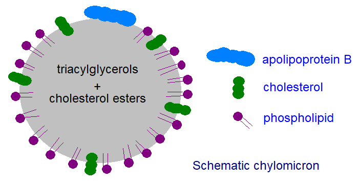
Subsequently, enterocytes in the small intestine incorporate triacylglycerols into lipoprotein complexes called chylomicrons. In brief, these consist of a core of triacylglycerols together with some cholesterol esters, stabilized and rendered compatible with an aqueous environment by a surface film consisting of phospholipids, free cholesterol, and one molecule of a truncated form of apoprotein B (called apo B48). A cartoon structure of a chylomicron is shown in Figure \(\PageIndex{11}\).
These particles are secreted into the lymph and the plasma for transport to peripheral tissues, where they are stored or serve structural purposes. Adipose tissue, in particular, exports appreciable amounts of the enzyme lipoprotein lipase. This enzyme binds to the luminal membrane of endothelial cells facing the blood, where it rapidly hydrolyses triacylglycerols as they pass through the cell surface, releasing free fatty acids. Most are absorbed into adjacent adipocytes and re-utilized for triacylglycerol synthesis within the cell.
Chylomicron remnants eventually reach the liver, where hepatic lipase hydrolyses the remaining lipids at the external membranes, and the remnants are absorbed. The liver can use the fatty acids for various purposes, from oxidation to the synthesis of structural lipids. Still, a proportion is reconverted into triacylglycerols; some of this is stored as lipid droplets within the cells' cytoplasm (see the next section). In addition, the liver takes up phosphatidylcholine from high-density lipoproteins, and a large proportion of it is eventually converted to triacylglycerols. In a healthy liver, triacylglycerol levels are low (<5% of total lipids) because the rates of fatty acid uptake from plasma and de novo synthesis in the liver are balanced by rates of oxidation and secretion into plasma. In contrast, excessive accumulation of storage triacylglycerols is associated with fatty liver, insulin resistance, and type 2 diabetes.
Most newly synthesized triacylglycerols are exported into the plasma as very-low-density lipoproteins (VLDL), consisting of a triacylglycerol and cholesterol ester core surrounded by phospholipids and free cholesterol, together with one molecule of full-length apoprotein B (100 kDa), apoprotein C, and sometimes apoprotein E. These particles are then transported to peripheral tissues, where they are hydrolyzed, and the free acids are absorbed. Eventually, the remnants are returned to the liver.
In the mammary gland, triacylglycerols are synthesized in the endoplasmic reticulum, and large lipid droplets are produced with a monolayer of phospholipids derived from this membrane. These are transported to the plasma membrane and bud off into the milk, surrounded by a phospholipid envelope, to form milk fat globules as food for the newborn. Thus, this process differs from the secretion of triacylglycerol-rich lipoproteins from other organs.
Triacylglycerol Synthesis and Catabolism (Lipolysis) in Adipocytes and Lipid Droplets
Adipose tissue and adipocytes are characterized by accumulations of triacylglycerols, the main energy store in animals, although they also cushion and insulate the body. Large fat deposits occur around internal organs, such as the liver, and subcutaneously, and each may respond differently to metabolic constraints. Thus, triacylglycerols stored in excess nutrients are mobilized for energy production during starvation. Adipose tissue also serves as a reserve of bioactive lipids, such as eicosanoids and lipid-soluble vitamins, and, when needed, provides structural components, including fatty acids, cholesterol, and retinol, for membrane synthesis and repair. By buffering against fatty acid accumulation that might exceed their capacity, non-adipose cells defend themselves against lipotoxicity while providing a rapid source of energy and essential metabolites by sensing and responding rapidly to changes in systemic energy balance. Brown and beige fat have special properties and are discussed below, while bone marrow adipocytes also have distinctive functions.
Similarly, within most other animal cells, including most cell types in the brain, a proportion of the fatty acids taken up from the circulation is converted to triacylglycerols as described above and incorporated into cytoplasmic lipid droplets (also termed 'fat globules', 'oil bodies', 'lipid particles' or 'adiposomes'). In adipocytes, lipid droplets can reach up to 200 μm in diameter, while other cell types contain smaller droplets of 50 nm. The triacylglycerol droplets, cholesterol esters, and other neutral lipids are surrounded by a protective monolayer that includes phospholipids, cholesterol, and hydrophobic proteins. The phospholipid component of the monolayer consists mainly of phosphatidylcholine and phosphatidylethanolamine derived from cytosolic leaflets of the endoplasmic reticulum and plasma membrane. Many proteins function directly in lipid metabolism, including acyltransferases, lipases, perilipins, caveolins, and the Adipose Differentiation Related Protein (ADRP, also known as adipophilin). Acting in concert with other cellular organelles, they participate in many metabolic processes, facilitating coordination and communication among organelles and serving as vital hubs of cellular metabolism.
Cytosolic lipid droplets with similar metabolic activities are found in most eukaryotic cells, including those of the fruit fly Drosophila melanogaster. Many aspects of triacylglycerol processing and regulation parallel those in humans. They are also present in the cytoplasm of some prokaryotes and in the plastids and other plant organelles (see below).
Lipid droplet assembly
This process occurs in subdomains of the endoplasmic reticulum, where at least one isoform of each enzyme in triacylglycerol biosynthesis, from acyl-CoA synthetases through glycerol-3-phosphate acyltransferases, is located, probably within a protein assembly or 'interactome'. As triacylglycerols accumulate, they reach a critical level when a spontaneous condensation or nucleation by phase separation occurs, leading to the formation of an oil blister within the hydrophobic bilayer region, attracting perilipins and other proteins that allow lipid droplets to grow further in patches of the membrane as lens-like swellings between the two membrane leaflets.
The protein seipin stabilizes nascent droplets with minimal membrane disruption and enables their maturation. Seipin monomers assemble into a decameric, cage-like structure and sit in the ER. This allows a space where triacylglycerol molecules interact with each other rather than with phospholipid acyl chains. Transmembrane protein segments probably aid this process. This enables phase separation of triacylglycerols, lens formation, and growth until seipin oligomerizes toward the cytoplasm, allowing the lens to form a budding lipid droplet.
Figure \(\PageIndex{12}\) presents a cartoon that shows the role of ER membrane seipins and other proteins in the assembly of lipid droplets.

In panel (A), seipin is enriched at the endoplasmic reticulum (ER)/lipid droplet (LD) contact sites and is crucial in triglyceride (TAG) flow from the ER to the LD. Seipin interacts with Perilipin1 (PLIN1) and several TAG synthetic enzymes, including glycerol-3-phosphate acyltransferase (GPAT3), 1-acyl-sn-glycerol-3-phosphate acyltransferase beta (AGPAT2), and LIPIN. No interaction with diacylglycerol acyltransferases (DGAT) has been formally reported. Magré J et al., Int J Mol Sci. 2022 Jan 11;23(2):740. doi: 10.3390/ijms23020740. Creative Commons Attribution (CC BY) license (https://creativecommons.org/licenses/by/4.0/
In panel (B), representing the fasting state, seipin is enriched at the ER/mitochondria (Mt) contact sites, also named mitochondria-associated membranes (MAM) and participates in ER/mitochondria calcium (Ca2+) flux and mitochondrial activity. Seipin is near MAM Ca2+ regulators IP3(inositol 1,4,5-trisphosphate) receptor (IP3R), voltage-dependent anion channel (VDAC), and sarco-/ER Ca2+ ATPase 2 (SERCA2), as well as glycerol-3-phosphate (G3P), lysophosphatidic acid (LPA), phosphatidic acid (PA), and diacylglycerol (DAG).
Figure \(\PageIndex{13}\) shows an interactive iCn3D model of the S. cerevisiae seipin Homo 10-mer flexible cage at lipid droplet formation sites (7RSL).

Figure \(\PageIndex{13}\): S. cerevisiae seipin homo 10-mer flexible cage at lipid droplet formation sites (7RSL). Click the image for a popup or use this external link: https://structure.ncbi.nlm.nih.gov/i...BjYLiQXVJjBpk8
The N-terminal transmembrane segment (TMS) on each monomer is shown in cyan, while the C-terminal TMS is magenta. These form the cage sides and top, anchoring the complex in the ER membrane. The side chains of a conserved switch sequence (F232xxGLR) on each monomer are shown in spacefill, CPK colors. The gray ribbons represent the part of the complex in the ER lumen.
Adjacent monomers in the homo-10-mer cage, even though they are identical in sequence, adopt different orientations for the two helices that enable a binary switch in the conformation of the complex. Two adjacent subunits, the A chain in blue and the B chain in orange, are shown, along with their overlap, in Figure \(\PageIndex{14}\).
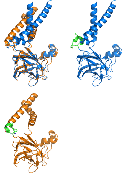
A highly conserved "switch" region is shown in green, with its highly conserved motif F232xxGLR shown as sticks with CPK colors. Note the large conformational change in the switch region, driven by the large conformational change of the two helices. This suggests two overall conformations of the complex: a closed form that allows TAG accumulation and an open form that allows lipid droplet budding and release. A possible model for the formation and budding of lipid droplets is illustrated in Figure \(\PageIndex{15}\). As in the figure above, the A (open) chain is shown in blue while the B (closed) chain is shown in orange.

Some surface proteins on lipid droplets can extend long helical hairpins of hydrophobic peptides deep into the lipid core. For example, perilipins constitute a family of at least five phosphorylated proteins that bind to animal droplets and share a common region, the so-called ‘PAT’ domain, named for the family's three original members, including perilipin and ADRP. Evolutionarily related proteins are found in more primitive organisms, including insects, slime molds, and fungi, but not in the nematode Caenorhabditis elegans. In mammals, perilipin A (or 'PLIN1' or, more accurately, the splice variant 'PLIN1a') is a well-established regulator of lipolysis in adipocytes, and it is believed to be involved in the formation of the large lipid droplets in white adipose tissue. The perilipins PLIN1 and PLIN2 function in triacylglycerol metabolism in tissues other than adipocytes, and PLIN2 is the main perilipin in hepatocytes; PLIN5 operates in tissues that oxidize fatty acids, such as the heart. Other surface proteins of lipid droplets are enzymes intimately involved in triacylglycerol metabolism. However, some suggest that cytoplasmic droplets may serve as storage organelles for hydrophobic proteins whose functions are elsewhere in the cell.
Lipolysis
When fatty acids are required by other tissues for energy or other purposes, they are released from the triacylglycerols by the sequential actions of three cytosolic enzymes at neutral pH, i.e., adipose triacylglycerol lipase (ATGL), hormone-sensitive lipase (HSL) and monoacylglycerol lipase, which cycle between the cytoplasmic surfaces of the endoplasmic reticulum and the surface layer of lipid droplets. Simply, ATGL hydrolyses triacylglycerols to diacylglycerols, which HSL hydrolyses to monoacylglycerols, before monoacylglycerol lipase hydrolyses these to complete the process. Lipolysis proceeds in a highly ordered manner, stimulated by cell-surface receptors via neurotransmitters, hormones, and autocrine/paracrine factors, which activate various intracellular signaling pathways and increase kinase activity. Perilipin (PLIN1) has been described as "the gatekeeper of the adipocyte lipid storehouse," regulating lipolysis by acting as a barrier in non-stimulated cells. However, during fasting, β-adrenergic stimulation activates cAMP-dependent protein kinase, which phosphorylates it, alters its shape, and reduces its hydrophobicity, thereby activating lipolysis. Perilipin A (an isoform) is the main regulatory factor in white adipose tissue. However, many other proteins interact with the three enzymes to modulate their activity, location, and stability.
The adipose triacylglycerol lipase, which initiates the process, was recently discovered. It is structurally related to the plant acyl-hydrolases in that it has a patatin-like domain in the N-terminal region (patatin is a non-specific acyl-hydrolase in potatoes). Specific transport mechanisms direct ATGL from the endoplasmic reticulum membrane to lipid droplets on the surface, in both the basal and activated states. This lipase is specific for triacylglycerols containing long-chain fatty acids, preferentially cleaving ester bonds in the sn-1 or sn-2 position (but not sn-3), and it yields diacylglycerols and free fatty acids as the main products, with low activity only towards diacylglycerols and none towards monoacylglycerols and cholesterol esters. However, it also has transacylase and phospholipase activities and hydrolyses retinol esters in hepatic stellate cells. Adipose triacylglycerol lipase can be activated at the same time as hormone-sensitive lipase and is now believed to be rate-limiting for the first step in triacylglycerol hydrolysis. Figure \(\PageIndex{16}\) shows the sequential hydrolysis of TAGs in adipocytes and lipid droplets.

Regulation of enzymatic activity is complex. For example, a lipid droplet protein designated Gene identification-58 (CGI-58 or ABHD5), is an important activating factor and is required for hydrolysis of fatty acids from position sn-1. In the resting state, this protein binds to perilipin (PLIN1). Under hormonal stimulation, however, perilipin is phosphorylated, leading to dissociation and interaction of CGI-58 with phosphorylated ATGL, initiating the first step in triacylglycerol hydrolysis. Mutations in adipose triacylglycerol lipase or CGI-58 are believed to cause a human syndrome known as ‘neutral lipid storage disease’. A second protein (G0S2) inhibits the enzyme.
Hormone-sensitive lipase in various isoforms is a structurally unique member of the large Ser-lipase/esterase family of enzymes in animals. Insulin and noradrenaline regulate it via a mechanism that ultimately involves phosphorylation by cAMP-protein kinase (as with perilipin), increasing its activity and causing it to translocate from the cytosol to the lipid droplet to initiate the second step of hydrolysis. Various proteins further regulate its activity, including PLINs and fatty acid-binding proteins (FABPs). Hormone-sensitive lipase has broader substrate specificity than other neutral lipases. In addition to its activity toward triacylglycerols, it rapidly hydrolyzes diacylglycerols, monoacylglycerols, retinol esters, and cholesterol esters. Diacylglycerols are hydrolyzed ten times as rapidly as triacylglycerols. Within the triacylglycerol molecule, hormone-sensitive lipase preferentially hydrolyses ester bonds in the sn-1 and sn-3 positions, leaving free acids and 2‑monoacylglycerols as the main end products.
Monoacylglycerol lipase is believed to be the rate-limiting enzyme in lipolysis, i.e., the final step in triacylglycerol catabolism, releasing free glycerol and fatty acids, and is found in the cytoplasm, plasma membrane, and lipid droplets. It is specific for monoacylglycerols but lacks positional specificity and shows no activity against di- or triacylglycerols. Because it is the main enzyme responsible for deactivating the endocannabinoid 2-arachidonylglycerol and is highly active in malignant cancers, it attracts pharmaceutical interest. A further enzyme, α/β hydrolase containing-6 (ABHD6), is located on the inner leaflet of the plasma membrane and preferentially hydrolyses fatty acids at the sn-1 position over the sn-2 position of monoacylglycerols. It also hydrolyses lysophospholipids and bis(monoacylglycerol)-phosphate. This has been associated with the development of insulin resistance and the progression of cancer. Additional lipolytic enzymes, including carboxylesterases, are believed to operate against triacylglycerols in cytoplasmic lipid droplets in the liver.
Unesterified fatty acids released by the combined action of these three lipases are exported into the plasma for transport to other tissues as albumin complexes. In contrast, the released glycerol is transported to the liver for metabolism by either glycolysis or gluconeogenesis.
Eventually, the entire organelle, including its proteins, can be degraded through a process called autophagy (lipophagy), i.e., the delivery of the organelle to lysosomal compartments for degradation. This can occur through direct lysosomal invagination or, more often, through a multistep process involving the formation of double-membrane vesicles termed 'autophagosomes' around droplets, followed by lysosomal fusion and degradation of the triacylglycerols by lysosomal acid lipase. This process helps regulate cellular lipid levels in various tissues and disease conditions, especially during starvation. It is relevant to tumorigenesis and cancer metastasis, as well as to neurodegenerative and neuroinflammatory diseases. Although lipophagy is mechanistically distinct from lipolysis, the two cross-talk.
Lipases carry out hydrolysis reactions (nucleophilic substitution with water as the nucleophile), alcoholysis reactions (with an ROH as the nucleophile), and even transesterification reactions with other fatty acid esters. Lipases are critical enzymes, yet good structures are still unavailable for hormone-sensitive lipase, for example. Much is known about lipase structure and activity. Lipases have a catalytic Ser-His-Asp triad like serine proteases, which also catalyze hydrolysis reactions. However, lipase substrates are nonpolar and may be part of larger structures, such as lipid droplets. As such, the lipases work at the interface between aqueous and non-aqueous environments. They are interfacially activated.
Lipases appear to have low activity in aqueous solutions but higher activity in less polar solvents. Many appear to have two conformations: a closed one with lower activity and poor lipid substrate binding, and an open one with higher activity and activation by nonpolar solvents. An alpha-helical lid appears to close off the active site in the closed conformation. In a less polar solvent or in bulk lipids, such as lipid droplets, the alpha-helical lid moves, exposing the enzyme's hydrophobic binding site to substrate interactions.
Figure \(\PageIndex{17}\) shows an interactive iCn3D model comparing the structure of a Thermomyces lanuginosus lipase in its closed (1DT3) and open conformation (1EIN).
_and_is_open_conformation_(1EIN).png?revision=1&size=bestfit&width=232&height=174)
Figure \(\PageIndex{17}\): The structure of a Thermomyces lanuginosus lipase in its closed (1DT3) and its open conformation (1EIN). Click the image for a popup or use this external link: https://structure.ncbi.nlm.nih.gov/icn3d/share.html?C9yEwy6SR6MZ9Q1S7
The closed structure (1DT3) is shown in magenta, and the open (1EIN) is in cyan. The active-site triad Ser146, His258, and Asp201 is shown as CPK-colored sticks, labeled for each. The lid is shown in yellow with its side chains as sticks. Toggle between the two states using the "a" key. Note how the lid covers the active site in the closed (1DT3) structure. You will have to rotate the molecule to see the optimal orientation. Also, close out menu boxes for a larger and more centered display.
Endocrine functions
Not only does the adipocyte store energy, but it is also an endocrine organ that manages energy flow by producing the hormone leptin, which signals through the adiponectin receptors (AdipoR1 and AdipoR2)) and binds to the non-signaling interacting protein T‑cadherin. This stimulates signaling cascades that communicate with other tissues through the secretion of cytokines and other mediators. Adipocytes tightly regulate leptin synthesis. However, the stomach, placenta, and mammary gland also produce it, and its principal function is to inform other tissues about the state of fat stores. In so doing, it regulates food intake and energy expenditure. Leptin was initially described as an anti-obesity hormone. Still, it serves as an adiposity signal, playing a vital role in maintaining adipose tissue mass to ensure survival under negative energy balance and to protect against both adiposity deficit and excess. Lipid droplets play a role in this process, as perilipin is required for sensing.
Adiponectin is a potent insulin sensitizer and suppressor of cell death and inflammation, directly promoting anti-diabetic and anti-atherosclerotic outcomes. Insulin is the main hormone that affects metabolism, and its plasma membrane receptor is located in caveolae. Proinflammatory cytokines can stimulate lipolysis and induce insulin resistance, leading to adipose tissue dysfunction and systemic metabolic disruption. Thus, adipose tissue metabolism has profound effects on whole-body metabolism, and defects in these processes can have severe implications for such serious pathological conditions as diabetes, obesity, cardiovascular disease, fatty liver disease, and cancer in humans. Developing specific inhibitors for hormone-sensitive lipase is hoped to improve the treatment of such metabolic complications. Because caveolae contain the proteins caveolins (and presumably sphingolipids) and are particularly abundant in adipocytes, modulate fatty acid flux across the plasma membrane, and participate in signal transduction and membrane trafficking, they play a major role in this aspect of lipid metabolism.
White fat acts as an endocrine organ and can release a variety of hormones, including adipokines (analogous to cytokines, chemokines, and lymphokines), adipsin, and leptin, as well as tumor necrosis factor α (TNF-α), adiponectin, resistin, and RBP4.
Functions other than energy management
Lipid droplets accumulate in many cell types beyond adipocytes, including leukocytes, epithelial cells, hepatocytes, and even astrocytes, especially during infections, cancer, and other inflammatory conditions. They are important for storing and releasing hydrophobic vitamins, signaling precursors, and other lipids unrelated to energy homeostasis, while reducing the risk of lipotoxicity. Conversely, excessive fatty acid accumulation is associated with lipotoxicity, endoplasmic reticulum stress, and mitochondrial damage and dysfunction, so lipid storage in lipid droplets must be balanced for health.
Various enzymes are associated with lipid droplets, including protein kinases involved in many aspects of lipid metabolism, such as cell signaling, membrane trafficking, and regulating the production of inflammatory mediators such as eicosanoids. Lipolysis enables the secretion of lipid species termed lipokines (more generally 'adipokines') from adipocytes that may signal in a hormone-like fashion to other tissues, thereby modulating gene expression and physiological function, including food intake, insulin sensitivity, insulin secretion, and related processes. These include palmitoleic acid (9-16:1) and fatty acid esters of hydroxy fatty acids (FAHFA), though researchers have studied the circulating proteins adiponectin and leptin more intensively. Adiponectin is a powerful insulin sensitizer and suppressor of apoptosis and inflammation with anti-diabetic and anti-atherosclerotic functions, often operating through its effects on sphingolipids. At the same time, leptin exerts most of its effects in the brain, triggering behavioral, metabolic, and endocrine responses to regulate the body's fuel reserves.
Indeed, lipid droplets in all cell types are now suggested to be essential for cellular stress responses, including autophagy, inflammation, and immunity, and to act as hubs that integrate metabolic and inflammatory processes. Through their lipolytic machinery, they regulate fatty acid availability to activate signaling pathways and produce oxylipins from polyunsaturated fatty acids. For example, triacylglycerols in cytoplasmic lipid droplets of human mast cells, which are potent mediators of immune reactions and influence many inflammatory diseases, have a high content of arachidonic acid, which can be released by adipose triacylglycerol lipases as a substrate for the production of specific eicosanoids when the cells are stimulated appropriately. Active metabolism in lipid droplets is important for monocyte differentiation and essential for the sustained functional activity of differentiated macrophages, especially in inflammation.
During apoptosis, triacylglycerols enriched in polyunsaturated fatty acids accumulate in lipid droplets, possibly as a protective mechanism against membrane damage caused by oxidative stress and hydroperoxide formation. Triacylglycerols in skin lipid droplets are a highly specific source of linoleic acid required to form O-acylceramides, which are essential for epidermal barrier function. In dividing cells of humans and rodents, an organelle called the midbody contains a unique triacylglycerol, a single molecular species consisting of three fatty acids: 16:1-12:0-18:1 (12:0 is rarely detected in human lipids), but its function is not known.
Vitamin E (tocopherols) and vitamin A in the form of retinyl esters are stored in cytoplasmic lipid droplets, and the latter are present in appreciable concentrations in the stellate cells of the liver, for example. In endocrine cells of the gonads and adrenals, cholesterol esters stored in lipid droplets are an important source of cholesterol for the mitochondrial biosynthesis of various steroid hormones. In addition to providing a reservoir of fatty acids for membrane remodeling in the cell's nucleus, lipid droplets can sequester transcription factors and chromatin components and generate the lipid ligands for certain nuclear receptors.
Lipid droplets participate in protein degradation and glycosylation in addition to their role in lipid biochemistry. Pathogenic viruses and bacteria, such as Mycobacterium tuberculosis, can manipulate their metabolism, with unfortunate consequences for the host. Still, they also serve as reservoirs for proteins that fight intracellular pathogens. Consequently, such lipid droplets and their associated enzyme systems may serve as markers of disease states and as targets for pharmaceutical intervention.
Insects
In insects, the fat body is a multifunctional tissue and the main metabolic organ. It integrates signals that control the immune system, molting, metamorphosis, and synthesis of hormones that regulate innumerable aspects of metabolism. In fat tissue, lipids, carbohydrates, and proteins serve as substrates and products in many energy-production pathways. Fat also serves as a reserve that can be mobilized at appropriate life stages (diapause, metamorphosis, and flight). In innate and acquired humoral immunity, the fat body produces bactericidal proteins and polypeptides, such as lysozyme. It is also important in the early stages of an insect's life because it promotes vitellogenin production, the yolk protein needed for oocyte development.
Brown Adipose Tissue
Most adipose tissue depots ('white fat') serve primarily as storage and endocrine organs, providing a reservoir of nutrients for release when the food supply is low. However, a second specialized form of adipose tissue, brown fat, is multilocular, highly vascularized, and rich in mitochondria and the iron-containing pigments that transport oxygen and give the tissue its color and name. Brown adipocytes arise from progenitor cells closer to those of skeletal muscle than white adipocytes. In humans, these depots tend to be located in specific anatomical regions, such as the subcutaneous areas around the neck, where their function may be to supply warm venous blood directly to the spinal cord and brain, and elsewhere to the heart, kidney, pancreas, and liver. Brown fat can oxidize fat so rapidly that it generates heat (“non-shivering thermogenesis”), and it is especially important in young animals and those recovering from hibernation.
In brief, during cold exposure, the release of noradrenaline and stimulation of β-adrenergic receptors in the nervous system initiate a catabolic program that begins with rapid breakdown of cellular triacylglycerol stores, release of unesterified fatty acids, and transient activation of a co-activator of peroxisome proliferator-activated receptor gamma (PPARγ). These trigger a signaling process that enables the efficient β-oxidation of fatty acids to produce heat. The key molecule is believed to be uncoupling protein-1 (UCP1), which acts as a valve that uncouples electron transport in the respiratory chain from ATP production, releasing chemical energy as heat rather than ATP. Its actions are illustrated in Figure \(\PageIndex{18}\).
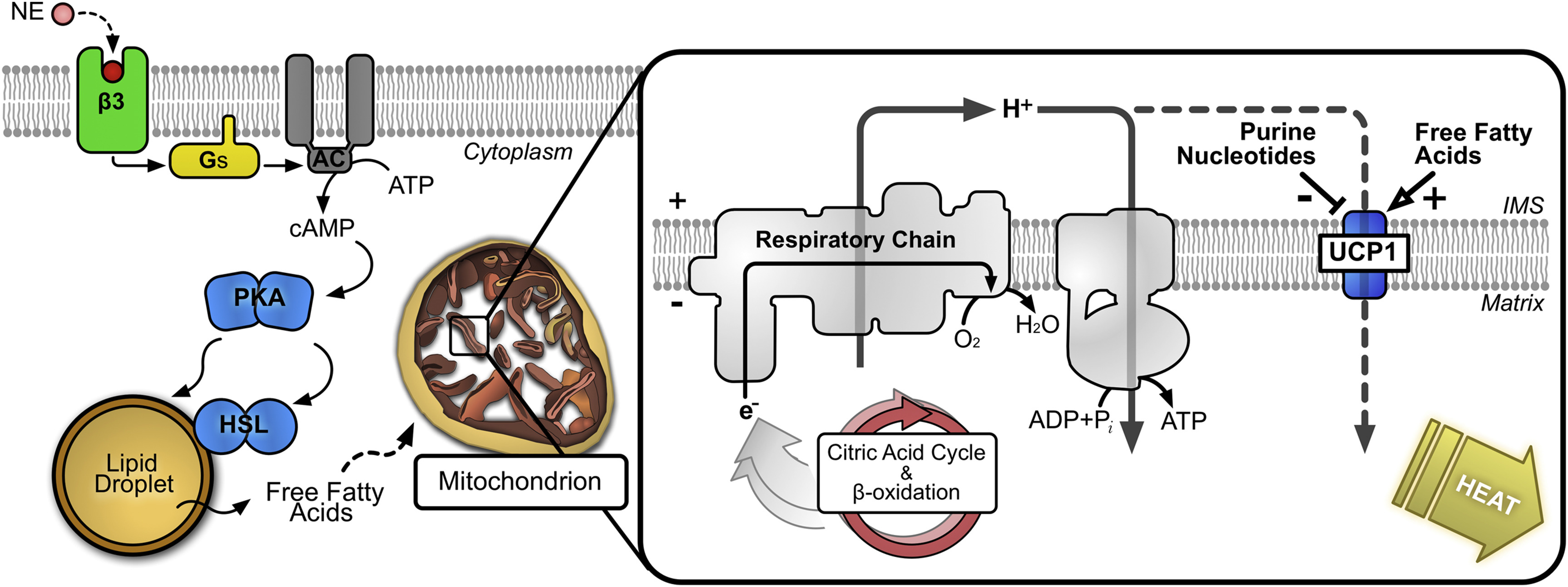
Figure \(\PageIndex{18}\): Role of UCP1 in generation of heat. https://commons.wikimedia.org/wiki/F...n_the_cell.jpg. Creative Commons Attribution-Share Alike 4.0 International

This is an example of a futile cycle that releases heat. Although many aspects of the mechanism are uncertain, UCP1-mediated proton conductance is clearly highly regulated and inducible. It is activated by free long-chain fatty acids and inhibited by purine nucleotides. Fatty acids are the substrate for thermogenesis and act as self-regulating second messengers. The mitochondrial phospholipid cardiolipin, which is intimately involved in oxidative phosphorylation, is indispensable for stimulating and sustaining thermogenic fat function. Upon activation of brown adipose tissue, its dense vasculature increases delivery of fatty acids and glucose to brown adipocytes, warming the blood passing through the tissue.
Long-chain fatty acids bind to UCP1 and may be transported across the membrane in exchange for a proton. Alternatively, it may simply bind and activate proton transport. Figure \(\PageIndex{19}\) shows an interactive iCn3D model of the AlphaFold predicted structure of human UCP1 (P25874).

Figure \(\PageIndex{19}\): AlphaFold predicted structure of human UCP1 (P25874). Click the image for a popup or use this external link: https://structure.ncbi.nlm.nih.gov/i...FkfrEqWE3D2dX6
Imagine a bilayer oriented perpendicular to the gray helices, representing the transmembrane helical segments that embed the protein in the inner mitochondrial membrane. The spacefill Ile150 denotes the matrix domain. Two key residues (K56, K29) involved in binding the carboxyl end of the fatty acids are shown on the matrix side of the protein, in CPK-colored sticks and labeled. The black faces of the helices in the inner membrane region show the most probable binding site for the fatty acid acyl chain. The lighter yellow/orange regions have greater predicted uncertainty in the AlphaFold computational model.
Evidence suggests that acylcarnitines produced in the liver from fatty acids released from white adipose tissue in response to cold exposure are transported in plasma to brown adipose tissue and can serve as a substrate for thermogenesis. Indeed, a wide array of circulating lipids, including free fatty acids and triacylglycerols, contribute to thermogenic potential.
Acyl-CoA synthetases and acyl-CoA thioesterases determine the availability of substrates for β-oxidation and, consequently, thermogenic capacity. Synthesis of the lipokine (or 'batokine') octadecanoid 12,13-dihydroxy-9Z-octadecenoic acid (12,13-diHOME), is induced by cold also, and this stimulates the activity of brown adipose tissue by promoting the uptake of fatty acids, acting via G-protein-coupled receptors. It increases cardiac function and cardiomyocyte respiration via enhanced calcium cycling. Similarly, n‑3 polyunsaturated fatty acids may promote adaptive thermogenesis, for example, through the activity of the 12‑lipoxygenase metabolite and batokine 12‑hydroxyeicosapentaenoic acid (12-HEPE), which improves glucose metabolism by increasing glucose uptake into adipocytes and skeletal muscle. (FAHFA) are relevant in this context. Peroxisomal synthesis of plasmenyl-phospholipids is believed to regulate adipose tissue thermogenesis by mediating mitochondrial fission.
In hibernating mammals, brown adipose tissue is metabolically important, and even in laboratory animals such as mice, it can consume about 50% of dietary lipids and glucose when exposed to cold. Similarly, in humans, cold acutely induces brown adipose tissue activity. The sympathetic nervous system stimulates it, and the relevance of this tissue to human metabolism is becoming apparent. For example, some suggest that brown adipose tissue can function as an endocrine organ, secreting endocrine factors ('batokines') that may help reduce cardiovascular risk. For obvious reasons, researchers are working to determine whether sustained pharmaceutical activation of brown fat could benefit human disease states, including obesity, diabetes, and cardiovascular disease.
Such research has been stimulated by the observation that clusters of distinct adipocytes with thermogenic capacity and their storage function can be present in white adipose tissue and emerge in response to various physiological signals, especially reactive oxygen species. They are called beige or brite adipocytes and arise from multipotent pre-adipocytes. Brown adipose tissue is a discrete organ in animals, whereas beige adipose tissue is interspersed with white adipose tissue, and the two forms have different developmental origins. In adult humans, most deposits once thought to be classical brown adipose tissue lack the genetic markers for this tissue and are now recognized as beige/brite fat. However, brown adipose tissue per se is present in significant amounts in human newborns and infants. However, beige adipocytes use the same machinery to generate heat by oxidizing fatty acids in response to β-adrenergic stimulation.
The uncoupling protein 1 (UCP1) appears to be expressed only in brown and beige fat cells and serves as a marker for those cells. ROS-induced modification of cysteine residues can regulate it transcriptionally and posttranscriptionally, producing sulfenyl groups. The more classical brown adipocyte arises developmentally from skeletal muscle-like cells, whereas beige adipocytes appear to develop to some extent from vascular smooth muscle cells. The beige cells can differentiate into more white adipocytes or more brown ones by the factors described in Figure \(\PageIndex{20}\).

Figure (\PageIndex{20}\): Factors altering beige adipose tissue. Paul Cohen and Bruce M. Spiegelman. Molecular Biology of the Cell, 27, 2017. https://doi.org/10.1091/mbc.e15-10-0749. Attribution–Noncommercial–Share Alike 3.0 Unported Creative Commons License (http://creativecommons.org/licenses/by-nc-sa/3.0).
In addition to fat cell type, the adipocyte's location and size also affect health risks. Obesity, cardiovascular disease, type II diabetes, and nonalcoholic fatty liver disease all depend on and are correlated with fat cell dysfunction. Adipose tissue's endocrine role plays a key role in the development of dysfunction. The main fat depots are subcutaneous adipose tissue (SAT) and visceral adipose tissue (VAT), the latter of which is associated with proinflammatory effects and greater health risks (type II diabetes, hypertension, metabolic syndrome, obesity, etc.). Figure (\PageIndex{21}\) below summarizes the health consequences as the size of the SAT and VAT adipocytes increases (in the direction of the red arrow).

Figure (\PageIndex{2}\): Adipocyte characteristics and cardiovascular risk. The diagram describes a proposed model based on adipocyte size/source, potential interactions with metabolic mediators, and pathophysiological effects. SAT, subcutaneous adipose tissue; VAT, visceral adipose tissue; HbA1c, glycated hemoglobin (a marker of elevated blood sugar associated with diabetes); RES, resistin; ADIPON, adiponectin; INS, insulin; HDLc; high-density lipoprotein cholesterol; FMD, flow-mediated dilation; CIMT, carotid intima media thickness. Suárez-Cuenca et al. Sci Rep 11, 1831 (2021). https://doi.org/10.1038/s41598-021-81289-2. Creative Commons Attribution 4.0 International License. http://creativecommons.org/licenses/by/4.0/.
The larger the adipocyte, the greater the inflammatory response. This condition, adipocyte hypertrophy, may be more important than obesity per se as a risk factor for inflammation, as indicated by elevated inflammatory cytokine levels and macrophage infiltration into adipose tissue. Under normal conditions, the adipose-derived hormone leptin suppresses appetite and reduces liver and muscle fat. Higher fat storage is associated with increased blood leptin levels. It also correlates with larger lymphocytes.
In contrast, the adipose hormone adiponectin has positive health effects, including increased insulin sensitivity, reduced inflammation, and adipocyte formation from stem cells (adipogenesis). The larger the fat cell, the lower the serum adiponectin levels. However, obesity may more strongly influence the link between fat cell size and adiponectin levels.
Other Functions of Triacylglycerol Depots
Subcutaneous depots cushion joints and insulate against cold in many terrestrial animals, as in the pig, which is surrounded by a layer of fat. It is especially true for marine mammals such as seals. Adipocytes embedded in the skin differ from the general subcutaneous depots and support hair follicle growth and skin regeneration. They may also have a defensive role, both as a physical barrier and by responding metabolically to bacterial infection.
In marine mammals and fish, fat depots are less dense than water, which helps them float. As a result, they expend less energy swimming. More surprisingly, triacylglycerols, together with the structurally related glyceryl ether diesters and wax esters, are the main components of the sonar lens used in echolocation by dolphins and toothed whales. Triacylglycerols are distinctive in containing two molecules of 3‑methylbutyric (isovaleric) acid and one long-chain fatty acid. The relative concentrations of the various lipids in an organ in the head of the animals (termed the ‘melon’) appear to be arranged anatomically in a three-dimensional topographical pattern to enable them to focus sound waves.
In cold climates, many insects do not feed over winter and must manage their energy stores to meet the energetic demands of development and reproduction in the spring. Some insect species tolerant of freezing produce triacylglycerols containing acetic acid, which remain liquid at low temperatures; interaction with water may also play a role in cryoprotection.
Triacylglycerol Metabolism in Plants and Yeasts
Fruit and seed oils are major agricultural products with appreciable economic and nutritional value to humans. The mesocarp of fruits is a highly nutritious energy source that attracts animals that help disperse seeds, and in plants such as oil palm and olive trees, a high proportion of the fruit flesh contains triacylglycerols. Similarly, triacylglycerols are the main storage lipids in seeds and can comprise up to 60% of their weight. Fruit lipids are not intended for plant use per se and are stored in lipid droplets as large, irregular structures that break down readily. Still, seed lipids are required for plant embryo development, so their metabolism is particularly important. However, triacylglycerol biosynthesis and metabolism are also required for pollen viability and to maintain lipid homeostasis in chloroplasts (see the note on plastoglobules below).
Seed oils: In seeds and other plant tissues, biosynthesis of fatty acids takes place in plastids, and these are stored in the form of triacylglycerols in lipid droplets with a coherent surface layer of proteins and lipids in the embryo (e.g., Arabidopsis, soybean, or sunflower) or endosperm (e.g., castor bean) tissues of seeds. In addition to the common range of fatty acids synthesized in plastids, mainly palmitate and oleate, some plant species produce novel fatty acids, including medium- and very-long-chain components and those with oxygenated and other functional moieties. A specific means of diverting these to seeds for triacylglycerol production exists to prevent disruption of the plant membranes. Seed development occurs in three stages: rapid cell division with no accumulation of storage material, rapid deposition of triacylglycerols and other energy-rich metabolites, and finally, desiccation.
During the period of oil accumulation in seeds, the newly formed ACP esters of fatty acids are first hydrolyzed by two different classes of acyl-ACP thioesterases at the inner plastid envelope membrane, before the unesterified fatty acids are transported to the endoplasmic reticulum (ER), by a family of fatty acid export proteins (FAX) of which there are seven isoforms in Arabidopsis, two of which (FAX2 and FAX4) are highly expressed during the early stage of seed development. Unesterified fatty acids are shuttled across the plastid outer envelope, probably via vectorial acylation by long-chain acyl-CoA synthases, which catalyze the formation of CoA esters.
In the ER, triacylglycerols and membrane lipids are synthesized by the Kennedy and other pathways described above. In yeast and plants, 1,2‑diacyl-sn-glycerol esterification is the only committed step in triacylglycerol production, and this occurs by mechanisms that can be both dependent and independent of acyl-CoA esters. The acyl-CoA-dependent route is catalyzed by diacylglycerol:acyl-CoA acyltransferases (DGATs), which use acyl-CoA and diacylglycerols as substrates; two membrane-bound isoenzymes (DGAT1 and DGAT2) and a cytosolic isoenzyme (DGAT3) are known. DGAT1 is a key enzyme involved in triacylglycerol formation in developing seeds. DGAT2 is especially important in plant species with unusual fatty acid compositions. In Arabidopsis, DGAT3 has some specificity for polyunsaturated fatty acids in seed development. Figure \(\PageIndex{22}\) summarizes TAG synthesis in plant ER.
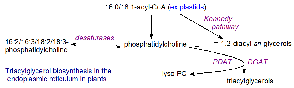
In addition, a substantial proportion of triacylglycerol biosynthesis in some plant species is synthesized by flux through the membrane phospholipid phosphatidylcholine, produced by what are sometimes inaccurately termed the 'eukaryotic and prokaryotic pathways' with differing positional distributions, in which diacylglycerols are generated from phosphatidic acid by the action of a phosphatidate phosphatase as an intermediate. In the acyl-CoA-independent reaction, the direct transfer of one fatty acid from phosphatidylcholine to diacylglycerol by the action of the phospholipid:1,2-diacyl-sn-glycerol-acyltransferase (PDAT) enzyme also occurs, with the formation of lysophosphatidylcholine as a byproduct, which can be re-esterified for further reaction. As phosphatidylcholine undergoes extensive remodeling and its fatty acid components are subject to modification, for example, by desaturation to form linoleic and linolenic acids, the compositions and especially the positional distributions of triacylglycerols produced in this way can be very different from those synthesized by the ‘classical’ pathways. Phosphatidylcholine may also act as a carrier for trafficking acyl groups between organelles and membrane subdomains, and an assembly of interacting enzymes may facilitate the transfer of polyunsaturated fatty acids from this phospholipid to triacylglycerols in seeds. Both sterol and sphingolipid biosynthesis are important factors for efficient seed oil production.
As triacylglycerol synthesis continues, oil droplets accumulate between endoplasmic reticulum leaflets. They are surrounded by a monolayer of phospholipids, sterols, and proteins, which in Arabidopsis include oleosins, a caleosin, a steroleosin, a putative aquaporin, and a glycosylphosphatidylinositol-anchored protein. Oleosins are the most abundant of these (~65%). They are small proteins (15-30 kDa) with cytosolic-facing N- and C-termini and a large hydrophobic domain that targets them to lipid droplets, where they play an important role in controlling their size and stability. Eventually, lipid droplets “bud off” from the endoplasmic reticulum with their monolayer of phospholipids and proteins, and they are released into the cytosol by a yet-to-be-defined mechanism.
Water is absorbed at the onset of germination, and esterases/lipases are activated. Lipolysis begins at the surface of oil bodies, where oleosins, the most abundant structural proteins, are believed to assist in lipase docking and control the size and stability of lipid droplets in seeds. Several esterases/lipases have been cloned from various plant species and possess a conserved catalytic triad of Ser, His, and Asp or Glu, somewhat different from the animal lipases, as in patatin (an especially abundant lipolytic protein in potatoes), which can hydrolyze triacylglycerols but not phospho- or galactolipids. The most important of these is believed to be the 'sugar-dependent lipase 1 (SDP1)', a patatin-like lipase similar in function to the mammalian adipose triacylglycerol lipase discussed above, and located on the surface of the oil body. This is active mainly against triacylglycerols to generate diacylglycerols but presumably works in conjunction with di- and monoacylglycerol lipases to generate free fatty acids and glycerol.
Seed lipid droplets exist near glyoxysomes (broadly equivalent to peroxisomes). These membrane-bound organelles contain most of the enzymes required to oxidize fatty acids derived from triacylglycerols via acetyl-CoA to four-carbon compounds, such as succinate, which are then converted to soluble sugars to provide germinating seeds with energy to fuel seedling growth and produce shoots and leaves. In addition, they supply structural elements before the seedlings develop the capacity to photosynthesize. How lipolysis products are transported to the glyoxysomes for further metabolism remains to be determined. Still, a specific ‘ABC’ transporter is required to import fatty acids into the glyoxysomes in Arabidopsis. Two long-chain acyl-CoA synthetases on the inner face of the peroxisome membrane convert the free acids to their coenzyme A esters before entry into the β‑oxidation pathway. An intricate regulatory network controls these processes, involving transcription factors that crosstalk with signaling events from seed maturation through embryo development. After about two days of germination, the glyoxysomes begin to break down, but β-oxidation can continue in peroxisomes in leaf tissue.
Lipid droplets - plastoglobules: Triacylglycerol-rich lipid droplets (LD) have been observed in most cell types in vegetative tissues of plants as well as in seeds, and although their origin and function are poorly understood, they contain all the enzymes required for triacylglycerol metabolism together with phospholipases, lipoxygenases, and other oxidative enzymes. Rather than oleosins, these lipid droplets in plants and algae contain a family of ubiquitously expressed 'LD-associated proteins' on the surface, along with a monolayer of phospholipids (mainly phosphatidylcholine), galactolipids such as sulfoquinovosyldiacylglycerol, and, in some species, betaine lipids. As in yeast and humans, seipins (three in Arabidopsis) are necessary for normal LD biogenesis. Again, LDs form within the endoplasmic reticulum bilayer and pinch off into the cytoplasm. Abiotic stresses can induce lipid membrane remodeling via lipase activity, leading to toxic lipid intermediates. Cells can sequester these in lipid droplets to limit membrane damage and potentially prevent cell death. While they are believed to be involved mainly in stress responses, lipid droplets may have other specialized roles, for example, in anther and pollen development, where triacylglycerols serve as a source of fatty acids for membrane biosynthesis. Fatty acids derived from triacylglycerols in lipid droplets are believed to undergo peroxisomal β‑oxidation to produce the ATP required for stomatal opening and many other purposes.
In addition, lipid droplets that have been termed 'plastoglobules' are produced by a localized accumulation of triacylglycerols and other neutral lipids between the membrane leaflets of the thylakoid cisternae and then pinch off into the stroma, where they are involved in a wide range of biological functions from biogenesis to senescence via the recruitment of specific proteins. During senescence, for example, lipid droplets accumulate rapidly in A. thaliana leaves. It may have a more direct function in reproductive tissues by recruiting and transporting proteins for organ formation and successful pollination. Antifungal compounds such as 2-hydroxy-octadecatrienoic acid and other oxylipins are produced from α-linolenic acid in these organelles, and it has been suggested that these organelles function as intracellular factories, producing stable metabolites from unstable intermediates by efficiently concentrating enzymes and hydrophobic substrates. Plastoglobules are also implicated in the biosynthesis and metabolism of vitamins E and K.
Microalgae: Triacylglycerol metabolism in lipid droplets in microalgae is intensively studied because of their potential for nutraceutical and biodiesel production. Similar processes seem to occur in higher plants, but with a simpler genome that encodes fewer redundant proteins. In the unicellular green microalga Chlamydomonas reinhardtii, researchers have characterized key lipid droplet proteins, lipases, and β-oxidation enzymes.
Yeasts: Lipid droplets in yeast are a highly dynamic and functionally diverse hub that ensures stress resistance and cell survival by promoting membrane and organelle homeostasis. Because most key biosynthetic and catabolic enzymes involved in triacylglycerol metabolism are conserved between yeasts and mammals, yeasts are useful models for studying triacylglycerol production. The size and triacylglycerol content of lipid droplets in yeasts change appreciably in different stages of growth and development, and Saccharomyces cerevisiae contains a single phosphatidic acid phosphatase (Pah1), which has an essential role in this process. During vegetative growth, multiple protein kinases phosphorylate cytosolic Pah1, enabling phospholipid synthesis rather than triacylglycerol synthesis. As cells enter stasis, Pah1 is dephosphorylated and translocates to the endoplasmic reticulum, promoting triacylglycerol synthesis for storage in lipid droplets. Some fatty acids derived from phospholipids are utilized for triacylglycerol biosynthesis at the inner nuclear membrane, which is important for nuclear integrity.
Triacylglycerol Metabolism in Prokaryotes
Awareness of the role of this lipid class in the pathogenesis of Mycobacterium tuberculosis and its relationship with antibiotic biosynthesis in Streptomyces coelicolor has stimulated research into triacylglycerol biosynthesis in bacteria. For example, triacylglycerols are believed to serve as an energy reserve for long-term survival of M. tuberculosis during the persistence phase of infection and to detoxify unesterified fatty acids. Increasing numbers of bacterial species, including genera such as Mycobacterium, Nocardia, Rhodococcus, Micromonospora, Dietzia, and Gordonia, are now known to produce triacylglycerols (sometimes wax esters), which can be stored in lipid droplets within the organisms. The first three steps in triacylglycerol biosynthesis are catalyzed by GPAT, LPAT, and PAP enzymes, which are comparable to those in other organisms. However, it has become apparent that the DGAT can be a dual-function CoA-dependent acyltransferase known as wax ester synthase/diacylglycerol acyltransferase, which accepts a broad diversity of acyl-CoA substrates for esterification of diacylglycerols or long-chain fatty alcohols for the synthesis of triacylglycerols or wax esters, respectively, depending on which intermediates are present in the organisms. Bacteria that lack this enzyme cannot produce these non-polar lipids.
Summary
(Summary written by Claude, Sonnet 4.6, Anthropic)
This chapter provides a comprehensive account of triacylglycerol (TAG) biosynthesis, storage in lipid droplets, mobilization by lipolysis, intestinal digestion and transport, and the broader physiological roles of fat in animals, plants, and microorganisms.
Triacylglycerol biosynthesis follows four main pathways, of which the sn-glycerol-3-phosphate (Kennedy) pathway accounts for more than 90% of liver TAG production. In this pathway, sn-glycerol-3-phosphate (derived from glycolysis, glycerol kinase, or de novo glyceroneogenesis from pyruvate) is acylated at the sn-1 position by glycerol-3-phosphate acyltransferase (GPAT) to form lysophosphatidic acid (LPA), then acylated at sn-2 by 1-acyl-sn-glycerol-3-phosphate acyltransferase (AGPAT) to form phosphatidic acid (PA) — the central branch-point intermediate for both TAG and phospholipid synthesis. PA is then dephosphorylated by a lipin phosphatidate phosphatase (PAP), generating sn-1,2-diacylglycerol (DAG); this step, catalyzed by Mg²⁺-dependent lipins (1, 2, and 3 in mammals) with a HAD-family DxDxT catalytic motif, is a key regulatory branch point: DAG can be channeled to TAG synthesis (by DGAT) or to phosphatidylcholine and phosphatidylethanolamine synthesis. The final committed step is catalyzed by diacylglycerol acyltransferases: DGAT1 (an MBOAT-family integral ER membrane enzyme that accepts acyl-CoA through a cytosolic tunnel while DAG enters from within the bilayer, using His415 as the catalytic nucleophile, with broad substrate specificity including monoacylglycerols, long-chain alcohols, and retinol) and DGAT2 (associated with ER, lipid droplet surfaces, and mitochondria, primarily esterifying endogenously synthesized fatty acids and physically tethering the ER to nascent lipid droplets to direct newly synthesized TAGs into storage). Additional pathways include the DHAP pathway (using dihydroxyacetone-phosphate from glycolysis as the glycerol backbone, important in peroxisomes and ER), the monoacylglycerol pathway (predominant in intestinal enterocytes after a meal, where absorbed 2-monoacylglycerol is reacylated by MGAT and DGAT1), and a transacylation pathway (DAG transacylase-catalyzed transfer between diacylglycerols, important in post-synthetic remodeling).
GPAT1 (outer mitochondrial membrane) is activated by insulin and inhibited by AMPK, serving as a regulatory gateway: high GPAT1 activity favors TAG synthesis over β-oxidation of the same fatty acyl-CoA pool. Lipins additionally function as transcriptional coactivators in the nucleus — lipin-1α activates nuclear receptors to regulate lipid metabolism gene expression, while lipin-1β induces lipogenic genes (FAS, SCD, DGAT) — linking TAG synthesis capacity to transcriptional programs. In yeast, the Pah1 phosphatase (the lipin ortholog) is phosphorylation-regulated to partition lipid flux between phospholipid membrane synthesis during vegetative growth and TAG storage during stationary phase.
Cytoplasmic lipid droplets are now recognized as bona fide organelles with dynamic metabolic functions. They form within ER subdomains where TAG concentration reaches a critical level, triggering phase separation that forms an oil lens between the membrane leaflets. The seipin decameric cage — assembled from identical monomers that adopt two distinct conformations, creating a binary conformational switch (closed, accumulating TAGs; open, permitting lipid droplet budding) mediated by the conserved F232xxGLR switch motif — stabilizes nascent droplets and enables their maturation and release into the cytoplasm. Mature lipid droplets are bounded by a phospholipid monolayer (predominantly phosphatidylcholine and phosphatidylethanolamine) decorated with hydrophobic proteins: perilipins in mammals (PLIN1–5, with PLIN1 being the dominant regulator in white adipose tissue), oleosins in plants, and related proteins in fungi and insects. Beyond energy storage, lipid droplets serve diverse roles: sequestering toxic lipid intermediates and reactive lipid species; storing hydrophobic vitamins (A, E, K); providing arachidonic acid for eicosanoid production in immune cells; regulating transcription factors; and participating in autophagy (lipophagy) and pathogen defense.
Lipolysis proceeds by the sequential action of three lipases on the lipid droplet surface, tightly coordinated by hormonal and signaling inputs. In the resting adipocyte, perilipin A (PLIN1) physically shields the droplet from lipase access. Upon β-adrenergic stimulation (epinephrine/noradrenaline during fasting or exercise), cAMP rises, and PKA phosphorylates both PLIN1 (causing a conformational change and exposing the droplet surface) and HSL (increasing its activity and promoting translocation from the cytosol to the droplet). ATGL, constitutively present on lipid droplets, initiates lipolysis by hydrolyzing TAGs at sn-1 or sn-2 to give DAG and free fatty acids; its activity requires co-activator CGI-58/ABHD5, which is released from PLIN1 upon phosphorylation and inhibited by G0S2. HSL then hydrolyzes DAG (ten-fold more rapidly than TAG) preferentially at sn-1 and sn-3, yielding 2-monoacylglycerol. Monoacylglycerol lipase completes lipolysis, releasing glycerol and the final fatty acid; this enzyme also degrades the endocannabinoid 2-arachidonylglycerol and is highly upregulated in cancers. Released fatty acids enter the circulation complexed with albumin for delivery to other tissues; glycerol is transported to the liver for gluconeogenesis or glycolysis. Lipases — including HSL and the plant patatin-like enzymes — share an α/β-hydrolase Ser-His-Asp catalytic triad and are interfacially activated: they exist in a low-activity closed conformation with an α-helical lid occluding the active site, transitioning to an open, active conformation upon contact with lipid–water interfaces.
Dietary fat digestion begins in the stomach (gastric lipase), but quantitatively important hydrolysis occurs in the duodenum, where pancreatic lipase/colipase — recruited by cholecystokinin-stimulated bile salt release — cleaves the sn-1 and sn-3 positions of emulsified TAGs, releasing fatty acids and 2-monoacylglycerol. Intestinal enterocytes absorb these products and reassemble them into TAGs via the monoacylglycerol pathway, package them into chylomicrons (apo B48 + phospholipids + TAG + cholesterol ester core), and secrete them into lymph for delivery to peripheral tissues. Lipoprotein lipase on endothelial cell surfaces hydrolyzes TAG in passing chylomicrons; chylomicron remnants are taken up by the liver, where the lipids are processed, stored (as cytoplasmic lipid droplets in a healthy liver at <5% of total lipids), or re-exported in VLDL (apo B100 coat). Excessive hepatic TAG accumulation produces fatty liver, insulin resistance, and type 2 diabetes. The positional specificity of dietary TAGs affects fatty acid uptake; palmitate at sn-2 of milk fat benefits infants but increases atherogenic potential in adults.
The chapter concludes by discussing the specialized functions of adipose tissue subtypes. White adipose tissue serves as the primary energy store and endocrine organ, secreting leptin (appetite suppression, fuel reserve signaling) and adiponectin (insulin sensitization, anti-inflammatory); adipocyte hypertrophy — a greater risk factor than obesity per se — correlates with increased leptin, decreased adiponectin, elevated inflammatory cytokines, and macrophage infiltration. Brown adipose tissue is specialized for non-shivering thermogenesis: β-adrenergic stimulation initiates TAG lipolysis, releasing fatty acids that both fuel and activate UCP1, which uncouples mitochondrial electron transport from ATP synthesis to generate heat. Beige/brite adipocytes emerge in white fat depots in response to cold, reactive oxygen species, and other signals, express UCP1, and function thermogenically by the same mechanism as classical brown fat. In plants, TAG synthesis and lipid droplet biogenesis in seeds occur in the ER by closely analogous pathways, with seipin homologs, oleosins, and patatin-like lipases playing roles functionally equivalent to their mammalian counterparts; seed TAGs are mobilized during germination by SDP1 (a patatin-like lipase analogous to mammalian ATGL), and the released fatty acids are β-oxidized in glyoxysomes/peroxisomes to provide energy and carbon skeletons for seedling growth.
This page titled 21.2: Biosynthesis of Triacylglycerols was last modified on Tue, 15 Sep 2026 14:55:52 GMT and is shared under a not declared license and was authored, remixed, and/or curated by Henry Jakubowski and Patricia Flatt.
The book could not be loaded just now.