Opening the book…
Extracellular vesicles, or EVs, are particles released by cells and enclosed  by a lipid membrane. They can carry proteins, lipids, and nucleic acids. Conditioned medium also contains nonvesicular particles and dissolved molecules. A collected fraction may therefore contain EVs together with other materials that affect later measurements.
An exosome is an EV formed through a particular pathway involving endosomal compartments inside cells. Size alone cannot establish this origin. Routine separation methods often collect mixed populations, and one marker does not prove exosome identity. Use EV and describe the source and measured properties when the formation pathway is unknown.
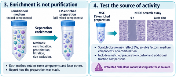
Enrichment increases the share or concentration of a selected material in a preparation. It does not mean complete purification. Centrifugation, precipitation, filtration, and size-exclusion methods separate materials in different ways. Each method retains some components and loses others. Describe an MSC small-particle preparation by how it was made and the checks actually performed.
A treatment study requires both a well-described preparation and relevant controls. If an MSC EV-enriched sample changes NHDF scratch closure, the activity could arise from vesicles, co-collected soluble factors, background medium components, or a combination. Comparing only treated NHDFs with untreated NHDFs cannot distinguish these sources. A matched preparation control and additional fraction comparisons help test the explanation.
Characterization measures properties that help describe the preparation.  Particle concentration and size distribution are useful, but a particle measurement may also detect non-EV material. Combine measurements of physical properties with EV-associated components and likely contaminants. Protein concentration alone cannot establish EV number or purity.
Describe the treatment amount explicitly. Volume, total protein, particle number, and source-culture equivalents are different ways to express input. A preparation containing 10 µg total protein may not contain the same EV amount as another preparation with 10 µg total protein. Retain the input measure and its limits rather than calling it simply “one EV dose.”
Attribution: Images AI-generated using ChatGPT Modified by Emalee Mackenzie, Irvine Valley College. Licensed under CC BY-NC-SA
This page titled 14.3: Extracellular Vesicles and What Enrichment Means was last modified on Wed, 21 Oct 2026 01:35:36 GMT and is shared under a CC BY-NC-SA 4.0 license and was authored, remixed, and/or curated by Emalee MacKenzie.
The book could not be loaded just now.