Opening the book…
Inherited disorders can arise when chromosomes behave abnormally during meiosis. Chromosome disorders can be divided into two categories: abnormalities in chromosome number and chromosome structural rearrangements. Because even small segments of chromosomes can span many genes, chromosomal disorders are characteristically dramatic and often fatal.
Disorders in Chromosome Number
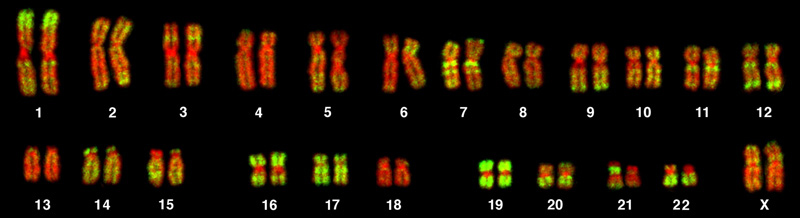
The isolation and microscopic observation of chromosomes forms the basis of cytogenetics and is the primary method by which clinicians detect chromosomal abnormalities in humans. A karyotype is the number and appearance of chromosomes, including their length, banding pattern, and centromere position. To obtain a view of an individual’s karyotype, cytologists photograph the chromosomes and then cut and paste each chromosome into a chart, or karyogram (Figure 7.16).
Nondisjunctions, Duplications, and Deletions
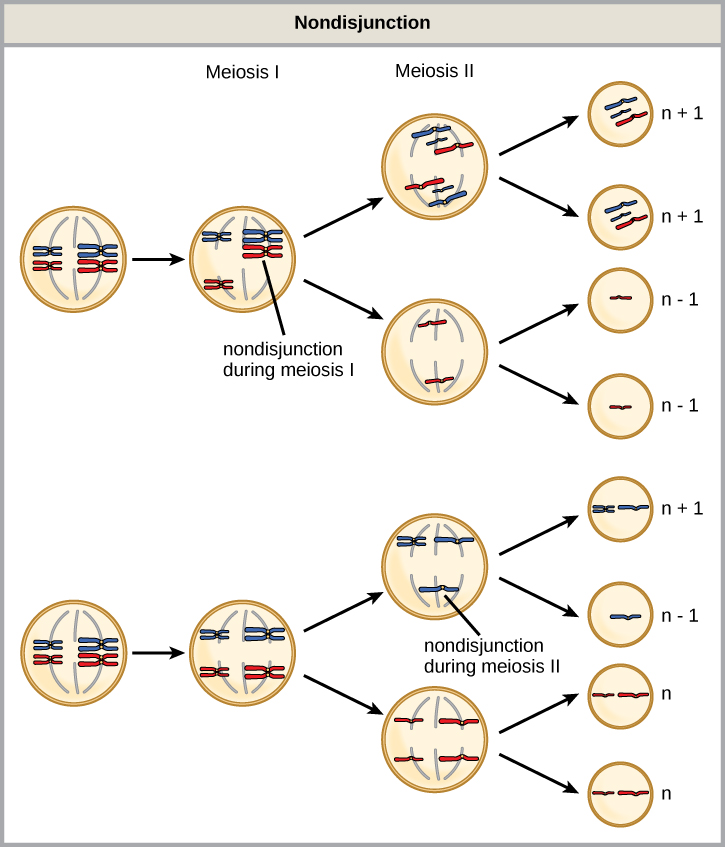
Of all the chromosomal disorders, abnormalities in chromosome number are the most easily identifiable from a karyogram. Disorders of chromosome number include the duplication or loss of entire chromosomes, as well as changes in the number of complete sets of chromosomes. They are caused by nondisjunction, which occurs when pairs of homologous chromosomes or sister chromatids fail to separate during meiosis. The risk of nondisjunction increases with the age of the parents.
Nondisjunction can occur during either meiosis I or II, with different results (Figure 7.17). If homologous chromosomes fail to separate during meiosis I, the result is two gametes that lack that chromosome and two gametes with two copies of the chromosome. If sister chromatids fail to separate during meiosis II, the result is one gamete that lacks that chromosome, two normal gametes with one copy of the chromosome, and one gamete with two copies of the chromosome.
An individual with the appropriate number of chromosomes for their species is called euploid; in humans, euploidy corresponds to 22 pairs of autosomes and one pair of sex chromosomes (XX is female and XY is male in humans). An individual with an error in chromosome number is described as aneuploid, a term that includes monosomy (loss of one chromosome) or trisomy (gain of an extraneous chromosome). Monosomic human zygotes missing any one copy of an autosome invariably fail to develop to birth because they have only one copy of essential genes. Most autosomal trisomies also fail to develop to birth; however, duplications of some of the smaller chromosomes (13, 15, 18, 21, or 22) can result in offspring that survive for several weeks to many years. Trisomic individuals suffer from a different type of genetic imbalance: an excess in gene dose. Cell functions are calibrated to the amount of gene product produced by two copies (doses) of each gene; adding a third copy (dose) disrupts this balance. The most common trisomy is that of chromosome 21, which leads to Down syndrome. Individuals with this inherited disorder have characteristic physical features and developmental delays in growth and cognition.
Humans display dramatic deleterious effects with autosomal trisomies and monosomies. Therefore, it may seem counterintuitive that human females and males can function normally, despite carrying different numbers of the X chromosome. In part, this occurs because of a process called X inactivation. Early in development, when female mammalian embryos consist of just a few thousand cells, one X chromosome in each cell inactivates by condensing into a structure called a Barr body. The genes on the inactive X chromosome are not expressed. The particular X chromosome (maternally or paternally derived) that is inactivated in each cell is random, but once the inactivation occurs, all cells descended from that cell will have the same inactive X chromosome. By this process, females compensate for their double genetic dose of X chromosome.
In so-called “tortoiseshell” cats, X inactivation is observed as coat-color variegation (Figure 7.18). Females heterozygous for an X-linked coat color gene will express one of two different coat colors over different regions of their body, corresponding to whichever X chromosome is inactivated in the embryonic cell progenitor of that region. When you see a tortoiseshell cat, you will know that it has to be a female.

In an individual carrying an abnormal number of X chromosomes, cellular mechanisms will inactivate all but one X in each of her cells. As a result, X-chromosomal abnormalities are typically associated with mild mental and physical defects, as well as sterility. If the X chromosome is absent altogether, the individual will not develop.
Several errors in sex chromosome number have been characterized. Individuals with three X chromosomes, called triple-X, appear female but express developmental delays and reduced fertility. The XXY chromosome complement, corresponding to one type of Klinefelter syndrome, corresponds to male individuals with small testes, enlarged breasts, and reduced body hair. The extra X chromosome undergoes inactivation to compensate for the excess genetic dosage. Turner syndrome, characterized as an X0 chromosome complement (i.e., only a single sex chromosome), corresponds to a female individual with short stature, webbed skin in the neck region, hearing and cardiac impairments, and sterility.
An individual with more than the correct number of chromosome sets (two for diploid species) is called polyploid. For instance, fertilization of an abnormal diploid egg with a normal haploid sperm would yield a triploid zygote. Polyploid animals are extremely rare, with only a few examples among the flatworms, crustaceans, amphibians, fish, and lizards. Triploid animals are sterile because meiosis cannot proceed normally with an odd number of chromosome sets. In contrast, polyploidy is very common in the plant kingdom, and polyploid plants tend to be larger and more robust than euploids of their species.
Chromosome Structural Rearrangements
Cytologists have characterized numerous structural rearrangements in chromosomes, including partial duplications, deletions, inversions, and translocations. Because these alterations change the physical architecture of the chromosome rather than changing the total number of individual chromosomes, they can have highly varied effects on an organism's development, phenotype, and fertility.
Duplications and Deletions
Duplications and deletions fundamentally alter gene dosage, meaning they disrupt the normal balance of cellular proteins by changing the number of copies of specific genes available in the genome. These dosage imbalances often produce offspring that survive but exhibit distinct physical and mental abnormalities. Deletions occur when a part of the chromosome is completely lost. The loss of genetic material can eliminate essential genes entirely. For example, Cri-du-chat (from the French for “cry of the cat”) is a syndrome associated with severe nervous system abnormalities and identifiable physical features that results from a deletion of most of the small arm (the p arm) of chromosome 5.
Duplications happen when a segment of the chromosome is duplicated. The addition of genetic material introduces extra copies of genes. While this can disrupt metabolic and developmental pathways, gene duplications are also crucially important in evolutionary biology, as the "extra" copy can mutate and acquire new functions over time without depriving the organism of the original gene's function.
Inversions and Translocations
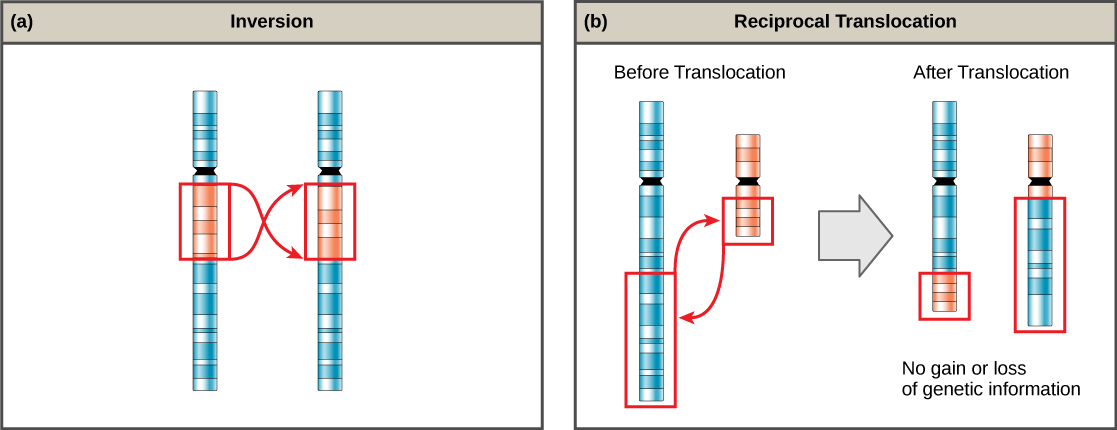
A chromosome inversion is the detachment, 180° rotation, and reinsertion of part of a chromosome. Inversions can be classified as either pericentric (including the centromere) or paracentric (occurring outside the centromere). Unless the break-and-rejoin points directly disrupt a vital structural gene sequence or regulatory element, inversions simply alter the linear orientation of genes. Consequently, they are likely to have much milder phenotypic effects on the individual than aneuploid errors or large deletions.
A translocation occurs when a segment of a chromosome dissociates and reattaches to a different, nonhomologous chromosome. Translocations can be reciprocal, in which segments are cleanly exchanged between two nonhomologous chromosomes without any net loss or gain of genetic material, or non-reciprocal, where a segment moves from one chromosome to another without an exchange. Even a balanced reciprocal translocation can have severe clinical effects if it fuses two distinct genes together. A notable example is the Philadelphia chromosome, a reciprocal translocation between chromosomes 9 and 22 that creates an abnormal fusion gene responsible for chronic myelogenous leukemia (CML).
Not all structural rearrangements of chromosomes produce nonviable, impaired, or infertile individuals. In rare instances, such a change can result in the evolution of a new species. In fact, an inversion in chromosome 18 appears to have contributed to the evolution of humans. This inversion is not present in our closest genetic relatives, the chimpanzees.
The chromosome 18 inversion is believed to have occurred in early humans following their divergence from a common ancestor with chimpanzees approximately five million years ago. Researchers have suggested that a long stretch of DNA was duplicated on chromosome 18 of an ancestor to humans, but that during the duplication it was inverted (inserted into the chromosome in reverse orientation.
A comparison of human and chimpanzee genes in the region of this inversion indicates that two genes—ROCK1 and USP14—are farther apart on human chromosome 18 than they are on the corresponding chimpanzee chromosome. This suggests that one of the inversion breakpoints occurred between these two genes. Interestingly, humans and chimpanzees express USP14 at distinct levels in specific cell types, including cortical cells and fibroblasts. Perhaps the chromosome 18 inversion in an ancestral human repositioned specific genes and reset their expression levels in a useful way. Because both ROCK1 and USP14 code for enzymes, a change in their expression could alter cellular function. It is not known how this inversion contributed to hominid evolution, but it appears to be a significant factor in the divergence of humans from other primates.1
Key Terms
Karyotype
The complete set of chromosomes of an individual, including their number, size, shape, and banding pattern.
Nondisjunction
The failure of homologous chromosomes or sister chromatids to separate properly during meiosis or mitosis, resulting in cells with abnormal chromosome numbers.
Aneuploid
A cell or organism with an abnormal number of chromosomes due to the gain or loss of one or more chromosomes.
Monosomy
A type of aneuploidy in which one chromosome of a homologous pair is missing (2n – 1).
Trisomy
A type of aneuploidy in which an extra copy of a chromosome is present (2n + 1).
Gene Dosage
The number of copies of a particular gene present in a cell or organism, which influences the amount of gene product produced.
Down Syndrome (Trisomy 21)
A genetic disorder caused by the presence of an extra copy of chromosome 21.
X Inactivation
The process in female mammals in which one X chromosome is randomly condensed and transcriptionally silenced to equalize gene expression between males and females.
Barr Body
A condensed, inactive X chromosome found in the nuclei of most female mammalian cells.
Polyploidy
The presence of more than two complete sets of chromosomes in a cell or organism.
Chromosomal Rearrangement
A structural alteration of a chromosome resulting from chromosome breakage and rejoining.
Deletion
The loss of a segment of a chromosome, resulting in the removal of genetic material.
Duplication
A chromosomal mutation in which a segment of DNA is copied one or more additional times.
Inversion
A chromosomal rearrangement in which a chromosome segment breaks, rotates 180°, and reinserts into the chromosome.
Pericentric Inversion
An inversion that includes the centromere.
Paracentric Inversion
An inversion that does not include the centromere.
Translocation
A chromosomal rearrangement in which a chromosome segment moves to a different, nonhomologous chromosome.
Reciprocal Translocation
A translocation in which chromosome segments are exchanged between two nonhomologous chromosomes.
Check Your Understanding
- What is nondisjunction, and during which stages of meiosis can it occur?
- Compare nondisjunction occurring during Meiosis I with nondisjunction occurring during Meiosis II.
- What is the difference between euploidy and aneuploidy?
- Compare monosomy and trisomy.
- Why are most autosomal monosomies incompatible with life?
- What is X inactivation, and why is it necessary in female mammals?
- What is a Barr body?
- Explain why individuals with abnormal numbers of X chromosomes often have milder symptoms than individuals with autosomal aneuploidies.
- Why are tortoiseshell cats almost always female?
- What is polyploidy?
- Why are triploid animals usually sterile?
- Compare deletions and duplications.
- How can changes in gene dosage affect phenotype?
- What causes Cri-du-chat syndrome?
- Compare pericentric and paracentric inversions.
- Why do many inversions have relatively mild effects on phenotype?
- What is a translocation?
- Compare reciprocal and nonreciprocal translocations.
- What is the Philadelphia chromosome, and why is it medically important?
Footnotes
- 1 V Goidts, et al., “Segmental duplication associated with the human-specific inversion of chromosome 18: a further example of the impact of segmental duplications on karyotype and genome evolution in primates,” Human Genetics, 115 (2004):116–22.
Contributors and Attributions
Samantha Fowler (Clayton State University), Rebecca Roush (Sandhills Community College), James Wise (Hampton University). Original content by OpenStax (CC BY 4.0; Access for free at https://cnx.org/contents/b3c1e1d2-83...4-e119a8aafbdd).
This page titled 2.1.6: Errors in Meiosis was last modified on Thu, 20 Aug 2026 01:22:08 GMT and is shared under a CC BY 4.0 license and was authored, remixed, and/or curated by OpenStax.
The book could not be loaded just now.