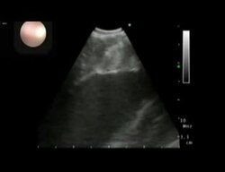
The-effectiveness-of-rigid-pericardial-endoscopy-for-minimally-invasive-minor-surgeries-cell-1749-8090-7-117-S1
Kimura T, Miyoshi S, Okamoto K, Fukumoto K, Tanimoto K, Soejima K, Takatsuki S, Fukuda K
Low resolution (312×240): this is the archive's own copy. Where the browser can use the graphics chip, Enhance picture in the player's settings draws it sharper.
Make a clip
CC BY 2.0 · Kimura T, Miyoshi S, Okamoto K, Fukumoto K, Tanimoto K, Soejima K, Takatsuki S, Fukuda K
Up to 60 seconds, at most the picture size of the copy it is cut from. The clip carries the title, the credit, the licence and a link back here. It is made in this browser, and nothing is sent to this site.
Or send the moment itself: https://invisiblenetwork.ai/watch/archive/commons/55213391?play=1&t=0
Parts
About
Video: rigid pericardial endoscopy. A rigid pericardial endoscope was inserted from the epicardial fossa to the pericardial space. Atrial appendages, coronary sinus and pulmonary veins were clearly identified. As the demonstration of cell transplantation, silicon beads, indocyanide green, ultrasonographic and fluoroscopic contrast medium were injected 20 times. Transient ventricular tachycardia did not deteriorate hemodynamic conditions. The use of an ultrasonography-attached needle was successful in avoiding intraventricular injection. For pacemaker implantation, the left ventricular free wall was clearly visualized in the left recombinant position. The intraventricular pacemaker lead was successfully implanted, and the epicardial sutureless lead and the suture trial were attempted. For epicardial ablation, the left pulmonary vein was identified and clamped by rigid forceps for the minimally invasive Maze procedure.
Human-made No record of this work says a model made it. What this means.
More from Wikimedia Commons
 Short
Making a silver chain
 Short
Jennifer Doudna Nobel Prize medal presentation
 Short
¿Qué es Wikipedia?
 Short
Wie entstehen Fossilien (CC by 4.0)
 Short
Rudy Mancuso & Maia Mitchell - Magic (Official Music Video)
 Documentary
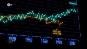
Die wichtigsten Klimafaktoren (ZDF, Terra X) 720p 50FPS
 Short
Curiosity's Seven Minutes of Terror
 Short
Wikilovesmonuments.org
 Short
Wikipedia - FactsMatter2016
 Short
President Obama on Death of Osama bin Laden no watermark
 Short
Cassini's Grand Finale
 Cartoon