
MRI-Based-Localisation-and-Quantification-of-Abscesses-following-Experimental-S.-aureus-Intravenous-pone.0154705.s008
Allen E, van Diemen P, Yamaguchi Y, Lindemann C, Soilleux E, Rollier C, Hill F, Schneider J, Wyllie D
Make a clip
CC BY 4.0 · Allen E, van Diemen P, Yamaguchi Y, Lindemann C, Soilleux E, Rollier C, Hill F, Schneider J, Wyllie D
Up to 60 seconds, at most the picture size of the copy it is cut from. The clip carries the title, the credit, the licence and a link back here. It is made in this browser, and nothing is sent to this site.
Or send the moment itself: https://invisiblenetwork.ai/watch/archive/commons/52232345?play=1&t=0
Parts
About
MRI video showing images of kidneys from the sortase A vaccination experiment. The video is of kidneys reconstructed in one plane. The video goes from the top to the bottom of the agar tube containing kidneys which was used for analysis. The groups are as shown in Fig 7, with the addition of three non-infection control kidneys, designated C. For example, in row 8 the kidneys are laid out as in S1 Fig. The group assignations are shown in S2 Table.
Human-made No record of this work says a model made it. What this means.
More from Wikimedia Commons
 Short
Making a silver chain
 Short
Jennifer Doudna Nobel Prize medal presentation
 Short
¿Qué es Wikipedia?
 Short
Wie entstehen Fossilien (CC by 4.0)
 Short
Rudy Mancuso & Maia Mitchell - Magic (Official Music Video)
 Documentary
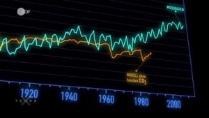
Die wichtigsten Klimafaktoren (ZDF, Terra X) 720p 50FPS
 Short
Curiosity's Seven Minutes of Terror
 Short
Wikilovesmonuments.org
 Short
Wikipedia - FactsMatter2016
 Short
President Obama on Death of Osama bin Laden no watermark
 Short
Cassini's Grand Finale
 Cartoon