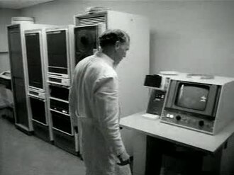
Belangrijke ontwikkeling in röntgenonderzoek dankzij 'computer-tomograaf' Weeknummer, 77-12 - Open Beelden - 21994
Nederlands Instituut voor Beeld en Geluid
Low resolution (320×240): this is the archive's own copy. Where the browser can use the graphics chip, Enhance picture in the player's settings draws it sharper.
Make a clip
CC BY-SA 3.0 nl · Nederlands Instituut voor Beeld en Geluid
Up to 60 seconds, at most the picture size of the copy it is cut from. The clip carries the title, the credit, the licence and a link back here. It is made in this browser, and nothing is sent to this site.
Or send the moment itself: https://invisiblenetwork.ai/watch/archive/commons/12107522?play=1&t=0
Parts
About
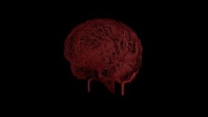
Newsreels in which Dutch subjects of a certain week are presented. X-ray research greatly improved by the so-called computer tomograph. With the help of this device it's now possible to use X-ray photography for soft tissue, like the brain. This X-ray technique has already been used for other body parts in the Saint Lucas hospital in Amsterdam. SHOTS: a man of whom X-rays of a.o. his brain are taken in hospital with the help of a computer tomograph; the X-ray tube of the computer tomograph; X-rays on the monitor; the working of the computer tomograph is explained with an instruction film.
Human-made No record of this work says a model made it. What this means.
- Archive
- Wikimedia Commons, in Wikimedia Commons
- Licence or status
- CC BY-SA 3.0 nl, as the Wikimedia Commons states it.
- Rights statement
- Creative Commons Attribution-Share Alike 3.0 nl
- Credit
- Nederlands Instituut voor Beeld en Geluid
- Subtitles
- English
- Who can watch
- The archive gives no rating. Invisible Network shows it to Teens profiles and older, judged from its collection and subjects. How films are placed
More from Wikimedia Commons
 Short
Making a silver chain
 Short
Jennifer Doudna Nobel Prize medal presentation
 Short
¿Qué es Wikipedia?
 Short
Wie entstehen Fossilien (CC by 4.0)
 Short
Rudy Mancuso & Maia Mitchell - Magic (Official Music Video)
 Documentary
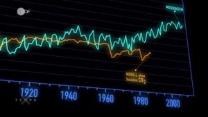
Die wichtigsten Klimafaktoren (ZDF, Terra X) 720p 50FPS
 Short
Curiosity's Seven Minutes of Terror
 Short
Wikilovesmonuments.org
 Short
Wikipedia - FactsMatter2016
 Short
President Obama on Death of Osama bin Laden no watermark
 Short
Cassini's Grand Finale
 Cartoon