|

- Image credit:
-
“Histology of Simple Squamous and Simple Cuboidal Epithelia" by Jennifer Lange, slide provided by the Regents of the University of Michigan Medical School under CC BY-NC-SA 4.0 © 2022.
|
- Answer Key (click to show/hide answers)
-
A - lumen
B - apical side
C - simple squamous epithelium
D - basal side
E - simple cuboidal epithelium
|
|
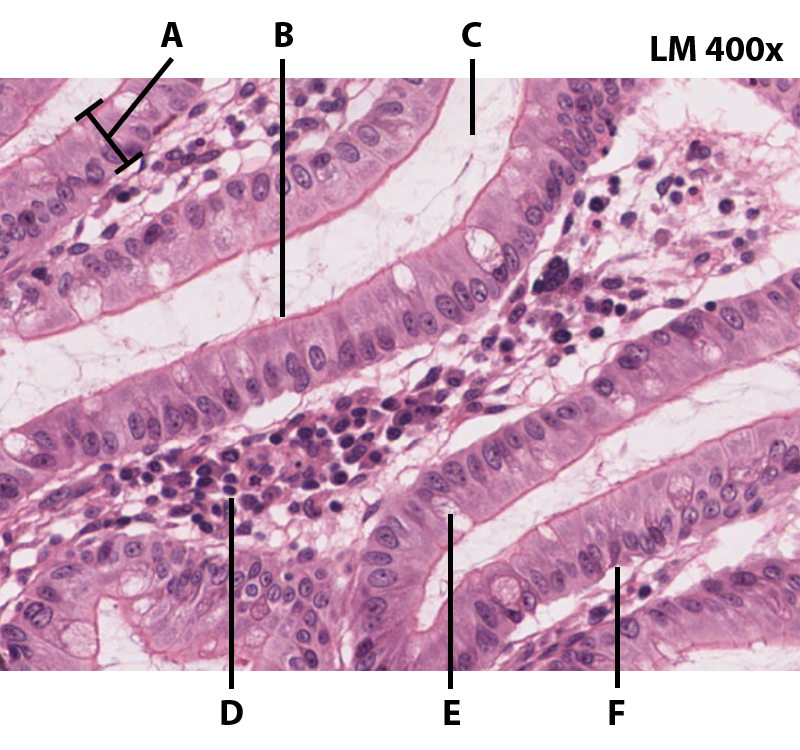
- Image credit:
-
“Histology of Simple Columnar Epithelium" by Jennifer Lange, slide provided by the Regents of the University of Michigan Medical School under CC BY-NC-SA 4.0 © 2022.
|
- Answer Key (click to show/hide answers)
-
A - simple columnar epithelium
B - apical side
C - lumen
D - connective tissue of lamina propria
E - goblet cell
F - basal side
|
|

- Image credit:
-
“Histology of Stratified Squamous Epithelium" by Jennifer Lange, slide provided by the Regents of the University of Michigan Medical School under CC BY-NC-SA 4.0 © 2022.
|
- Answer Key (click to show/hide answers)
-
A - connective tissue of the lamina propria
B - basal side/basement membrane
C - apical side
D - lumen
E - stratified squamous epithelium
|
|

- Image credit:
-
"Pseudostratified Columnar Epithelium Histology" by Jennifer Lange is licensed under CC BY-NC-SA 4.0, Micrograph provided by Berkshire Community College Bioscience Image Library is in the Public Domain.
|
- Answer Key (click to show/hide answers)
-
A - cilia
B - apical side
C - pseudostratified columnar epithelium
D - goblet cell
E - basal side/basement membrane
F - connective tissue of lamina propria
|
|

- Image credit:
-
“Histology of Transitional Epithelium" by Jennifer Lange, slide provided by the Regents of the University of Michigan Medical School under CC BY-NC-SA 4.0 © 2022.
|
- Answer Key (click to show/hide answers)
-
A - apical side
B - lumen
C - basal side/basement membrane
D - transitional epithelium
E - connective tissue of the lamina propria
|
|

- Image credit:
-
"Areolar Connective Tissue Histology" by Jennifer Lange is licensed under CC BY-NC-SA 4.0, Micrograph provided by Berkshire Community College Bioscience Image Library is in the Public Domain.
|
- Answer Key (click to show/hide answers)
-
Areolar Connective Tissue
A - ground substance
B - fibroblast
C - collagen fiber
D - elastin fiber
|
|

- Image credit:
-
“Histology of Adipose Connective Tissue" by Jennifer Lange, slide provided by the Regents of the University of Michigan Medical School under CC BY-NC-SA 4.0 © 2022.
|
- Answer Key (click to show/hide answers)
-
Adipose Connective Tissue
A - adipocyte
|
|

- Image credit:
-
"Reticular Connective Tissue Histology" by Jennifer Lange is licensed under CC BY-NC-SA 4.0, Micrograph provided by Berkshire Community College Bioscience Image Library is in the Public Domain.
|
- Answer Key (click to show/hide answers)
-
A - ground substance
B - reticulin fibers
|
|

- Image credit:
-
“Histology of Dense Regular Connective Tissue" by Jennifer Lange, slide provided by the Regents of the University of Michigan Medical School under CC BY-NC-SA 4.0 © 2022.
|
- Answer Key (click to show/hide answers)
-
Dense Regular Connective Tissue
A - fibroblast
B - collagen fiber (bundle)
C - ground substance
|
|

- Image credit:
-
“Histology of Dense Irregular Connective Tissue" by Jennifer Lange, slide provided by the Regents of the University of Michigan Medical School under CC BY-NC-SA 4.0 © 2022.
|
- Answer Key (click to show/hide answers)
-
Dense Irregular Connective Tissue
A - fibroblast
B - collagen fiber (bundle)
C - ground substance
|
|

- Image credit:
-
“Histology of Elastic Connective Tissue" by Jennifer Lange, slide provided by the Regents of the University of Michigan Medical School under CC BY-NC-SA 4.0 © 2022.
|
- Answer Key (click to show/hide answers)
-
Elastic Connective Tissue
A - ground substance
B - elastic fiber
C - fibroblast
|
|

- Image credit:
-
“Histology of Elastic Cartilage" by Jennifer Lange, slide provided by the Regents of the University of Michigan Medical School under CC BY-NC-SA 4.0 © 2022.
|
- Answer Key (click to show/hide answers)
-
Elastic Cartilage
A - elastic fiber
B - ground substance
C - chondrocyte
D - lacuna
E - perichondrium
|
|

- Image credit:
-
“Histology of Fibrocartilage" by Jennifer Lange, slide provided by the Regents of the University of Michigan Medical School under CC BY-NC-SA 4.0 © 2022.
|
- Answer Key (click to show/hide answers)
-
Fibrocartilage
A - ground substance
B - chondrocyte
C - collagen fiber
|
|

- Image credit:
-
“Histology of Hyaline Cartilage" by Jennifer Lange, slide provided by the Regents of the University of Michigan Medical School under CC BY-NC-SA 4.0 © 2022.
|
- Answer Key (click to show/hide answers)
-
Hyaline Cartilage
A - chondrocyte
B - lacuna
C - perichondrium
D - extracellular matrix
|
|

- Image credit:
-
“Histology of Smooth Muscle" by Jennifer Lange, slide provided by the Regents of the University of Michigan Medical School under CC BY-NC-SA 4.0 © 2022.
|
- Answer Key (click to show/hide answers)
-
Smooth Muscle Longitudinal Section
A - nucleus
B - endomysium
C - myofiber
|
|

- Image credit:
-
"Skeletal Muscle Histology - Longitudinal Section" by Jennifer Lange is licensed under CC BY-NC-SA 4.0, Micrograph provided by Berkshire Community College Bioscience Image Library is in the Public Domain.
|
- Answer Key (click to show/hide answers)
-
Skeletal Muscle Longitudinal Section
A - endomysium
B - myofiber
C - nucleus (peripheral)
D - striations
|
|

- Image credit:
-
"Cardiac Muscle Histology" by Jennifer Lange; micrographs provided by Virginia Commonwealth University under CC BY-NC-SA 4.0.
|
- Answer Key (click to show/hide answers)
-
Cardiac Muscle Tissue
A - nucleus (central)
B - endomysium
C - intercalated disc
D - myofiber
|
|

- Image credit:
-
"Nervous Tissue Histology - Spinal Cord Smear" by Jennifer Lange is licensed under CC BY-NC-SA 4.0, Micrograph provided by Berkshire Community College Bioscience Image Library is in the Public Domain.
|
- Answer Key (click to show/hide answers)
-
Nervous Tissue - Grey Matter, Spinal Cord Smear
A - neuron
B - neuroglia
-
-
-
-
-
-
|
|

- Image credit:
-
"Neuron Model" by Jennifer Lange is licensed under CC BY-NC-SA 4.0.
|
- Answer Key (click to show/hide answers)
-
Red - soma/cell body
Blue - axon
Purple - myelin sheath/neurolemmocytes
Green - dendrites
Orange - axon/synaptic terminals
A - nucleus
B - axon hillock
C - neurofibril node (of Ranvier)
|