Opening the book…
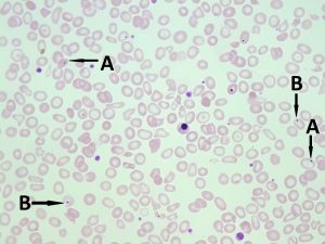
- A peripheral blood smear with Howell-Jolly bodies. A. shows Howell-Jolly bodies. B. shows platelets on top of a red blood cell. Note the clear space surrounding the platelet. 50x oil immersion. From MLS Collection, University of Alberta, https://doi.org/10.7939/R30R9MK3C
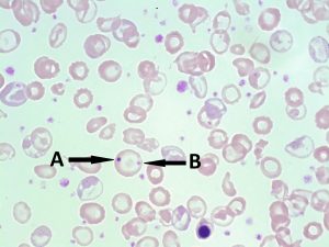
- A peripheral blood smear with a Howell-Jolly body and a platelet on the same red blood cell. A. shows Howell-Jolly body. B. shows platelets on top of a red blood cell. 100x oil immersion. From MLS Collection, University of Alberta, https://doi.org/10.7939/R3B853Z9R
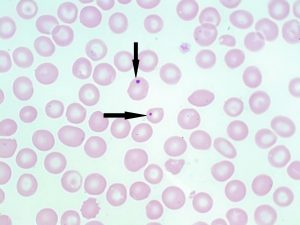
- A peripheral blood smear with Howell-Jolly bodies (indicated with arrows). 100x oil immersion. From MLS Collection, University of Alberta, https://doi.org/10.7939/R3028PV21
Appearance:
Under Wright/Romanowksy stains, Howell-Jolly Bodies appear as dark blue/purple round inclusions located at the periphery of the RBC. They usually present as a single inclusion inside the cell. Howell-Jolly Bodies are also visible under supravital stains.1-4
Inclusion composition:2,3
Nuclear fragments/remnants made up of DNA 1-4
Associated Disease/Clinical States:
Thalassemia
Megaloblastic Anemia
Myelodysplastic Syndrome
Post-splenectomy
References:
1. Landis-Piwowar K, Landis J, Keila P. The complete blood count and peripheral blood smear evaluation. In: Clinical laboratory hematology. 3rd ed. New Jersey: Pearson; 2015. p. 154-77.
2. Jones KW. Evaluation of cell morphology and introduction to platelet and white blood cell morphology. In: Clinical hematology and fundamentals of hemostasis. 5th ed. Philadelphia: F.A. Davis Company; 2009. p. 93-116.
3. Fritsma GA. Bone marrow examination. In: Rodak’s hematology clinical applications and principles. 5th ed. St. Louis, Missouri: Saunders; 2015. p. 253-68.
4. Ford J. Red blood cell morphology. Int J Lab Hematol [Internet]. 2013 Mar 9 [cited 2018 Jul 12];35(3):351–7. Available from: https://doi.org/10.1111/ijlh.12082
This page titled 3.7: Howell-Jolly Bodies was last modified on Sat, 02 Sep 2023 08:16:24 GMT and is shared under a CC BY-NC 4.0 license and was authored, remixed, and/or curated by Valentin Villatoro and Michelle To (Open Education Alberta) via source content that was edited to the style and standards of the LibreTexts platform.
The book could not be loaded just now.