Opening the book…
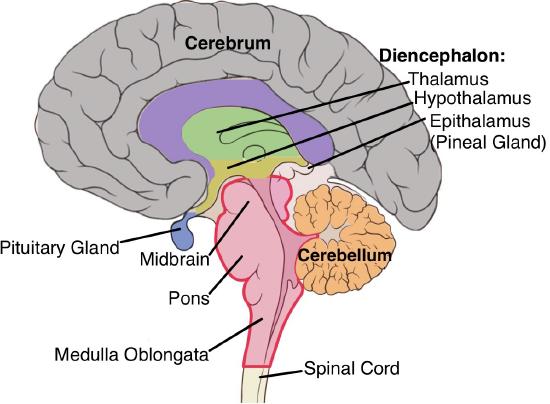
The adult brain is described in terms of four major regions: the cerebrum, the diencephalon, the brainstem, and the cerebellum (Figure 8). The cerebrum is the largest part of the brain responsible for advanced cognitive processes, including thought, perception, and voluntary motor functions. The diencephalon comprises the thalamus, hypothalamus, and pineal gland. The thalamus relays sensory information to the cerebral cortex, which is crucial in perception and consciousness. Situated beneath the thalamus, the hypothalamus regulates essential bodily functions, including temperature, hunger, and hormonal secretion. Positioned at the back of the brain, the cerebellum is essential for motor coordination, balance, and precision. Comprising the medulla, pons, and midbrain, the brainstem controls vital life functions such as breathing, heartbeat, and sleep-wake cycles. Extending from the brainstem, the spinal cord is a vital pathway for transmitting sensory and motor information between the brain and the rest of the body.
The CNS has a distinctive arrangement of neurons. These neurons form clusters, where cell bodies and dendrites create one aspect, while adjacent groups of axons present a contrasting feature. This phenomenon has led to the terms gray matter and white matter. Gray matter encompasses regions where cell bodies and dendrites predominate, while white matter refers to the areas where bundles of axons are visible to the naked eye. The pale appearance of white matter arises from the presence of myelin sheaths encasing axons. These protective sheaths, rich in lipid content, confer distinct coloration and the ability to facilitate rapid signal transmission (Figure 9). Notably, neuron cell bodies and axons are often visually particular. In the CNS, a localized aggregation of neuron cell bodies is termed a nucleus. At the same time, in the PNS, a similar grouping is called a ganglion (plural = ganglia).

The nomenclature diverges further when considering bundles of axons. Within the CNS, these bundled axons are termed tracts, signifying their role in conveying information. Conversely, these axonal clusters in the PNS are called nerves, indicating their function in transmitting signals to and from various body parts.
The Cerebrum
The prominent gray overlay of the human brain, resembling a substantial portion of its overall mass, corresponds to the cerebrum (Figure 10). This exterior is intricately folded to form the cerebral cortex (derived from the Latin term bark of a tree), and underlying this convoluted surface lies the remainder of the structure. Numerous advanced neurological functions, including memory, emotion, and consciousness, emanate from the intricate workings of the cerebral domain. The cerebral cortex constitutes the outer gray matter layer. A pronounced separation known as the longitudinal fissure delineates the two halves of the cerebrum, resulting in distinct right and left cerebral hemispheres. Deeper within this structure, the white matter of the corpus callosum is the primary conduit for interhemispheric communication within the cerebral cortex.
Deeply embedded nuclei termed the basal nuclei, also called basal ganglia, significantly contribute to cognitive processing and hold paramount importance in planning movements. The basal forebrain houses nuclei crucial to learning and memory. The limbic structure, a segment of the cerebral cortex intertwined with the limbic system, encompasses an assembly of regions dedicated to emotion, memory, and behavioral functions.
Cerebral Cortex

A continuous stratum of gray matter envelops the cerebrum and encircles both forebrain hemispheres, known as the cerebral cortex. This tenuous yet extensive domain of intricately folded gray matter is accountable for sophisticated neural functions. A ridge formed by one of these convoluted elevations is identified as a gyrus (plural = gyri), while the groove delineating two gyri is designated a sulcus (plural = sulci). The configuration of these tissue folds signifies specific territories within the cerebral cortex.
The cerebral surface can be mapped based on prominent gyri and sulci. Employing these distinctive landmarks, the cortex can be segregated into four principal zones or lobes (Figure 10). Among these markers is the lateral fissure, which restricts the temporal lobe from other sectors. The parietal and frontal lobes are situated superior to the lateral sulcus, segregated by the central sulcus. The posterior domain of the cortex corresponds to the occipital lobe. However, no conspicuous anatomical boundary delineates it from the parietal or temporal lobes on the lateral aspect of the brain. The absence of explicit anatomical demarcation between these lobes aligns with the interrelated functional nature of these regions.
The cerebrum is pivotal in orchestrating intricate cognitive functions such as memory, learning, language, and conscious awareness. These complex processes are meticulously distributed across discrete cortical regions, each with a dedicated responsibility for specific functions. The cerebral cortex encompasses a multitude of domains critical for sensory processing. These contain the primary cortical sectors specialized in processing somatosensory, visual, auditory, and gustatory stimuli, as well as association areas that facilitate information integration within these sensory domains. In the intricate organization of the cerebral cortex, sensory processing primarily occurs within the parietal, temporal, and occipital lobes, whereas the frontal lobe predominates in governing motor functions. Moreover, distinct regions within the cortex are dedicated to language processing, forming a finite collection further refined by their different roles in various aspects of linguistic function.
During the early 1900s, German neuroscientist Korbinian Brodmann conducted an extensive investigation into the microscopic structure of the cerebral cortex. His analysis led to the subdivision of the cortex into 52 distinct regions based on histological characteristics. This classification, termed Brodmann's areas, remains integral in characterizing cortical anatomical differences (Figure 11), which closely align with functional disparities. For instance, primary visual perception is attributed to areas 17 and 18 within the occipital lobe. Brodmann's areas 41 and 42 within the superior temporal lobe correspond to primary auditory sensation. Given the involvement of the temporal lobe in the limbic system, it bears significance in memory formation, as memories often emerge as recalled sensations. Memories of movement are the recollections of sensory feedback arising from those movements.15

The parietal lobe predominantly handles somatosensation—the general bodily sensations. The postcentral gyrus, or primary somatosensory cortex, situated posterior to the central sulcus, houses Brodmann's areas 1, 2, and 3. This cortical region processes various tactile senses, encompassing touch, pressure, pain, tickle, itch, vibration, proprioception, and kinesthesia, signifying body position and movement. Conversely, anterior to the central sulcus, the frontal lobe is chiefly associated with motor functions (Figure 12). The precentral gyrus, the primary motor cortex, orchestrates movement execution. Cells within this cortex relay commands to spinal cord cells, inducing skeletal muscle movement. Adjacent to this area lies the premotor area or motor association area, which is responsible for movement planning. Broca's area, predominantly in the left hemisphere, controls language production and speech-related activities.

The prefrontal lobe, positioned anteriorly, encompasses cognitive functions that underlie personality, short-term memory, and consciousness. The prefrontal cortex plays a pivotal role in shaping individual personality, largely influencing intention and plan execution. A renowned historical case involving Phineas Gage in 1848 helps us understand the significance of the prefrontal cortex. Following a traumatic incident where a metal spike penetrated his prefrontal cortex (Figure 13), Gage underwent marked personality alteration, transitioning from a diligent, amiable individual to an irritable and sluggish one. Despite these drastic changes, Gage managed to function as a stagecoach driver, illustrating the brain's potential for recovery even from substantial trauma.

Beyond the primary and association cortical domains, additional regions recognized as integrative areas occupy the interstitial spaces between distinct sensory or motor functional parts. These zones integrate multisensory information or engage in more intricate processing of sensory and motor inputs. Proprioceptive input derived from arm and hand movements interfaces with visual stimuli to orchestrate the execution of actions.
Cortical Processing
The precentral gyrus serves as the primary motor cortex. Pioneering neurosurgeon Walter Penfield significantly contributed to our foundational comprehension of the primary motor cortex by using electrical stimulation on the surface of the cerebrum. Penfield's technique involved delicately probing the cortex while patients remained under local anesthesia, facilitating real-time observation of their responses to stimulation. This approach initially fostered the assumption that the precentral gyrus could directly induce muscle movements. Contemporary knowledge, however, reveals that the primary motor cortex receives inputs from diverse areas instrumental in movement planning. Its direct output then triggers the activation of spinal cord neurons, culminating in the regulation of skeletal muscle activity.
The primary motor cortex delineates a somatotopic arrangement of the body, giving rise to the representation of a motor homunculus. The terminology homunculus originates from the Latin term denoting a little man, embodying a graphical portrayal of the human body superimposed on a designated sector of the cerebral cortex. Notably, neurons governing the musculature of the lower legs and feet inhabit the medial expanse of the precentral gyrus. At the same time, the thighs, trunk, and shoulders are localized at the apex of the longitudinal fissure. The hand and face regions are positioned on the lateral aspect of the gyrus. Furthermore, this spatial distribution disproportionately emphasizes muscles endowed with extensive innervation. Specifically, greater cortical representation is accorded to muscles implicated in intricate and elegant actions, such as those governing the fingers and lower face. In contrast, muscles responsible for more coarse movements, like the back and buttock muscles, occupy comparatively more minor domains on the motor cortex (Figure 14).19

Adjacent to the primary motor cortex lies the premotor cortex, Broca's area, and the frontal eye fields, all closely related to planning specific movement types. These areas encompass judgment, abstract reasoning, and working memory. Precursors to devising particular movements contain the assessment of whether these actions should be executed, exemplified by decisions such as whether to snooze the alarm or not.
Parallel to the primary motor cortex, the positions of somatosensory receptors on the body are transcribed onto the somatosensory cortex. This representation is often portrayed through a sensory homunculus (Figure 15).

Within the somatosensory cortex, the medial aspect of the gyrus positioned within the longitudinal fissure represents the external genitals, feet, and lower legs. As the gyrus curves outward from the fissure, extending along the parietal lobe's surface, the somatotopic arrangement proceeds sequentially through the thighs, hips, trunk, shoulders, arms, and hands. Approaching the lateral sulcus, the head and face representation is lateral to the finger representation. This organization on the topographical map follows a medial-to-lateral arrangement, progressing from the lower to the upper body. Furthermore, axons transmitting sensory information from the head and neck extend to the thalamus and run adjacent to the upper body fibers. The connections via the thalamus uphold this topographical order, ensuring the preservation of anatomical information. Notably, this correspondence does not lead to a scaled-down body replica; instead, it accentuates more responsive regions, such as the fingers and lower face. Areas of the body with lower sensitivity, such as the shoulders and back, are allocated smaller areas within the cortex.
Language and Speech
Language constitutes a distinctive facet of neurological function within the human realm. An illustrative instance of multimodal integrative territories is linked to language functionality. Wernicke's area is situated adjacent to the auditory association cortex at the terminus of the lateral sulcus anterior to the visual cortex. Broca's area lies in the lateral domain of the frontal lobe, positioned anteriorly to the motor cortex sector linked to the head and neck. This terminology originates from a French surgeon and anatomist who examined patients incapable of vocalizing speech. These individuals exhibited intact speech comprehension but the inability to articulate speech sounds, indicating compromised or underdeveloped Broca's areas. The interconnection between these two domains is facilitated by tracts extending between the posterior temporal lobe and the lateral sector of the frontal lobe, referred to as arcuate fasciculus (Figure 16).22

The Left and the Right Brain
The notion that individuals are either "right-brained" or "left-brained" represents an oversimplified perspective on a nuanced concept involving cerebral hemispheres. Lateralization of function does exist, attributing language function predominantly to the left hemisphere and spatial and nonverbal reasoning to the right hemisphere. However, it is important to recognize that various pervasive functions, such as language, are globally distributed throughout the cerebrum. This misconception has found support in studies involving split-brain patients. For individuals with intractable epilepsy, a severe neurological condition, one approach is to surgically sever the corpus callosum, thereby isolating the two cerebral hemispheres. Subsequently, a split-brain patient might struggle to formulate verbal responses based on sensory inputs processed in the right hemisphere. This leads to the misconception that language function resides solely in the left hemisphere. Nevertheless, documented cases highlight language function loss resulting from damage to the right hemisphere. Damage to the left hemisphere is typically categorized as aphasia, a speech function deficit, while damage to the right hemisphere can impact language usage. Impaired comprehension of figurative language, like jokes, irony, or metaphors, is associated with right hemisphere damage. Moreover, nonverbal elements of speech, including facial expressions and body language, can be influenced by right hemisphere damage, leading to flattened emotional expression in speech.
Ambidexterity denotes the ability to use both left and right hands equally and represents a renowned form of cross-dominance. Ambidextrous individuals are rare, comprising only 1% of the population. More commonly, individuals identified as ambidextrous were initially left-handed and learned ambidexterity either intentionally or during circumstances such as childhood or occupations that emphasize or require right-handed behaviors. The prevalence of right-handed tools and objects leads many left-handers to adopt right-handed usage, thus enhancing motor skills in their non-dominant hands. Right-handers might become ambidextrous due to their dominant right hand or arm injuries.24
Basal Nuclei
The basal nuclei, interchangeably termed basal ganglia, encompass a confluence of nuclei within the cerebrum that oversee the appraisal of cortical processing in relation to the broader operational state of the nervous system. Positioned deep within the cerebrum, the caudate, putamen, and globus pallidus constitute the principal components of basal nuclei responsible for orchestrating movement regulation (Figure 17). Caudate and putamen together form the striatum. Additional structures include the subthalamic nuclei and the substantia nigra. Thus, the basal nuclei within the cerebrum establish functional connectivity with a subset of brainstem nuclei, collectively forming a motor pathway.
Within the basal nuclei, two distinct streams of information processing operate. The entire input to the basal nuclei emanates from the cortex into the striatum. The direct pathway of the basal nuclei is a key circuit that facilitates movement by promoting the activation of the motor cortex. It begins with excitatory signals from the motor cortex to the striatum, where specific neurons are activated. These neurons then inhibit the internal segment of the globus pallidus and the substantia nigra, which normally send inhibitory signals to the thalamus, an example of disinhibition. By inhibiting the globus pallidus and substantia nigra, the direct pathway reduces their inhibitory effect on the thalamus, allowing it to send stronger excitatory signals back to the motor cortex. This results in the promotion and initiation of movement. In a healthy brain, the direct pathway works alongside the indirect pathway to regulate motor activity, ensuring movements are smooth and properly controlled.
The indirect pathway of the basal ganglia is a critical circuit in the brain that helps control and fine-tune movement by inhibiting excessive or unwanted motions. It starts when excitatory signals from the motor cortex activate neurons in the striatum, which then inhibit neurons in the external segment of the globus pallidus. This reduction in activity leads to increased activity in the subthalamic nucleus, which in turn stimulates the internal segment of the globus pallidus. The stronger inhibitory signals are sent from the internal segment of the globus pallidus to the thalamus, reducing its ability to excite the motor cortex and thus suppressing movement. In conditions like Parkinson’s disease, disruptions in this pathway can lead to excessive inhibition of movement, contributing to symptoms like slowness and difficulty initiating actions. The indirect pathway works in balance with the direct pathway to ensure smooth and controlled motor function. An illustrative video (http://openstaxcollege.org/l/basalnuclei1) visually expounds on the basal nuclei and the pathway that underlies information processing within the cerebrum. As depicted, the direct pathway represents a shorter route through the system, culminating in heightened cortical activity and amplified motor responses.25

Parkinson's disease, a hypokinetic condition affecting the basal nuclei with a specific impact on the substantia nigra, provides an illustration of the consequences of the direct and indirect pathways. In this disorder, neurons within the substantia nigra undergo degeneration, impeding dopamine release into the striatum. Without this modulatory influence, the basal nuclei predominantly rely on the indirect pathway. As elucidated earlier, the resulting hypokinetic disorder stems from diminished or absent direct pathway activation, which is pivotal for enhancing cortical motor commands. 27
The Limbic System
The limbic system consists of structures arranged around the edge of the cerebrum, including the amygdala, hippocampus, and cingulate gyrus. It is connected to the hypothalamus and deals with emotional responses and memory functions. The limbic lobe demonstrates interconnections with the hypothalamus, exerting a regulatory influence over its activity contingent on emotional states. For instance, during anxiety or fear, the amygdala, situated in the medial portion of the temporal lobe, transmits signals to the hypothalamus. This communication triggers the activation of the sympathetic fight-or-flight response. Simultaneously, under the impetus of input from the amygdala, the hypothalamus prompts the release of stress hormones via its governance of the endocrine system (Figure 18).28

The Diencephalon
The diencephalon, a region of the brain that preserves its designation from embryonic development, is called the "through brain" due to its connecting role. It functions as an intermediary between the cerebrum and the broader nervous system, except the olfactory system, which is responsible for the sense of smell. All other brain components, the spinal cord, and the PNS transmit information to and from the cerebrum through the diencephalon. This region also serves as the conduit for output originating from the cerebrum.
Situated beneath the cerebrum, the diencephalon comprises the walls of the third ventricle. The thalamus and the hypothalamus constitute the primary divisions of the diencephalon (Figure 19). Additional structures include the epithalamus, housing the pineal gland, and the subthalamus, encompassing the subthalamic nucleus, which is integrated within the basal nuclei.
Epithalamus
The epithalamus is a small part of the diencephalon, located near the posterior segment of the third ventricle. It plays a crucial role in regulating circadian rhythms, endocrine functions, and emotional responses. Its primary structure, the pineal gland, secretes melatonin, which helps regulate the sleep-wake cycle. Melatonin is derived from serotonin. Secretion of melatonin exhibits modulation contingent upon the prevailing environmental light levels. When light stimulates the retina, a nerve impulse is sent to the suprachiasmatic nucleus in the hypothalamus, a key regulator of biological rhythms. Subsequently, the neural signal proceeds through the spinal cord and culminates in the pineal gland, suppressing melatonin production. Consequently, blood melatonin levels decline, fostering wakefulness. Conversely, during diminishing light conditions, as typically experienced in the evening hours, melatonin production escalates, elevating its levels in the blood and instigating drowsiness. Given its pivotal role in governing the body's innate circadian rhythms, melatonin can be harnessed to induce natural sleep patterns. Hence, melatonin finds utility in managing jet lag and serving as a sleep aid.30 Additionally, the epithalamus connects the limbic system with other brain regions, influencing emotional processing and behavioral responses.
Thalamus
The thalamus assembles nuclei that mediate communication between the cerebral cortex and the peripheral structures, spinal cord, or brainstem. All sensory data, excluding olfaction, traverse the thalamus before reaching the cerebral cortex. Axons originating from peripheral sensory receptors or intermediate nuclei establish synaptic connections within the thalamus, from which thalamic neurons project directly to the cerebrum. Beyond its role in relaying information, the thalamus also undertakes information processing. For instance, the specific segment of the thalamus that receives visual input influences the prioritization of significant visual stimuli and the allocation of attention.

The cerebrum reciprocally exchanges information with the thalamus, predominantly conveying motor directives. This engagement necessitates coordination with the cerebellum and various nuclei within the brainstem. Simultaneously, the cerebrum engages with the basal nuclei, an interaction that entails intricate connections with the thalamus. The cerebral cortex dispatches signals to the thalamus, which subsequently impacts the outcomes orchestrated by the basal nuclei.
Hypothalamus
Inferior and marginally anterior to the thalamus lies the hypothalamus, constituting the second prominent part of the diencephalon (Figure 19). Composing an assemblage of nuclei such as the arcuate nucleus or ventromedial nucleus, the hypothalamus primarily undertakes the task of governing homeostasis. The hypothalamus is the primary regulator of the autonomic nervous system functions and the activities of the endocrine system, exerting influence via the anterior pituitary gland. Several subdivisions of the hypothalamus participate in processes of memory and emotion, integrated within the broader framework of the limbic system. Additionally, the hypothalamus encompasses numerous distinct centers responsible for functions like regulating body temperature and synchronizing circadian rhythms.32
Brainstem
The brainstem, comprising the midbrain, pons, and medulla oblongata, acts as a vital conduit between the cerebral hemispheres and the spinal cord, facilitating the transmission of sensory and motor signals. The midbrain integrates sensory inputs from various modalities like vision, hearing, and touch while contributing to motor control. It houses cranial nerves III and IV, which are involved in eye movements and proprioception. Serving as a bridge between different brain regions, the pons plays a crucial role in transmitting sensory and motor information, particularly to and from the cerebellum. It houses cranial nerves V-VIII as well as nuclei vital for regulating respiration and sleep. The medulla oblongata governs essential autonomic functions like heart rate, blood pressure, and respiration and contains nuclei responsible for reflex actions such as coughing and swallowing. Several cranial nerves, including IX, X, XI, and XII, originate from the medulla, contributing to functions related to the head, neck, and specialized senses.

Midbrain
The midbrain, originating as one of the earliest divisions of the embryonic brain, occupies a compact space between the thalamus and the pons, housing the superior and inferior colliculi. The inferior colliculus plays a pivotal role in the auditory processing circuitry of the brainstem. Neurons within this structure transmit signals to the thalamus, which in turn relays auditory information to the cerebrum, enabling the conscious perception of sound. In contrast, the superior colliculus processes sensory inputs related to visual-spatial information and coordinates eye and head movement. Cranial nerves III (oculomotor) and IV (trochlear) originate from the midbrain, controlling extrinsic ocular movements. This intricate arrangement underscores the midbrain's significance in orchestrating sensory perception and motor responses essential for adaptive behavior.
Pons
"Pons," originating from the Latin word meaning "bridge," aptly describes its role as the primary connection between the cerebellum and the brainstem. Housing respiratory regulatory centers such as the apneustic and pneumotaxic centers alongside those of the medulla oblongata, the pons collectively contributes to the intricate control of breathing processes. Furthermore, within this structure reside cranial nerves V (trigeminal), VI (abducens), VII (facial), and VIII (vestibulocochlear), underscoring its importance in facilitating various sensory and motor functions, including facial sensation, eye movement, and hearing. The pons serves as a crucial nexus within the brainstem, orchestrating essential physiological processes and mediating communication between different regions of the CNS.
Medulla Oblongata
Often referred to as the medulla, this area corresponds to the myelencephalon during embryonic brain development (Figure 2). The prefix "myel" refers to the abundant white matter in this region, particularly its outer aspect, which seamlessly extends into the spinal cord's white matter. The brainstem features an extensive gray matter region known as the reticular formation or reticular activating system (RAS), distributed throughout, influencing processes such as sleep-wake cycles, cerebral activity, and attentiveness. The medulla houses vital centers crucial for cardiac, respiratory, and vasomotor functions, regulating autonomic processes like heart rate, blood pressure, and respiration. In maintaining cardiovascular homeostasis, the medulla coordinates autonomic nervous system responses, including adjustments in blood pressure. Additionally, it plays a crucial role in responding to descending inputs from the hypothalamus, amplifying sympathetic division activity in the cardiovascular system, particularly during periods of anxiety or stress. Cranial nerves IX (glossopharyngeal) through XII (hypoglossal) originate within the medulla, further emphasizing its significance in regulating functions associated with the head, neck, and specialized senses.34
Reticular Activating System (RAS) or Reticular Formation
The reticular formation, a dispersed aggregation of gray matter spanning the entirety of the brainstem, is intricately linked to the modulation of sleep-wake cycles, encompassing broad cerebral activity and attention mechanisms. Functionally, this network processes and consolidates incoming sensory information, pivotal in numerous visceral and autonomic operations. Ascending fibers originating within the reticular formation transmit signals upwards to rouse and invigorate the cerebral cortex, enhancing the overall state of cortical vigilance. Concomitantly, descending fibers from the cerebral cortex can activate the RAS and concurrently wield influence over spinal cord activities through interactions with reticular formation cells. Notably, the reticular formation exhibits heightened sensitivity to the effect of general anesthetics, suggesting that drugs predominantly inhibit this region to induce the anesthetic state. It is also the site of origin of the descending analgesic pathways, where the neurons reach the spinal cord and block the transmission of pain signals to the brain via the endogenous opioid system.35
Cerebellum
The cerebellum shares similar gyri and sulci as the cerebrum (see Figure 21). Its primary function is to integrate data from the cerebrum with sensory signals from the PNS through the spinal cord. The cerebellum contributes approximately 10 percent of the brain's total mass. The arbor vitae, or "tree of life," manifests as a distinct branching pattern of white matter. Its proximity to the pons underscores its integration into the brainstem, emphasizing its central position in coordinating bodily movements. The central regions of the cerebellum include the vermis and flocculonodular lobe (Figure 22). They are pivotal in aligning visual information, equilibrium, and proprioceptive feedback to sustain balance and synchronize movements like walking or gait, accomplished through the descending output from the red nucleus. Conversely, the lateral hemispheres primarily plan motor functions through inputs from the frontal lobe that return via thalamic projections to the premotor and motor cortices. Processing in the central regions is oriented towards movements of the axial musculature, while the lateral regions govern activities of the appendicular musculature. The vermis predominantly receives input from the dorsal columns and spinocerebellar pathways.


Efferent fibers stemming from the cerebrum give rise to branches that interface with neurons in the pons. These neurons project into the cerebellum, supplying a duplicate of the motor directives sent to the spinal cord. Peripheral sensory information, introduced through cranial or spinal nerves, is replicated within the inferior olive nucleus, situated in the medulla oblongata. Fibers from this nucleus infiltrate the cerebellum and are juxtaposed with the efferent instructions from the cerebrum. For instance, if the primary motor cortex in the frontal lobe dispatches a directive to initiate walking through the spinal cord, a copy of this directive is transmitted to the cerebellum. Sensory feedback concerning muscle and joint status, proprioceptive particulars regarding walking movements, and equilibrium sensations converge on the cerebellum via the inferior olive. The cerebellum evaluates these inputs, and if any inconsistency arises between the initial cortical directive and the sensory feedback, it issues a corrective order to rectify the variance. The cerebellar output is channeled to the midbrain, which, in turn, transmits a descending input to the spinal cord to correct the information sent to skeletal muscles.
The cerebellum plays a crucial role in procedural memory, particularly in motor skills acquisition. Classical conditioning, famously demonstrated by Ivan Pavlov's experiments with dogs, provides a notable model for understanding the cerebellum's involvement in learning. In Pavlov's experiments, dogs learned to associate a neutral stimulus (such as a bell) with a biologically significant stimulus (such as food), leading to the conditioned response of salivation upon hearing the bell alone. This form of associative learning involves the cerebellum, among other brain regions. The cerebellum is heavily interconnected with areas involved in motor control and coordination, making it well-suited for the formation and storage of motor memories.38
This page titled 3.8: The Brain was last modified on Thu, 10 Sep 2026 16:31:58 GMT and is shared under a CC BY-NC-SA 4.0 license and was authored, remixed, and/or curated by Chaya Gopalan (Consortium of Academic and Research Libraries in Illinois (CARLI)) .
The book could not be loaded just now.