Opening the book…
Simple somatic reflexes do not include the higher centers discussed for conscious or voluntary aspects of movement. Reflexes can be spinal or cranial, depending on the nerves and central components involved. A few examples are discussed in detail here.
An example of a monosynaptic reflex is the stretch reflex, such as the patellar reflex, in which muscle spindles serve as the primary sensory receptors (Figure 36).

Muscle spindles are sensory receptors that are distributed throughout the skeletal muscle. They are made of collections of specialized muscle fibers called intrafusal muscle fibers, which are situated parallel to the skeletal muscle fibers, referred to as extrafusal muscle fibers. Each muscle spindle has its own afferent and efferent supply. The gamma motor neuron is its efferent neuron, whereas the afferent fiber is its afferent neuron (Figure 36). Alpha motor neurons are the efferent neurons that supply the extrafusal muscle fibers.63
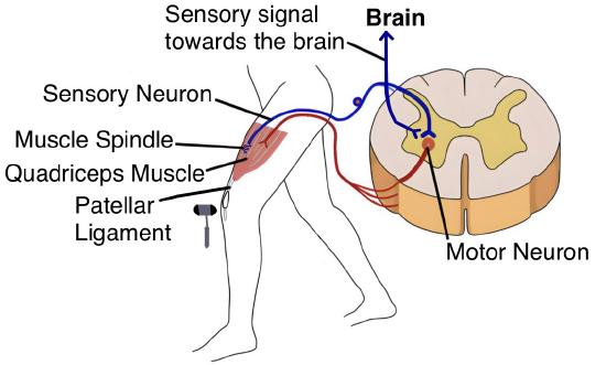
When the patellar tendon is tapped (Figure 37), skeletal muscle fibers within the quadriceps femoris muscle are stretched. Their intrafusal muscle fibers are also elongated, which increases the firing rate of afferent nerve fibers. The afferent neuron establishes a direct synapse with the alpha motor neuron, resulting in muscle contraction. The muscle spindles, inducing the muscle to contract, thus evoke the characteristic knee-jerk reaction. For a visual understanding of the role of the muscle spindle, refer to this YouTube video: http://www.youtube.com/watch?v=F871bBWS4oY.
The Golgi Tendon Reflex operates as a disynaptic or bisynaptic mechanism that opposes the stretch reflex. Golgi tendon organ is a proprioceptive sensory receptor designed to detect variations in muscle tension. These organs are positioned at the junctions where skeletal muscle fibers meet their corresponding tendons. During active muscle contraction, the Golgi tendon organs are stimulated, prompting the transmission of action potentials through their afferent fibers. Subsequently, these sensory neurons engage with inhibitory interneurons in the spinal cord. This engagement leads to the inhibition of alpha-motor neurons, ultimately bringing about the relaxation of the initially contracting muscle.
The Flexor Withdrawal Reflex is polysynaptic. For instance, when an individual steps on a painful stimulus like a sharp, broken glass piece, nociceptors on the same side (ipsilateral) are activated. These nociceptors trigger the activation of motor neurons responsible for the contraction of leg muscles, causing flexion. In parallel, an inhibitory interneuron, initiated by a collateral branch of the nociceptor fiber, impedes the motor neurons governing extensor muscles, effectively counteracting any extension. Conversely, a mirrored response is observed on the opposing side (contralateral). Extensor muscles are engaged, while flexor muscles are suppressed, a phenomenon known as the crossed-extensor reflex (Figure 38). This orchestrated process ensures leg extension on one side to maintain balance in response to the painful stimulus.65
Further insights into this topic can be gained from this video: http://www.youtube.com/watch?v=RLe9koPfVoo.

Sensory Pathways
Specific regions of the CNS coordinate somatic processes using sensory inputs and motor outputs of peripheral nerves. The essential areas of the CNS that play a role in somatic processes can be separated into the spinal cord, brainstem, diencephalon, cerebral cortex, and subcortical structures.
A neural conduit transmitting peripheral sensory information to the CNS is termed an ascending pathway or ascending tract (Figure 39). Different sensory modalities traverse distinct routes within the CNS. Tactile and other somatosensory stimuli elicit responses from receptors situated in the skin, muscles, tendons, and joints throughout the body. Somatosensory stimuli originating below the neck propagate through the sensory pathways in the spinal cord, whereas those emerging from the head and neck region follow the trajectories of cranial nerves, explicitly engaging the trigeminal system.
Typically, four distinct orders of neurons in the sensory pathways play a pivotal role in processing sensory information (Figure 39). First-order neurons, or primary afferent neurons, are the initial receptors for transduced signals. These neurons have their cell bodies situated within the dorsal root ganglia of the spinal cord or in the ganglia of cranial nerves. Second-order neurons in the spinal cord or brainstem receive input from one or more primary afferent neurons. They play a crucial role in transmitting this sensory information to the thalamus. In some instances, the axons of second-order neurons may undergo decussation (crossing over) within the spinal cord or brainstem before ascending to the thalamus. Third-order neurons are strategically located within the relay nuclei of the thalamus. It is from this central hub that encoded sensory information begins its journey towards the cerebral cortex. Fourth-order neurons are the final destination for this sensory data, residing within the appropriate sensory areas of the cerebral cortex. Once here, the processed information gives rise to the conscious perception of the stimulus, allowing us to experience and interpret the sensory input.
The dorsal column pathway, also known as the dorsal column-medial lemniscus pathway (DCML; Figure 39), is a fundamental component of the sensory system. It is responsible for transmitting sensory information related to fine touch, proprioception (the sense of body position), and vibratory sensations from the periphery to the brain. The DCML pathway is organized as follows:
- Primary Afferent Neurons: Sensory receptors, such as Meissner's corpuscles and Pacinian corpuscles for touch and Merkel cells for fine touch, are located throughout the body. The primary afferent neurons, also known as first-order neurons, have cell bodies located in the dorsal root ganglia (in the spinal cord) or cranial nerve ganglia (in the brainstem).
- Dorsal Columns: These ascending white matter tracts in the spinal cord consist of two main components: the fasciculus gracilis (located more medially) and the fasciculus cuneatus (located laterally). These columns carry sensory information from the lower body (gracilis) and upper body (cuneatus) respectively. The primary afferent neurons enter the nucleus gracilis and nucleus cuneatus within the medulla and synapse with second-order neurons.
- Second-order Neurons: Second-order neurons receive input from primary afferent neurons in the nucleus gracilis and nucleus cuneatus within the medulla. They then send their axons across to the opposite side in a process known as decussation. This crossing over ensures that sensory information from one side of the body is processed by the contralateral (opposite) hemisphere of the brain.
- Medial Lemniscus: The axons of the second-order neurons ascend through the brainstem in a bundle known as the medial lemniscus. This pathway ensures the transmission of sensory information to the thalamus.
- Third-order Neurons: The medial lemniscus neurons synapse in the thalamus. Third-order neurons in the thalamus receive these sensory signals and project them further into the cerebral cortex. Ultimately, the sensory information reaches the appropriate areas of the cerebral cortex, such as the primary somatosensory cortex (postcentral gyrus), where it is consciously perceived.
The DCML pathway enables us to discern between textures, recognize the position of our limbs without visual input, and perceive vibrations from objects or tools we handle. Dysfunction in this pathway can impair fine motor skills and spatial awareness.
The spinothalamic pathway, also known as the anterolateral system, is a component of our sensory system that helps detect and respond to noxious or potentially harmful stimuli. It is responsible for transmitting sensory information related to pain, temperature, and crude touch from the periphery to the brain. This pathway is essential for perceiving and responding to potentially harmful or noxious stimuli. The details of the spinothalamic pathway are described below:
- Primary Afferent Neurons (First-Order Neurons): Nociceptors and thermoreceptors are distributed throughout the body and are mainly concentrated in the skin and mucous membranes. The cell bodies of primary afferent neurons are located in the dorsal root ganglia (in the spinal cord) or cranial nerve ganglia (in the brainstem).
- Dorsal Horn of the Spinal Cord: The axons of primary afferent neurons enter the spinal cord and synapse with second-order neurons in the dorsal horn of the spinal cord. The axons of the second-order neurons (carrying pain and temperature information) cross over to the opposite side of the spinal cord soon after entering.
- Anterolateral Funiculus: The axons of the second-order neurons, which have now crossed over, ascend through the anterolateral funiculus, a white matter tract on the contralateral side of the spinal cord. Unlike the dorsal column pathway, where specific nuclei in the brainstem process sensory information, the spinothalamic pathway ascends through the spinal cord, reaching the reticular formation. From there, it projects to various thalamic nuclei.
- Third-Order Neurons: Third-order neurons from the thalamus project sensory signals further into the cerebral cortex, specifically the somatosensory cortex (postcentral gyrus), where it is consciously perceived. This perception enables us to sense pain, changes in temperature, and crude touch.

Parallel to their origin from dorsal root ganglion cells, these two systems share similarities. The dorsal column system predominantly mediates tactile sensations and proprioception, while the spinothalamic tract pathway predominantly mediates pain and temperature sensations. Furthermore, both systems feature contralateral second-order neurons projecting across the midline to the opposite side of the brain or spinal cord. In the dorsal column system, this crossing-over occurs in the brainstem; in the spinothalamic pathway, it transpires at the same spinal cord level where the sensory information entered. The third-order neurons within both pathways follow the same pattern. In both instances, the second-order neuron establishes synaptic connections within the thalamus, with the thalamic neuron subsequently projecting to the somatosensory cortex. The thalamus acts as a pivotal relay station for sensory input, encompassing a multitude of special senses and ascending somatosensory tracts. Each distinct sensory system is routed through a specific nucleus within the thalamus. The thalamus is an obligatory intermediary for most sensory tracts destined for the cerebral cortex, where conscious sensory perception is initiated except for olfaction.68
Motor Pathways
The salient attribute of the somatic nervous system resides in its jurisdiction over skeletal musculature. Somatic sensory input imparts information to the nervous system concerning the external milieu, yet the subsequent reaction involves deliberate engagement of voluntary muscle actions. The term voluntary conveys the notion that a conscious intent governs the initiation of a given movement. Nonetheless, specific facets of the somatic system employ voluntary muscles in ways that bypass the intended direction. A case in point is the capacity for respiratory functions to transition into an unconscious regulatory mode, enabling the individual to direct attention elsewhere.
Within the cerebral cortex, the sequential handling of sensory perception undergoes a continuum from primary processing to associative processing, culminating in integration within multimodal cortical regions. This hierarchy of processing levels facilitates the assimilation of sensory perceptions into memory and, of greater significance, drives the generation of responses. The culmination of cortical processing, spanning primary, associative, and integrative sensory domains, triggers an analogous series of motor processing stages, generally orchestrated within distinct cortical regions.
Located at the far front of the frontal lobe, the prefrontal cortex handles high-level planning rather than direct muscle execution. It sends its motor plans to secondary motor regions, the premotor cortex and supplemental motor area, which refine the movements. Unlike the rest of the frontal lobe, stimulating the prefrontal cortex with electricity does not trigger muscle contractions, defining it as an executive rather than a motor region.
Adjacent to the primary motor cortex, two noteworthy regions play integral roles in planning and synchronizing movements. The premotor cortex occupies a more lateral position, whereas the supplemental motor area is situated in a medial and superior orientation. The premotor area regulates core muscle movements to sustain posture during actions, while the supplemental motor area assumes responsibility for movement planning and coordination. Moreover, the supplemental motor area governs sequential movements rooted in prior experiential knowledge, encompassing learned motor patterns. Neuronal activity within these regions is most pronounced in the lead-up to movement initiation. For instance, these areas may predetermine the preparatory motions for driving a car in anticipation of a traffic signal altering its state.
Cerebral motor output cascades downward into the brainstem and spinal cord to govern musculature via motor neurons. Upper motor neurons within the primary motor cortex establish synapses with lower motor neurons in the spinal cord or brainstem. Two descending pathways manifest as the corticospinal tract (Figure 40) and the corticobulbar tract. These designations reflect their cortical origin and targets, either the spinal cord or the brainstem, with bulbar denoting the brainstem as an enlarged region atop the spinal cord. These two pathways are referred to as direct or pyramidal pathways that assume responsibility for voluntary skeletal muscle movements. Each motor command originating in the primary motor cortex travels along the axons, eliciting the activation of upper motor neurons located in the cranial motor nuclei or the ventral horn of the spinal cord. Axons of the corticobulbar tract are ipsilateral, projecting from the cortex to the motor nucleus on the same side of the body. Conversely, the corticospinal tract axons predominantly cross the brainstem (lateral) or spinal cord (anterior) midline and synapse on the contralateral side of the body. Consequently, the right motor cortex governs the muscles on the left side of the body and vice versa.
The corticobulbar tract is a two-neuron upper motor neuron pathway that conducts voluntary motor commands from the primary motor cortex to the motor nuclei of cranial nerves located throughout the brainstem. Arising predominantly from the head and face representations of the precentral gyrus, these axons descend through the corona radiata and pass through the genu of the internal capsule before terminating in the midbrain, pons, and medulla. The corticobulbar tract innervates cranial nerves V, VII, IX, X, XI, and XII to control the skeletal muscles of mastication, facial expression, swallowing, phonation, and tongue movement. Functionally, most cranial nerve motor nuclei receive bilateral innervation which provides redundancy and preserves muscle function following unilateral cortical damage. The two clinically vital exceptions to this rule are the motor neurons governing the lower quadrant of the face (CN VII) and one of the muscles of the tongue (CN XII), both of which receive predominantly contralateral input.

The corticospinal tract descends through the deep white matter of the cerebrum, traversing the caudate nucleus and putamen as the internal capsule bundle. Subsequently, it navigates the midbrain as the cerebral peduncles en route through the pons and finally enters the medulla. In the medulla, these tracts form the pyramids, a substantial white matter tract (Figure 40). The pyramidal decussation at the medullary-spinal boundary marks the pivotal point where most corticospinal tract fibers cross to the opposite side. At this juncture, the tract bifurcates into two segments, each commanding distinct muscular domains. The lateral corticospinal tract is the largest part of the corticospinal tract, with 90 percent of the nerve fibers. It begins in the cerebral cortex, decussates in the medulla and extends down the contralateral side of the spinal cord. It deals with the voluntary movement of the contralateral limbs. The anterior corticospinal tract governs the musculature of the body trunk. These axons refrain from crossing in the medulla, maintaining an anterior trajectory as they traverse the brainstem and enter the spinal cord. Upon reaching their designated level, they cross to the opposite ventral horn to synapse with lower motor neurons. As these axons enter the ventral horn, they interface with their corresponding lower motor neurons, predominantly stationed in their medial sectors to regulate axial trunk muscles. Because bodily trunk movements encompass both sides, the anterior corticospinal tract does not exhibit strict contralateral control. Certain collateral branches of the tract extend into the ipsilateral ventral horn, overseeing synergistic muscles on the same side or inhibiting antagonistic muscles via interneurons. The anterior corticospinal tract harmonizes postural muscles for expansive body movements through bilateral influence, exhibiting both ipsilateral and contralateral contributions.70
Extrapyramidal Pathways
Outside the corticospinal tract, the descending neural pathways connecting the brain and the spinal cord collectively constitute the extrapyramidal system. This term stems from its distinction from the corticospinal pathway, which encompasses the pyramids in the medulla. Several pathways originating from the brainstem contribute to this system.

Figure 41 | Locations of Spinal Fiber Tracts | 71 This schematic illustrates the major fiber tracts within the spinal cord. The corticospinal tract originates from the motor cortex and descends through the spinal cord in the lateral columns. The dorsal column-medial lemniscus pathway is located in the dorsal columns of the spinal cord. The spinothalamic tract runs through the lateral and ventral columns of the spinal cord. Spinocerebellar tracts are found in the lateral and ventral columns. The rubrospinal tract originates in the red nucleus of the midbrain and descends through the lateral columns. Reticulospinal tracts arise from the brainstem reticular formation and extend through various parts of the spinal cord. Vestibulospinal tracts begin in the vestibular nuclei and descend through the lateral and ventral columns. Descending autonomic pathways are distributed throughout the spinal cord, influencing autonomic functions.
The tectospinal tract emanates from the midbrain and extends to the spinal cord, playing a crucial role in postural movements governed by the superior colliculus, a part of the midbrain. The terminology of this tract derives from an alternate designation for the superior colliculus, tectum. The reticulospinal tract connects the reticular activating system and the spinal cord (Figure 41). This tract influences trunk and proximal limb muscles associated with posture and locomotion, impacting muscle tone and autonomic functions.
The vestibulospinal tract interlinks the brainstem nuclei of the vestibular system with the spinal cord, enabling the modulation of posture, movement, and balance through equilibrium-related inputs from the vestibular system. The basal nuclei, pivotal for regulating CNS-initiated movements, exert influence over the extrapyramidal system, including its thalamic feedback to the motor cortex.
The orchestration of conscious muscle movements surpasses the simple issuance of a solitary command from the precentral gyrus to the relevant motor neurons. During any bodily activity, muscles relay feedback to the brain, and the brain constantly revises instructions dispatched to the muscles. The cerebellum plays a significant role in the motor system by comparing cerebral motor commands with proprioceptive feedback. Corticospinal fibers projecting to the ventral horn of the spinal cord have branches that synapse in the pons, which further project to the cerebellum. Additionally, proprioceptive sensations from the dorsal column system have a collateral projection to the medulla, extending to the cerebellum. These two streams of information are juxtaposed within the cerebellar cortex. Conflicts arising from disparities between motor commands from the cerebrum and body position information relayed by proprioceptors prompt the cerebellum to stimulate the red nucleus of the midbrain.
The red nucleus transmits corrective commands to the spinal cord via the rubrospinal tract. The tract's name originates from the word ruby, reminiscent of the term red.
Thus, the somatic nervous system exclusively governs skeletal muscles. Due to the necessity of reaching peripheral muscles, the axon assumes a relatively elongated trajectory. Cell body diameters may span hundreds of micrometers to accommodate the lengthy axon, with some axons extending up to a meter, such as lumbar motor neurons that innervate muscles in the first digits of the feet.72
This page titled 3.13: Reflexes was last modified on Sun, 13 Sep 2026 16:14:15 GMT and is shared under a CC BY-NC-SA 4.0 license and was authored, remixed, and/or curated by Chaya Gopalan (Consortium of Academic and Research Libraries in Illinois (CARLI)) .
The book could not be loaded just now.