Opening the book…
-
Murphy, K., & Weaver, C. (2017). Janeway's immunobiology (9th ed.). Garland Science.
-
Betts, J. G. et al. (2022). Figure 21.6 Bone Marrow. In Anatomy and Physiology 2e. OpenStax. https://openstax.org/apps/archive/20241024.164013/resources/11cd5e374f86452e82a0270efcf9b20006c54f1d
-
Betts, J. G. et al. (2022). Figure 21.7 Location, Structure, and Histology of the Thymus. In Anatomy and Physiology 2e. OpenStax. https://openstax.org/apps/archive/20241024.164013/resources/c95c7a3ea1b9ea2fe9e5a89b04f1934942bd209b
-
Betts, J. G. et al. (2022). Figure 21.8 Structure and Histology of a Lymph Node. In Anatomy and Physiology 2e. OpenStax. https://openstax.org/apps/archive/20241024.164013/resources/d9f9b664217273a2bf5476dbc0faa4da30f70508
-
Betts, J. G. et al. (2022). Figure 21.9 Spleen. In Anatomy and Physiology 2e. OpenStax. https://openstax.org/apps/archive/20241024.164013/resources/97d7f0ff10bba9f39b746b2bf4878452ea445558
-
Betts, J. G. et al. (2022). Figure 21.10 Locations and Histology of the Tonsils. In Anatomy and Physiology 2e. OpenStax. https://openstax.org/apps/archive/20241024.164013/resources/68aa4a228261336d73fe90e3f15bb504539fbb44
-
Betts, J. G. et al. (2022). Figure 21.11 Mucosa-associated Lymphoid Tissue (MALT) Nodule. In Anatomy and Physiology 2e. OpenStax. https://openstax.org/apps/archive/20241024.164013/resources/02a3e13299cf8ec7059498ac7cb531d0fd779b4a
-
Betts, J. G. et al. (2022). Figure 21.12 Cooperation between Innate and Adaptive Immune Responses. In Anatomy and Physiology 2e. OpenStax. https://openstax.org/apps/archive/20241024.164013/resources/86e277aca20c56d538a49a0684c03063ca1a2f6d
-
Levinson, W. (2020). Review of medical microbiology and immunology: A guide for students (16th ed.). McGraw-Hill Education.
-
OpenStax. (2016). Phagocytosis: A form of active transport [Image]. In Anatomy and Physiology (Version 8.25). Wikimedia Commons. Retrieved December 3, 2024, from https://upload.wikimedia.org/wikipedia/commons/8/85/0309_Phagocytosis.png
-
Marieb, E. N., & Hoehn, K. (2019). Human anatomy & physiology (11th ed.). Pearson.
-
Papayannopoulos, V. Neutrophil extracellular traps in immunity and disease. Nat Rev Immunol 18, 134–147 (2018). https://doi.org/10.1038/nri.2017.105
-
Cacofonie. (2007). Schematic diagram indicating the complementary activities of cytotoxic T cells and natural killer cells [Image]. Wikimedia Commons. Retrieved December 3, 2024, from https://upload.wikimedia.org/wikipedia/commons/b/b7/Missing_self.svg
-
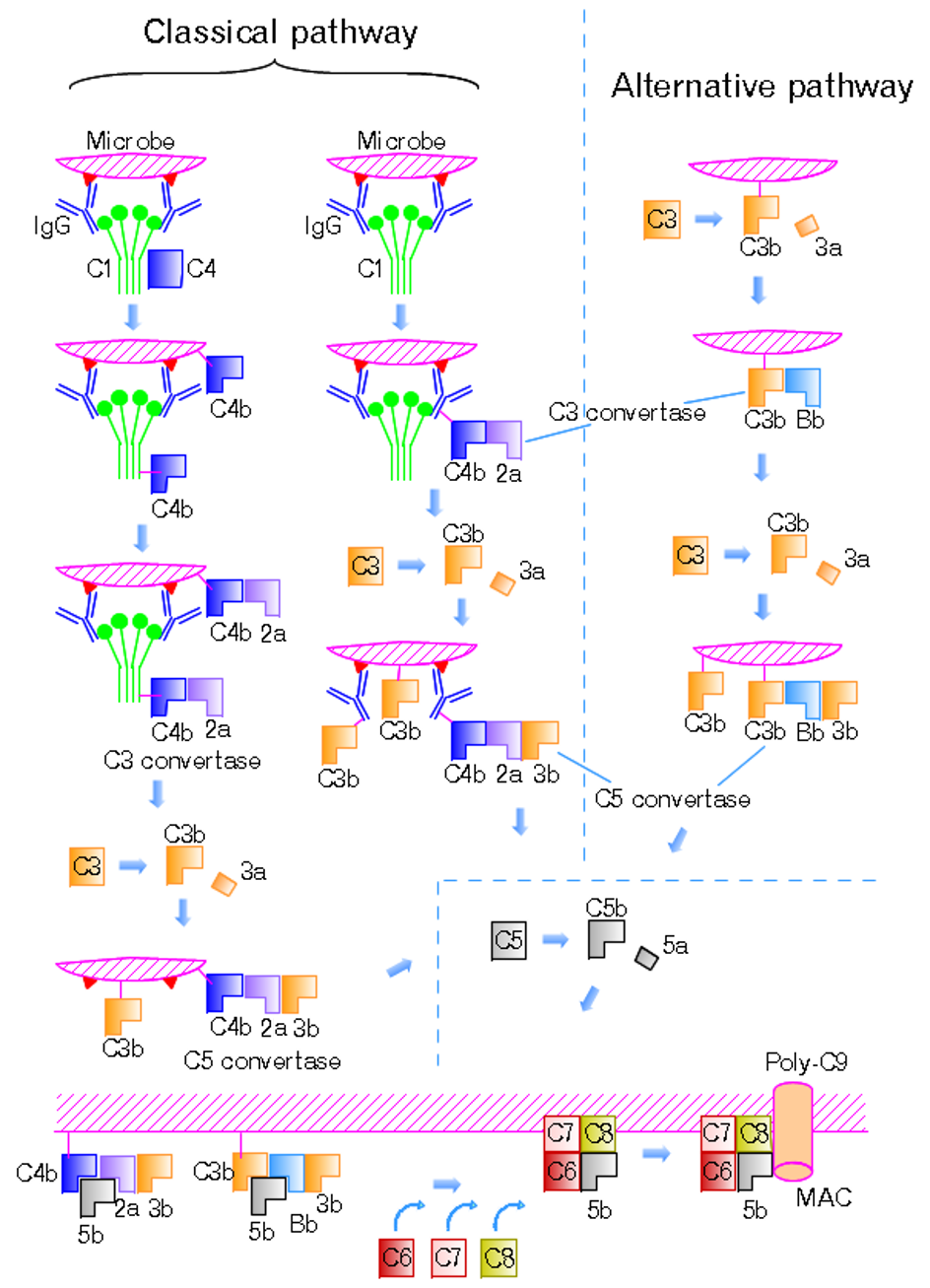
Tossh_eng. (2009). The classical and the alternative pathways with the late steps of complement activation [Image]. Wikimedia Commons. Retrieved December 3, 2024, from https://upload.wikimedia.org/wikipedia/commons/3/3a/Complement_pathways.png
-
Betts, J. G. et al. (2022). Figure 21.15 Alpha-beta T Cell Receptor. In Anatomy and Physiology 2e. OpenStax. https://openstax.org/apps/archive/20241024.164013/resources/4dfaa6804787ee7e22b513e991566e17093e41c4
-
Betts, J. G. et al. (2022). Figure 21.16 Antigenic Determinants. In Anatomy and Physiology 2e. OpenStax. https://openstax.org/apps/archive/20241024.164013/resources/b6d7be3aad087e0ebc2e4dc4dac94d929f73d94f
-
Betts, J. G. et al. (2022). Figure 21.17 Antigen Processing and Presentation. In Anatomy and Physiology 2e. OpenStax. https://openstax.org/apps/archive/20241024.164013/resources/80b14df7f466e9e7885f14546af68a5ad9a01142
-
Betts, J. G. et al. (2022). Figure 21.18 Differentiation of T Cells within the Thymus. In Anatomy and Physiology 2e. OpenStax. https://openstax.org/apps/archive/20241024.164013/resources/8f443571840bcdef5dc1b7e8d134a695a3c822a0
-
Rehua. (2012). T lymphocyte activation pathway [Image]. Wikimedia Commons. Retrieved December 3, 2024, from https://upload.wikimedia.org/wikipedia/commons/d/db/T_cell_activation.svg
-
Betts, J. G. et al. (2022). Figure 21.20 Pathogen Presentation. In Anatomy and Physiology 2e. OpenStax. https://openstax.org/apps/archive/20241024.164013/resources/5594300aedbab54ef46ecbd5e0d2660b3fba0d1e
-
Betts, J. G. et al. (2022). Figure 21.19 Clonal Selection and Expansion of T Lymphocytes. In Anatomy and Physiology 2e. OpenStax. https://openstax.org/apps/archive/20241024.164013/resources/1a4e976aaf6bdb5525663486eee9c8b1912ef8b6
-
Guerin, L. (2022). How a killer T cell destroys a cell infected with viruses [Image]. CK-12 Foundation. Retrieved December 3, 2024, from https://upload.wikimedia.org/wikipedia/commons/d/de/How_a_killer_T_cell_destroys_a_cell_infected_with_viruses.png
-
OpenStax College. (2013). Four chain structure of a generic antibody-IgG2 structures [Image]. In Anatomy & Physiology. Connexions. Retrieved December 3, 2024, from https://upload.wikimedia.org/wikipedia/commons/b/ba/2220_Four_Chain_Structure_of_a_Generic_Antibody-IgG2_Structures.jpg
-
Ciar. (2007). Class switch recombination [Image]. Wikimedia Commons. Retrieved December 3, 2024, from https://upload.wikimedia.org/wikipedia/commons/b/b3/Class_switch_recombination.png
-
Owen, J. A., Punt, J., & Stanford, S. (2019). Kuby immunology (8th ed.). W. H. Freeman.
-
Betts, J. G. et al. (2022). Figure 21.22 Five Classes of Antibodies. In Anatomy and Physiology 2e. OpenStax. https://openstax.org/apps/archive/20241024.164013/resources/fd92be58dabc00d374a8bd080cd5aae550f23505
-
Betts, J. G. et al. (2022). Figure 21.25 T and B Cell Binding. In Anatomy and Physiology 2e. OpenStax. https://openstax.org/apps/archive/20241024.164013/resources/03d40286081fd051a4e971c8ca4911467e6e7176
-
Betts, J. G. et al. (2022). Figure 21.23 Clonal Selection of B Cells. In Anatomy and Physiology 2e. OpenStax. https://openstax.org/apps/archive/20241024.164013/resources/05e3c5fcb2a46e4b7b2ba915fd41360e5d69eb22
-
Boone, B. (2013). A diagram of antibody receptor binding to ligand through processes of opsonization, agglutination, neutralization, and activation of complement [Image]. Wikimedia Commons. Retrieved December 3, 2024, from https://upload.wikimedia.org/wikipedia/commons/e/e4/Antibody_actions.jpg
-
Betts, J. G. et al. (2022). Figure 21.24 Primary and Secondary Antibody Responses. In Anatomy and Physiology 2e. OpenStax. https://openstax.org/apps/archive/20241024.164013/resources/3a6573e580de252fcb13565e3c1ab68526c7f074




14.8: References is shared under a CC BY-NC-SA license and was authored, remixed, and/or curated by LibreTexts.
The book could not be loaded just now.