Opening the book…
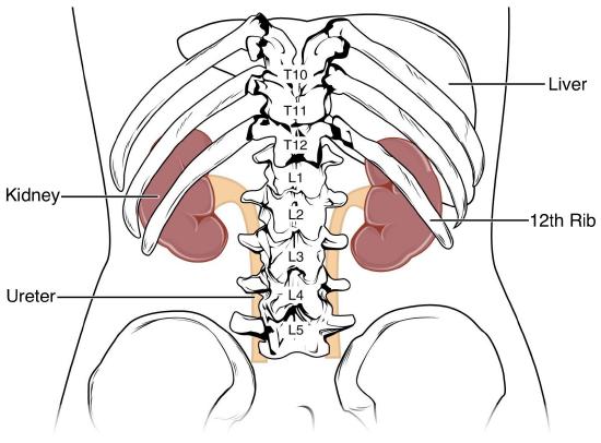
Figure 1 | The Kidneys | 1 An illustration of the posterior torso highlighting the position of the kidneys in relation to the vertebral column (T10-L2) and the 12th rib.
The kidneys are a pair of reddish-brown, bean-shaped organs. They are positioned behind the peritoneum, referred to as a retroperitoneal location. Although similar in function, they exhibit subtle variations in placement: the left kidney resides near the T12 to L3 vertebrae (Figure 1). In contrast, the right kidney sits somewhat lower due to its proximity to the liver. Each kidney weighs in the range of 125–175 grams for males and 115–155 grams for females. Their dimensions measure approximately 11–14 centimeters in length, 6 centimeters in width, and 4 centimeters in thickness.
A sturdy, fibrous tissue layer known as the renal capsule encapsulates each kidney, providing structural support and safeguarding the internal components. Adrenal glands are situated in the superior part of each kidney. The renal capsule encloses the adrenal gland along with the kidney on each side. Cushioning the kidneys is a layer of adipose tissue, which acts as a shock absorber, shielding them from harm.
This page titled 11.3: External Anatomy of the Kidney was last modified on Mon, 15 Jun 2026 23:38:28 GMT and is shared under a CC BY-NC-SA 4.0 license and was authored, remixed, and/or curated by Chaya Gopalan (Consortium of Academic and Research Libraries in Illinois (CARLI)) .
The book could not be loaded just now.