Opening the book…
State the difference between eukaryotic flagella and cilia. Briefly describe and state the function of eukaryotic flagella and cilia.
Flagellar arrangement schemes
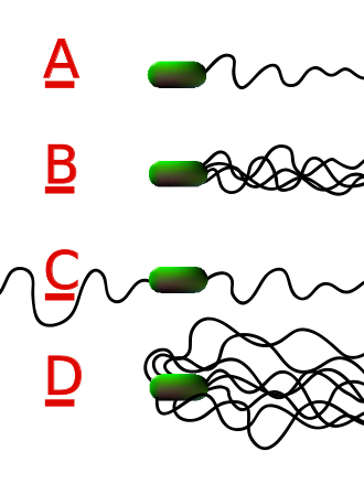
Different species of bacteria have different numbers and arrangements of flagella (Figure \(\PageIndex{7}\).7.1).
- Monotrichous bacteria have a single flagellum (e.g., Vibrio cholerae).
- Lophotrichous bacteria have multiple flagella located at the same spot on the bacteria's surfaces which act in concert to drive the bacteria in a single direction. In many cases, the bases of multiple flagella are surrounded by a specialized region of the cell membrane, the so-called polar organelle.
- Amphitrichous bacteria have a single flagellum on each of two opposite ends (only one flagellum operates at a time, allowing the bacteria to reverse course rapidly by switching which flagellum is active).
- Peritrichous bacteria have flagella projecting in all directions (e.g., E. coli).
Internal Structure
Flagella are long and few in number whereas cilia are short and numerous. Both consist of 9 fused pairs of protein microtubules with side arms of the motor molecule dynein that originate from a centriole. These form a ring around an inner central pair of microtubules that arise from a plate near the cell surface (Figure \(\PageIndex{7}\).2). The arrangement of microtubules is known as a 2X9+2 arrangement. This complex of microtubules is surrounded by a sheath continuous with the cytoplasmic membrane.
Summary
- Flagella are long and few in number whereas cilia are short and numerous.
- Both flagella and cilia consist of 9 fused pairs of protein microtubules with side arms of the motor molecule dynein that originate from a centriole. These form a ring around an inner central pair of microtubules that arise from a plate near the cell surface. This complex of microtubules is surrounded by a sheath continuous with the cytoplasmic membrane.
- Flagella and cilia function in locomotion. Cilia also function to move various materials that may surround a cell.
This page titled 7.7: Flagella and Cilia was last modified on Thu, 31 Aug 2023 18:55:06 GMT and is shared under a CC BY 4.0 license and was authored, remixed, and/or curated by Gary Kaiser via source content that was edited to the style and standards of the LibreTexts platform.
The book could not be loaded just now.