Opening the book…
Non-Vascular
Nonvascular plants, as their name would imply, lack the lignified vascular transport tissues found in higher plants. This lack of transport tissue places considerable constraints on these plants. Their size is limited by the need for all water and nutrients to diffuse from cell to cell. In general, the efficiency of cell-to-cell transport drop significantly a distance greater than five cells. Since a cell greater than five cells from a water source suffers from this limited transport potential, the plants stay small and close to the ground or water source.
The locations where they can survive are also limited. The plant body must be in contact with wet soil to prevent tissue desiccation. Their life cycle is also dependent on water being plentiful. The sperm of the gametophyte must swim to the egg for fertilization to occur. Without adequate water, the next generation would not be produced.
Another characteristic of the nonvascular plants is that they have a dominant gametophyte generation with a dependent sporophyte. This means that within the alternation of generation life cycle the haploid life form is typically larger and is present throughout the two-stage life cycle, while the sporophyte is only occasionally present and is completely dependent on the gametophyte for resources. For example, take the common moss – the green carpet like form that you often see is the haploid gametophyte generation (Fig. 2-14). The sporophyte only appears under certain conditions and is typical not green at maturity and does not contribute to the photosynthetic productivity of the moss. It depends on the gametophyte for water and carbohydrates. The sporophyte is in fact “rooted” into the gametophytic tissues.

There are three extant phyla of nonvascular plants: liverworts, hornworts, and mosses.
True mosses belong to the phyla Bryophyta (not to be confused with bryophytes – a generic term used to collectively refer to all nonvascular plants). There are approximately 12,000 species. We should point out that the use of the “moss” can be misleading. Many plants that are commonly called mosses are not really mosses. For example, clubmoss, spike moss, deer moss, and Spanish moss are not true mosses. (Spanish moss for example is a flowering plant, kin to the pineapple). Just another example of why common names can be problematic.
When you look at a true moss, the “grass” green carpet is the gametophyte stage. It is haploid and produces games via mitosis (as mentioned earlier). The gametophyte is responsible for the photosynthetic productivity for the plant and is the part in contact with the water supply.
In Fig. 2-14, you can see the reddish stalk-like structure with the green tip growing out of the top of the gametophytes. These represent the sporophyte stage. They are not photosynthetic and consist of a foot (unseen here) that anchors the sporophyte into the gametophyte’s tissues, a seta that elevated the capsule for better spore release, and the capsule where the spores are produced via meiosis.
Most mosses are short, typically 0.2-10 cm, but there are a few larger ones such as Dawsonia, the tallest moss in the world that can grow up to 50 cm in height. The larger species can overcome the diffusion limitations because they have two types of transport tissue (Fig. 2-15). These tissues are fundamentally different than true vascular tissues in that they lack lignin, but their transport function is similar. Within the axis of the moss, hydroids help with water transport while leptoids move carbohydrates. Their lack of lignin however makes them less rigid than true vascular tissue and thus does not provide much in the way of structural support.

Marchantiophyta is the liverworts phylum. Like the mosses they have a gametophyte-dominant life cycle. There are about 9,000 species. The various species take one of two forms and are known as either thallus liverworts or leafy liverworts. The thallus form grows like a flattened, leafless ribbon while the leafy species look like flattened mosses. They are small, usually 2-20 mm wide and less than 10 cm long.

The sporophyte is hidden on the bottom side of a gametophore and has the same structure as the moss sporophyte but with a reduced seta (Fig. 2-16). The first photomicrograph shows a cross section through an archegonia containing gametophore, with the A line pointing to a single archegonium (gametangium producing an egg). If the egg gets fertilized by a sperm, it will develop into the sporophyte (seen in the second photomicrograph). The capsule is note by the line of letter B.
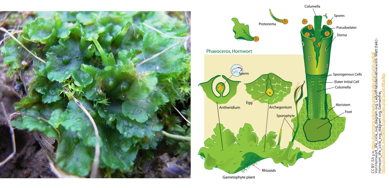
Anthocerophyta is the hornworts phylum and the sporophyte for this phylum is more advanced than that seen in the other nonvascular phyla (Fig. 2-17). At the base of the sporophyte is a meristem that allows the sporophyte to elongate indefinitely. It also produces pseudoelaters to help with spore dispersal. Although the sporophyte is photosynthetic, it is still dependent on the gametophyte. All research attempts at excising the sporophyte have failed.
Seedless Vascular Plants
The development of vascular tissue allowed for taller plant growth due to increased transport efficiency and structural support. These benefits allowed vascular plants to out compete the nonvascular plants. The taller growth of these plants allowed them to intercept sunlight, shading out the nonvascular plant in some cases. Along with the development of vascular tissues can the formation of leaves and roots. Roots helped the vascular plants exploit areas where water was accessible by roots but not necessarily at the surface. Thus, drier environment could be colonized.

All vascular plants have dominant sporophyte lifecycles. In the seedless vascular plants, the gametophyte and the sporophyte are independent of each other although the gametophyte is reduced (Fig. 2-18). In the photograph, the sporophyte of a fern can be seen (Bird’s Nest fern). In the photomicrograph on the right, the gametophyte of a fern is seen in the background while a new forming sporophyte emerges from the gametophyte archegonia.
As the name implies, vascular plants have vascular tissues. The transport of water and dissolved minerals is carried out by the Xylem tissue which includes dead transport cells that are lignified. Carbohydrates and other products of photosynthesis are distributed by the living transport cells of the phloem. Xylem tissue and phloem tissues are often found together in columns called vascular bundles. The arrangement of tissues within the bundles and of the bundles themselves varies with the type of plant and the organ being examined.

Only with the development of vascular tissues could true roots and leaves evolved. Branching of the plant axis occurred only after evolution of vascular tissues. With the branching of the vascular tissue came the ability to produce networks of roots and extensions of the stem that could form leaves. Two types of leaves developed – microphylls and megaphylls (Fig. 2-20). Microphylls (as seen in the first photograph) are small leaves with a single vascular bundle. Megaphylls (2nd photograph) are formed from webbing across numerous branches and thus multiple vascular bundles.

The vascular plants are divided into two groups: the seedless vascular plant and the seeded vascular plants. For the remainder of this segment, we will look at the seedless vascular plants. There are two groups of seedless vascular plants. The lycophytes (1300 species) and the monilophytes (9,000 species).
The lycophytes include the club and spike mosses as well as the quillworts. They differ from all other vascular plants by having microphylls instead of megaphylls. Lycopodium clavatum is an example of a club moss (Fig. 2-21). The sporangia of the club mosses are borne on the upper surface of specialized leaves arranged in a cone-like strobilus (plural: strobili) at the end of upright stems.

Monilophytes include the ferns, horsetails and whisk ferns; they are often referred to as the “ferns and fern allies.” These vascular plants reproduce via spores and have neither seeds nor flowers. They have branched stems and megaphylls. The megaphylls or fronds arise from a rhizome.
Some ferns have fronds that can become sporophylls (spore bearing leaves) as the plant matures while other ferns have specialized fronds, known as fertile fronds, which are responsible for housing the sporangia (Fig. 2-22). In Osmundastrum cinnamomeum (the cinnamon fern) both sterile and fertile fronds are produced. It is the rusty-brown color of the fertile fronds that give the organism its names.

In the ferns with non-specialized sporophylls, sori are often produced on the back side of the frond. Sori are clusters of sporangia that produce spores for asexual reproduction. Some sori are protected by a covering called an indusium which gives them a shiny appearance. The arrangement of sori is often used to identify/classify various ferns (Fig. 2-23).
The first image is a photograph of the sori on Athyrium filix-femina. The photomicrograph in the middle is of a cross section through a sori while the last image, is a close-up photomicrograph of the sporangia of the sori. Note the spores within the walled structure.

This page titled 2.3: Non-Vascular and Seedless-Vascular Plants was last modified on Thu, 12 Nov 2026 18:40:28 GMT and is shared under a CC BY-NC-SA 4.0 license and was authored, remixed, and/or curated by Stephanie Harvey.
The book could not be loaded just now.