Opening the book…
While there are many similarities in the DNA replication between bacteria and eukaryotes, the structural differences in the chromosomes necessitate some differences in the replication process. Bacterial DNA replication is one of the most thoroughly characterized processes in molecular biology, serving as a model for understanding replication in all organisms. Using the model bacterium Escherichia coli (E. coli) as our primary guide, this section tracks the complete life cycle of bacterial replication.
Replication can be divided into three phases: initiation, elongation, and termination. As stated in the previous section, E. coli has a single origin of replication (oriC) on its one chromosome, as do most prokaryotes. The oriC in E. coli is approximately 245 base pairs long and is rich in AT sequences. The transition into the initiation phase happens when the origin is activated. The primary driver of the initiation process is a specialized bacterial protein called DnaA. When metabolic conditions dictate that the cell is ready to divide, ATP-bound DnaA proteins locate and bind tightly to the oriC sequence. As more DnaA molecules accumulate, they bind to one another, oligomerizing into a tight, core-like spiral protein complex. The template DNA wraps around this DnaA core, introducing physical strain on the adjacent double helix. This forces the nearby, A-T rich region to pop open, creating the replication bubble.
Once the A-T rich region is open, helicase must be loaded onto the single-stranded template. Bacteria use two proteins for this. DnaB (helicase) is a ring-shaped moter that uses ATP to unzip the double helix. DnaB is a completely closed ring so it cannot clamp onto an intact strand on its own, so loader protein DnaC (helicase loader) is used as well. DnaC binds to DnaB, using ATP to pry the ring open. The DnaC loader then manually guides the open DnaB ring over the exposed single strands within the replication bubble. Two separate DnaB helicase rings are loaded onto the origin in a bidirectional configuration, with one facing clockwise and the other facing counterclockwise. Once the rings snap shut around the DNA, DnaC dissociates, leaving the functional helicases permanently locked onto their respective target strands.
With the helicases successfully loaded, the replication bubble transforms into two independent replication forks pointing in opposite directions. The DnaB helicases begin ecruiting the rest of the replication machinery to finalize the assembly of the replisome. Bacterial primase (DnaG) attaches directly to the moving helicase ring, preparing to synthesize the first RNA primers. DNA Polymerase III (DNA pol III) are loaded onto the strands via their sliding \(\beta \)-clamps, tethering themselves to the helicase complexes. At this point, the initiation complex is fully converted into a pair of replisomes, and the cell officially transitions from initiation into the elongation phase.
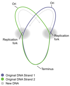
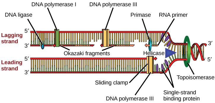
Once the replication forks break away from the oriC origin, the elongation stage begins, where replication occurs simultaneously along both sides of the circular chromosome in bidirectional replication. As the clockwise fork replicates down the right side of the circular chromosome, the counterclockwise fork duplicates the left side at exactly the same speed. Upstream of the synthesis site, the spinning DnaB helicase ring uses ATP to tear through the hydrogen bonds of the double helix, creating positive supercoils ahead of the fork so DNA gyrase (a bacterial Type II topoisomerase) continually rides ahead of the replisome, cutting both DNA strands, untwisting the overwound knots, and sealing the backbone back together.
Single stranded binding proteins coat the exposed single-stranded DNA templates, keeping them straight, accessible, and safe . One DNA Polymerase III tracks directly behind the helicase, continuously adding dNTPs to the \(3^{\prime }\) end of the leading strand. Concurrently, a second DNA Polymerase III synthesizes the lagging strand backward via the trombone model (Replication Fork Video below). In this configuration, primase (DnaG) periodically places an RNA primer near the fork crux, and the polymerase extends it into a 1,000 to 2,000 nucleotide Okazaki fragment before looping out, releasing the template, and resetting for the next cycle.
Because the lagging strand is made of alternating segments of RNA and DNA separated by physical gaps, it must edited before the replication cycle can conclude. In bacteria, this process is streamlined, relying on DNA Polymerase I and DNA Ligase. When DNA Polymerase III completes an Okazaki fragment, it dissociates when encountering the upstream primer of the previous fragment. DNA Polymerase I (Pol I) docks at this single-stranded nick. Using its unique \(5^\prime \rightarrow 3^\prime\) exonuclease activity, Pol I degrades the RNA primer directly ahead of it. Simultaneously, using its internal polymerase domain, it extends the \(3^{\prime }\) end of the new fragment, replacing every excised RNA nucleotide with a DNA nucleotide. Once Pol I has fully replaced the primer, it dissociates, leaving behind a filled gap but a structural break in the sugar-phosphate backbone. DNA Ligase forms a permanent, covalent phosphodiester bond between the adjacent \(3^\prime\text{-OH}\) and \(5^\prime\text{-phosphate}\) groups, connecting the fragments into an unbroken, continuous daughter strand. Bacteria also use another polymerase, DNA polymerase II, but it is primarily used for DNA repair.
Because bacterial chromosomes are closed circles, the two replication forks cannot simply run off the structural ends of the DNA. Instead, they must meet safely on the opposite side of the chromosome to cleanly complete genome duplication without damaging the molecule. If the clockwise and counterclockwise forks traveled at random speeds, they might collide unpredictably, potentially causing the replication machinery to stall, collapse, or break the chromosome. To prevent this, E. coli utilizes a specialized trap system consisting of Ter sites and Tus proteins. Ter sites are a cluster of short, directional DNA sequences (labeled TerA through TerJ) located roughly opposite to the oriC origin, while the Tus protein is a specialized replication termination protein that binds to these sequences.
The Tus-Ter complex acts as a one-way gate for advancing machinery. The complex has a permissive face and a non-permissive face. If an advancing replication fork arrives tracking in the proper direction, it hits the permissive face of the Tus-Ter complex; this orientation allows the fork to smoothly pass through, displacing the Tus protein and continuing its synthesis. However, if a fork arrives from the opposite direction, it hits the non-permissive face and induces a conformational change that locks the Tus protein tightly to the DNA backbone, trapping the replication fork. Because the Ter sites are arranged in two opposing sets of gates, they allow the two forks to enter the termination zone but prevent them from leaving it. Once the two forks are stopped at their meeting point, the remaining nucleotides are filled, the replisomes disassemble, and the two daughter chromosomes are completed.
Once the chromosome has been completely replicated, the two DNA copies move into two different cells during cell division. The process of DNA replication can be summarized as follows:
- DNA unwinds at the origin of replication.
- Helicase opens up the DNA-forming replication forks; these are extended in both directions.
- Single-strand binding proteins coat the DNA around the replication fork to prevent rewinding of the DNA.
- Topoisomerase binds at the region ahead of the replication fork to prevent supercoiling (over-winding).
- Primase synthesizes RNA primers complementary to the DNA strand.
- DNA polymerase III starts adding nucleotides to the 3′-OH (sugar) end of the primer.
- Elongation of both the lagging and the leading strand continues.
- RNA primers are removed and gaps are filled with DNA by DNA pol I.
- The gaps between the DNA fragments are sealed by DNA ligase.
Key Terms
oriC
The primary origin of replication in E. coli.
Initiation
The stage of replication during which replication machinery assembles and DNA unwinding begins.
Elongation
The stage during which new DNA strands are synthesized.
Termination
The stage during which replication forks meet and DNA synthesis is completed.
Bidirectional Replication
Replication proceeding in two directions from an origin.
Ter Sites / Tus Protein
Directional trap system responsible for safely catching and halting converging replication forks at the termination zone of a circular chromosome.
Check Your Understanding
- What is oriC?
- Why does bacterial replication begin at a specific origin?
- Why is the presence of an A-T rich region at the oriC locus evolutionary advantageous for the initiation of replication?
- What proteins participate in initiation?
- Why are primers required during replication?
- What happens during termination?
- What would occur within an E. coli cell during division if the Ter sites were completely deleted from its chromosome? Would replication still conclude? Why or why not?
References
Unless otherwise noted, images on this page are licensed under CC-BY 4.0 by OpenStax.
OpenStax, Concepts of Biology. OpenStax CNX. May 18, 2016 http://cnx.org/contents/s8Hh0oOc@9.10:2ousESf0@5/DNA-Replication
This page titled 1.2.3: DNA Replication in Bacteria was last modified on Thu, 20 Aug 2026 01:22:12 GMT and is shared under a CC BY 4.0 license and was authored, remixed, and/or curated by OpenStax.
The book could not be loaded just now.