From LibreTexts, under CC BY 4.0, as the source states it; its equations are written in TeX, which the PDF shows typeset. This page at LibreTexts.
6.1.2: Survey of Clinically Significant Yeasts
Opening the book…
Candida spp.
Disease: Candidiasis Other Information:
Most common species associated with infection is Candida albicans
Normally reside in the intestinal tract and mucous membranes without causing infection
Opportunistic pathogen that produces symptoms when overgrowth occurs
Candidiasis in the oral cavity or throat is referred to as “thrush,” while when in the vagina it is referred to as a “yeast infection”
Candida auris
Prior to ~2009 had not been reported to be able to grow at human body temperature; it has adapted to the human body temperature
Now reported to cause severe illness in hospitalized patients; with numbers of infections increasing annually
Does not respond to commonly used anti-fungal medications
“Candida albicans, magnification of 1200X.” Source: CDC/ Dr. Lucille K. Georg, CDC PHIL, Public Domain
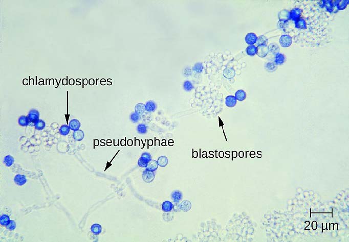
Candida albicans. Source: OpenStax CNX, Wikimedia Commons, CC BY 4.0
This image depicts a condition known as oral thrush, which manifested as a result of a Candida albicans infection. Note the yellowish-beige colored oral ulcers known as aphthae. Source: CDC, CDC PHIL, Public Domain
“This image depicted a frontal view of a Petri dish culture plate, which contained an unknown growth medium, that had been inoculated with a strain of Candida auris fungal organisms.” Source: Shawn Lockhart, CDC PHIL, Public Domain
“Candida auris cases reported prior to 2019 are categorized by the state where the specimen was collected. Most probable cases were identified when laboratories with current cases of C. auris reviewed past microbiology records for C. auris. Source: CDC, Wikimedia Commons, Public Domain
Cryptococcus neoformans
Transmission: Inhalation of fungal spores in the environment Disease: Cryptococcosis
Other Information:
Most infections occur in immunocompromised individuals, especially individuals with HIV/AIDS
This fungus often found in soils where bird droppings are present
Infection from inhalation leads to pneumonia, the fungus may spread to cause meningitis
At one time infections were primarily associated with individuals who worked closely with poultry
“This photomicrograph revealed some of the ultrastructural morphology exhibited by the fungal organism, Cryptococcus neoformans, one of the causes for the infection known as cryptococcosis.” Credit: CDC/ Dr. Leanor Haley, CDC PHIL, Public Domain
“This photomicrograph of a methenamine stained lung tissue specimen, was harvested from an acquired immunodeficiency syndrome (AIDS) patient with cryptococcosis. Histopathology reveals numerous extracellular Cryptococcus neoformans yeast form organisms within the alveolar space. Some of the yeast organisms showed narrow base budding, and characteristic variation in size.” Source: CDC/ Dr. Edwin P. Ewing, Jr., CDC PHIL, Public Domain
“This image depicted two test tube slant cultures, which contained an unidentified growth medium that had been inoculated with the fungal organism, Cryptococcus neoformans, which had been cultured from a sample, harvested from a cat. The colonial growth reflected the characteristic colonial morphology exhibited by C. neoformans, which included a creamy coloration, smooth surface, and mucoid appearance.” (Right) Source: CDC/ Dr. Libero Ajello, CDC PHIL, Public Domain
Pneumocystis jirovecii
Disease: Pneumocystic pneumonia Other Information:
Part of normal flora
Opportunistic pathogen
Prior to the AIDS epidemic cases were only observes in malnourished, premature infants and debilitated elderly patients; now it is also associated with AIDS patients
“Under a magnification of 1150X, this photomicrograph of a silver stained human lung tissue specimen, revealed histopathology encountered due to a case of Pneumocystis pneumonia (PCP), caused by the fungus Pneumocystis jirovecii, formerly known as P. carinii. In this view, numerous darkly-stained cysts were plainly visible, while intracystic bodies were not visible.” Source: CDC/ Lois Norman, M.S., CDC PHIL, Public Domain
This anteroposterior (AP) chest x-ray revealed radiologic evidence of pulmonary pneumocystosis in the form of bilateral pulmonary interstitial infiltrates. This infection was due to the presence of an opportunistic fungal infection, by the fungal organism Pneumocystis jirovecii, formerly known as Pneumocystis carinii. (This image was provided by Jonathan W.M. Gold, MD, Assoc. Dir. Special Microbiology Lab, Assist. Attending Physician, Memorial Sloan-Kettering Cancer Center and Assist. Prof. of Medicine, Cornell Univ. Med. College, New York). Source: CDC/ Jonathan W.M. Gold, MD, CDC PHIL, Public Domain