Opening the book…
A critical clue to understanding nucleic acid structure came from the work of Erwin Chargaff (1905-2002). When analyzing DNA from various sources, he found that the relative amounts of G, C, T and A nucleotides present varied between organisms but were very similar for organisms of the same type or species. On the other hand, the ratios of A to T and G to C were always equal to 1, no matter where the DNA came from. Knowing these rules, James Watson (1928-) and Francis Crick (1916-2004) built a model of DNA that used structural data from Rosalind Franklin (1920-1958) and fit what was known about the structure of nucleotides. Franklin got her data by pulling DNA molecules into oriented strands; fibers of many molecules aligned parallel to one another. By passing a beam of X-rays through these fibers she obtained a diffraction pattern; a pattern that defined key structural parameters that constrain any model of the molecule’s structure.[284] By making a model that was predicted to produce the observed X-ray data, Watson and Crick drew a number of conclusions about the structure of DNA molecules.[285]
 To understand their process, consider the chemical nature of a nucleotide and a nucleotide polymer (a nucleic acid) such as DNA. First the nucleotide bases in DNA (A, G, C and T) have a number of similar properties. Each nucleotide (←) has three hydrophilic regions: the negatively charged phosphate group, a sugar with a number of O–H groups, and the bases' hydrophilic edges, where the N–H and N groups lie. While the phosphate and sugar are three-dimensional moieties, the bases are flat, the atoms in the rings are all in one plane. The upper and lower surfaces of the rings are hydrophobic (non-polar) while the edge is hydrophilic. The same amphipathic factors that favor the assembly of lipids into bilayer membranes also influence nucleic acid structure. In their model Watson and Crick had the bases stacked on top of one another, hydrophobic surface next to hydrophobic surface, to reduce their interactions with water.
 Each base’s hydrophilic edge, with –C=O and –N-H groups can act as H-bond acceptors and donors. How were these hydrophilic groups arranged? With the two polynucleotide strands arranged in opposite orientations, that is, anti-parallel to one another: one from 5’ → 3’ and the other 3’ ← 5’; the bases attached to the sugar-phosphate backbone interact with one another in a highly specific way (←). An A can form two hydrogen bonding interactions with a T on the opposite, anti-parallel strand, while a G can form three hydrogen bonding interactions with a C. A key feature of this arrangement is that the lengths of the A::T and G:::C base pairs are almost identical. The hydrophobic surfaces of the bases are stacked on top of each other, while the hydrophilic sugar and phosphate groups are in contact with the surrounding aqueous solution. The repulsion between negatively charged phosphate groups is neutralized (or shielded) by the presence of positively charged ions present in the solution from which the X-ray measurements were made (and in the cell). This model also provided a direct explanation for why Chargaff’s rules are universal in double stranded DNA.
Each DNA polymer strand has a directionality to it, it runs from the 5’ phosphate group of the ribose/deoxyribose at one end to the 3’ hydroxyl group of the ribose/deoxyribose at the other end. Each nucleotide monomer is connected to the next through a phosphodiester linkage (→) involving its 5’ phosphate group attached to the 3’ hydroxyl of the existing strand. In their final model Watson and Crick depicted what is now known as B-form DNA. This is the usual structural form of DNA in a cell. Under different salt conditions DNA can form two other double helical forms, known as A and Z forms. A and B forms of DNA are "right-handed" helices, while the Z-form is a left-handed helix (←). We will not concern ourselves with these other forms of DNA, leaving that to more advanced courses, but you can imagine that they might well influence the types of intermolecular interactions that occur between DNA and other molecules, particularly proteins.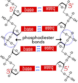
As soon as the Watson-Crick model of DNA structure was proposed its explanatory power was obvious. Because the A::T and G:::C base pairs are of the same length, the sequence of bases along the length of a DNA molecule, written, by convention in the 5’ to 3’ direction, has little effect on the overall three-dimensional structure of the molecule. That implies that essentially any sequence can be found, at least theoretically, in a DNA molecule. If information were encoded in the sequence of nucleotides along a DNA strand that information would be as stable as the DNA molecule itself. This is similar to the storage of information in various computer memory devices, that is, any type of information can be stored, because storage does not involve a dramatic change in the structure of the storage material. The structure of a flash memory drive is not dramatically different whether in contains photos of your friends, a song, a video, or a textbook. What matters is how the information is "encoded", most obviously in the specific sequence of nucleotides along a DNA strand.
At the same time, the double-stranded structure of DNA and the complementary nature of base pairing (A to T and G to C) suggested a simple model for information storage and replication. Pull the two strands of the molecule apart and build two new (anti-parallel) strands using the two original strands as templates. This model of DNA replication is facilitated by the fact that the two strands of the parental DNA molecule are held together by weak hydrogen bonding interactions; no covalent bonds need be broken to separate the strands from one another. In fact, at physiological temperatures DNA molecules  often open up over short stretches and then close again, a process known as DNA breathing.[286] This makes the replication of the information stored in the molecule conceptually straightforward, even though the actual biochemical process is complex, in part because of the importance of accurate replication. The existing strands determine the sequence of nucleotides on the newly synthesized strands. The newly synthesized strand can, in turn, direct the synthesis of a second strand, identical to the original strand. Finally, the double-stranded nature of DNA molecule means that any information within the molecule is stored in a redundant fashion. If one strand is damaged, that is its DNA sequence is lost or altered, the second undamaged strand can be used to repair the damage.
Questions to answer
- How is a DNA molecule structurally analogous to a lipid bilayer? Draw a diagram that reveals the similarities and note the most important differences.
- Which do you think is stronger (and why), an AT or a GC base pair?
- Why is the ratio of A to T the same in all organisms?
- Normally DNA exists inside of cells at physiological salt concentration (~140 mM KCl, 10 mM NaCl, 1 mM MgCl2 and some minor ions). What happens if you placed DNA into pure water?
- How many general types of mutation can you think of? How would they differ in their impact on the information encoded in a DNA molecule.
- Generate a model mechanism by which a DNA molecule could be accurately repaired, that is, without the loss of the information originally present within it.
Questions to ponder
- You isolated DNA from an organism, and you find it fails to obey Chargaff’s rule; what might you predict about the structure of its DNA?
References
[284] Fiber diffraction
[285] An interesting depiction of this process is provided by the movie “Life Story”
[286] Dynamic approach to DNA breathing: http://www.ncbi.nlm.nih.gov/pubmed/23345902
This page titled 7.5: Discovering the structure of DNA was last modified on Thu, 08 Jan 2026 23:36:48 GMT and is shared under a CC BY-NC-SA 4.0 license and was authored, remixed, and/or curated by Michael W. Klymkowsky and Melanie M. Cooper.
The book could not be loaded just now.