Opening the book…
- Define "negative stain" and their purpose in microbiology.
- Understand how negative stains work and stain specimen backgrounds instead of cell structures.
- Discuss key differences between simple and negative stains.
- Understand why negative stains are not prepared with heat fixation.
- Understand why negative stains are used for bacteria with capsules.
- Prepare microscope slides for negative staining with bacterial species whose cell shape and arrangement are unknown.
- Describe cell morphology.
Negative Stains
Similar to simple stains, negative stains are used to increase contrast when observing bacterial cells by microscope. Bacteria will be transparent and difficult to impossible to visualize without the assistance of simple or negative stains. However, simple stains physically stain the cell body using basic dyes. The goal of a negative stain is to protect the natural cell shape by staining the background, the microscope slide, and not the actual bacterial cell.
Negative stains still begin with the preparation of a thin, bacterial smear. These smears should be air dried only, though. A key difference between simple and negative stains is that negative stains do not use heat fixation. Heat fixation has the potential to alter or damage the natural cell shape of bacteria who have capsules. Capsules are optional protective outer layers that cover the cells of select bacterial species, like Bacillus megaterium. They help prevent phagocytosis and desiccation while also increasing cell adhesion and host colonization. Capsules are composed of thick, dense polysaccharide layers that have a neutral, non-ionic charge. This neutral charge does not react with the basic dyes used in simple staining and heat fixation can destroy the capsule, meaning simple stains can not be used to visualize capsule covered bacteria.
Instead, negative stains with acidic dyes are used for visualizing capsules or other cell types that are sensitive to extreme heat. Acidic dyes are composed of negatively charged ions with dye color. These negatively charged acidic dyes are not attracted to the neutral charge of the capsule and are also repelled by the natural negative charge of the cell body. Thus, the acidic dye will stain the surface of the background, typically a glass microscope slide. This stain will leave specimens white or transparent and specimens may appear to have a "halo"-like silhouette surrounding the cell. Common acidic dyes used for negative stains include: Nigrosin, Eosin, and Acid Fuchsin.

Figure 1. This figure illustrates how negative stains with acidic dyes can not bind to the bacteria's body. Bacteria have a natural negative charge that will repeal the negative charge of the acidic dye, causing the background to become stained while the bacteria remain clear/transparent.
Bacterial Cell Morphology
Negative stains can only be used to observe cell shape and arrangement. They can not be used to stain internal cell structures or differentiate between bacterial species. Further, negative stains only require 1 dye, no heat application, and no wash or rinse cycles and can not classified as differential stains. In this course, negative stains will still be used to observe variable coccus and bacillus cell shapes.
|
Simple Stain: Methylene Blue |
Negative Stain: Nigrosin Stain |
Differential Stain: Gram Stain |
|
|
 |
|
| E. coli stained with methylene blue (black arrows) and observed on a bladder epithelial cell. | C. neoformans yeast (red arrows) was stained with Nigrosin. The stain turns the background black while leaving the yeast cells clear/transparent. | Gram Stain mix of gram-negative E. coli (pink) and gram-positive S. aureus (purple). |

Table 1. Simple stains are good for observing overall cell shape and arrangement (cell morphology). Negative stains can also be used to observe cell morphology, but these stains will stain the background instead of the cell body. These stains are good for cells that are sensitive to heat. Differential stains can be used to identify differences in bacterial species, like the gram stain observes differences in the cell wall.
Cryptococcus, the Fungal Infection Diagnosed through Negative Staining
Cryptococcus is a genus of fungi that include two different species: C neoformans and C. gattii. These fungal species can typically be found in the soil and decaying wood up tropical or subtropical environments. However, when inhaled, these fungi have the potential to cause the disease, cryptococcosis, in humans. Disease progression first begins in the lungs, with most patients encountering respiratory ailments. If left untreated, the fungal spores can spread throughout the body, to the brain and spinal cord causing meningitis. The prevalence cryptococcosis tends to be the highest among people with weakened or compromised immune systems and is the most common case of adult meningitis in Sub-Saharan Africa.
A distinctive diagnostic characteristic of C. neoformans is its polysaccharide capsule. This capsule aids in immune evasion, allowing the fungus to survive in the host. Negative staining is a laboratory procedure that allows microbiologist to visualize this capsule. When performing the negative stain procedure, the background of the slide is stained black, while the microorganism's capsule remain unstained, appearing as a white halo around the organism. Identifying this capsule can be a quick way to diagnose a patient with cryptococcosis. When patient is exhibiting cryptococcosis symptoms, cerebrospinal fluid (CSF) samples are taken. Then by using a negative staining technique called India Ink staining, Cryptococcus can be then quickly identified by its large polysaccharide capsule.
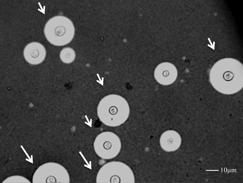
Figure 2. Adapted from Robertson et al. this imaged depicts a negative stain result from a patient's cerebral spinal fluid infected with Cryptococcus neoformans. C. neoformans can be easily identified by its capsule, which appears as a white halo from negative staining.
Attributions
"General Microbiology Lab Manual: Labs 3, Simple, Negative, and Gram Stain" by Nazzy Pakpour and Sharon Horgan, LibreTexts: Biology, Coastal State University, Easy Bay is licensed under CC BY-SA
"Microbiology Textbook: Chapter 2, How We See the Invisible World" by Openstax, Digital ISBN 13: 978-1-947172-23-4 is licensed under CC BY 4.0
"MB352 General Microbiology Laboratory 2021: Staining Techniques 4.3" by Joan Petersen and Susan McLaughlin , LibreTexts: Biology , Queeensborough Community College is licensed under CC BY-NC-SA
"Microbiology Laboratory Manual: Labs, 1.13 Capsule Stain" by Dr. Rosanna Hartline, LibreTexts: Biology, West Hills College Lemoore is licensed under CC BY-NC-SA 4.0
"Cryptococcus neoformans Ex Vivo Capsule Size Is Associated With Intracranial Pressure and Host Immune Response in HIV-associated Cryptococcal Meningitis" by Robertson et al., The Journal of Infectious Diseases, Volume 209, Issue 1, 1 January 2014, Pages 74–82,, Oxford Academic is licensed under CC BY 3.0
"Cryptococcosis: Clinical Overview of Cryptococcosis" by Center for Disease Control, CDC Materials Statement is in the Public Domain
Image Citations
Figure 1, Modified From:
- "General Microbiology Lab Manual: Labs 3, Simple, Negative, and Gram Stain" by Nazzy Pakpour and Sharon Horgan, LibreTexts: Biology, Coastal State University, Easy Bay is licensed under CC BY-SA
Table 1, Modified From:
- "UPEC adhered to BEC" by Stefan Walkowski is licensed under CC BY-SA 4.0.
- "Microbiology Textbook: Chapter 2, How We See the Invisible World" by Openstax, Digital ISBN 13: 978-1-947172-23-4 is licensed under CC BY 4.0
- "Gram stain 01.jpg" by Y tambe, WikiMedia Commons is licensed under CC BY-SA 3.0

Figure 1, Modified From:
- "Figure 2 from 'Cryptococcus neoformans Ex Vivo Capsule Size Is Associated With Intracranial Pressure and Host Immune Response in HIV-associated Cryptococcal Meningitis'" by Robertson et al., The Journal of Infectious Diseases, Volume 209, Issue 1, 1 January 2014, Pages 74–82, Oxford Academic Press is licensed under CC BY 3.0
5.3: Negative Stains is shared under a CC BY 4.0 license and was authored, remixed, and/or curated by LibreTexts.
The book could not be loaded just now.