From LibreTexts, under CC BY-NC-SA 4.0, as the source states it; its equations are written in TeX, which the PDF shows typeset. This page at LibreTexts.
20.6: Anatomical Atlas - Respiratory System
Opening the book…
A compendium of anatomical images: slides, models, and/or cadavers.
Models
Image credit
“Upper Respiratory System Regions" by Jennifer Lange, is licensed underCC BY-NC-SA 4.0.
Answer Key (click to show/hide answers)
A - Nasal bone
B - Nasal cartilage
C - Nose/nares
D - Nasal cavity
E - Pharynx
F - Larynx
G - Frontal sinus
H - Sphenoid sinus
Image credit
“Upper Respiratory System Structures" by Jennifer Lange, is licensed underCC BY-NC-SA 4.0.
Answer Key (click to show/hide answers)
A - Middle nasal concha
B - Inferior nasal concha
C - Superior nasal concha
D - Opening of auditory tube
E - Uvula
F - Hyoid bone
G - Epiglottis
H -Thyroid cartilage
I - Cricoid cartilage
J - Glottis
K - vocal folds/cords
Image credit
“Head Model External - Respriatory Organs Atlas" by Jennifer Lange, is licensed underCC BY-NC-SA 4.0. Labeled by John Polos
Answer Key (click to show/hide answers)
A - Nasal concha
B - Nasal meatus
C - Pharynx
D - Thyrohyoid membrane
E - Thyroid cartilage
F - Laryngeal prominence
Image credit
“Larynx Model- Respiratory Organs Atlas" by John Polos, is licensed underCC BY-NC-SA 4.0.
Answer Key (click to show/hide answers)
A - Hyoid bone
B - Thyroid cartilage
B1 - Laryngeal prominence
C - Epiglottis
D - Cricoid cartilage
E - Tracheal cartilage
F - Annular ligament
G - Vocal folds
H - Corniculate cartilage
I - Arytenoid cartilage
Image credit
“Torso Model - Respiratory Organs Atlas" by John Polos, is licensed underCC BY-NC-SA 4.0.
Answer Key (click to show/hide answers)
A - Larynx (thyroid cartilage)
B - Trachea
C - Primary bronchi (plural)
D - Secondary bronchi (plural)
E - Esophagus
Image credit
“Lung model external - Respiratory Organs Atlas" by Jennifer Lange, is licensed underCC BY-NC-SA 4.0. Labeled by John Polos
Answer Key (click to show/hide answers)
A - Larynx
B - Trachea
C - Apex of left lung
D - Apex of right lung
E - Superior lobe of right lung
F - Superior lobe of left lung
G - Horizontal fissure of right lung
H - Middle lobe of right lung.
I - Oblique fissure of right lung
J - Inferior lobe of right lung
K - Oblique fissure of left lung
L - Inferior lobe of left lung
M - Cardiac Notch of left lung
Image credit
“Lung model, deep - Respiratory Organs Atlas" by Jennifer Lange, is licensed underCC BY-NC-SA 4.0. Labeles by John Polos
Answer Key (click to show/hide answers)
A - Larynx
B - Trachea
C - Right primary bronchus
D - Left primary bronchus
E - Right secondary bronchi
F - Left secondary bronchi
Histology
Image credit
“Nasal cavity histology - Respiratory Organs Atlas" by John Polos, is licensed underCC BY-NC-SA 4.0.
Answer Key (click to show/hide answers)
A - Goblet cell
B - Pseudostratified columnar epithelium
C - Olfactory (Bowman's) glands
D - Artery
E - Vein
F - Bipolar cell nuclei
G - Nerve
H - Mucosal layer
I - Submucosal layer and lamina propria
Image credit
“Trachea histology - Respiratory Organs Atlas" by Jennifer Lange, is licensed underCC BY-NC-SA 4.0.
Answer Key (click to show/hide answers)
A - Submucosal glands/Tracheal glands
B - Mucosal layer/Pseudostratified columnar epithelium
C - Submucosal layer and lamina propria
D - Hyaline cartilage
E - Adventitia
Image credit
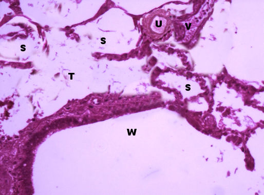
“Lung histology - Digestive Organs Atlas" by John Polos, is licensed underCC BY-NC-SA 4.0.
Answer Key (click to show/hide answers)
S - Alveoli
T - Alveolar duct
U - Small artery
V - Small vein
W - Respiratory bronchus
Cadaver
To view a larger version, right click (PC) or command click (Mac) and click open in new tab.
Image credit
“ Anterior Neck" provided by the regents of the University of Michigan and shared under CC 4.0 . Labeled by John Polos.
Answer Key (click to show/hide answers)
A - Hyoid bone
B - Thyrohyoid membrane
C - Thyroid cartilage
D - Laryngeal prominence
E - Cricoid cartilage
F - Trachea
Image credit
“ Posterior larynx" provided by the regents of the University of Michigan and shared under CC 4.0 . Labeled by John Polos.
Answer Key (click to show/hide answers)
A - Epiglottis
B - Thyroid cartilage/thyroid lamina
C - Cricoid cartilage
D - Trachea
Image credit
“ Thoracic cavity" provided by the regents of the University of Michigan and shared under CC 4.0 . Labeled by John Polos.
Answer Key (click to show/hide answers)
A - Larynx
B - Trachea
C - Right primary bronchus
D - Left primary bronchus
E - Right secondary bronchus (inferior)
Image credit
“ Anterior lungs" provided by the regents of the University of Michigan and shared under CC 4.0 . Labeled by John Polos.
Answer Key (click to show/hide answers)
A - Horizontal fissure of right lung
B - Cardiac notch of left lung
C - Lingula of left lung
Image credit
“ Lateral Lungs" provided by the regents of the University of Michigan and shared under CC 4.0 . Labeled by John Polos.
Answer Key (click to show/hide answers)
A - Apex right lung
B - Apex left lung
C - Oblique fissure right lung
D - Oblique fissure left lung
E - Horizontal fissure right lung
F - Base right lung
G - Base left lung