|

- Image Credit
-
"Ear Model - Atlas" by Jennifer Lange is licensed under CC BY-NC-SA 4.0.
|
- Answer Key (click to show/hide answers)
-
A - Vestibulocochlear nerve (CN VIII)
B - cochlea
C - semicircular canals
D - auditory (Eustachian) tube
E - tympanic membrane
F - external auditory canal
G - auricle
|
|
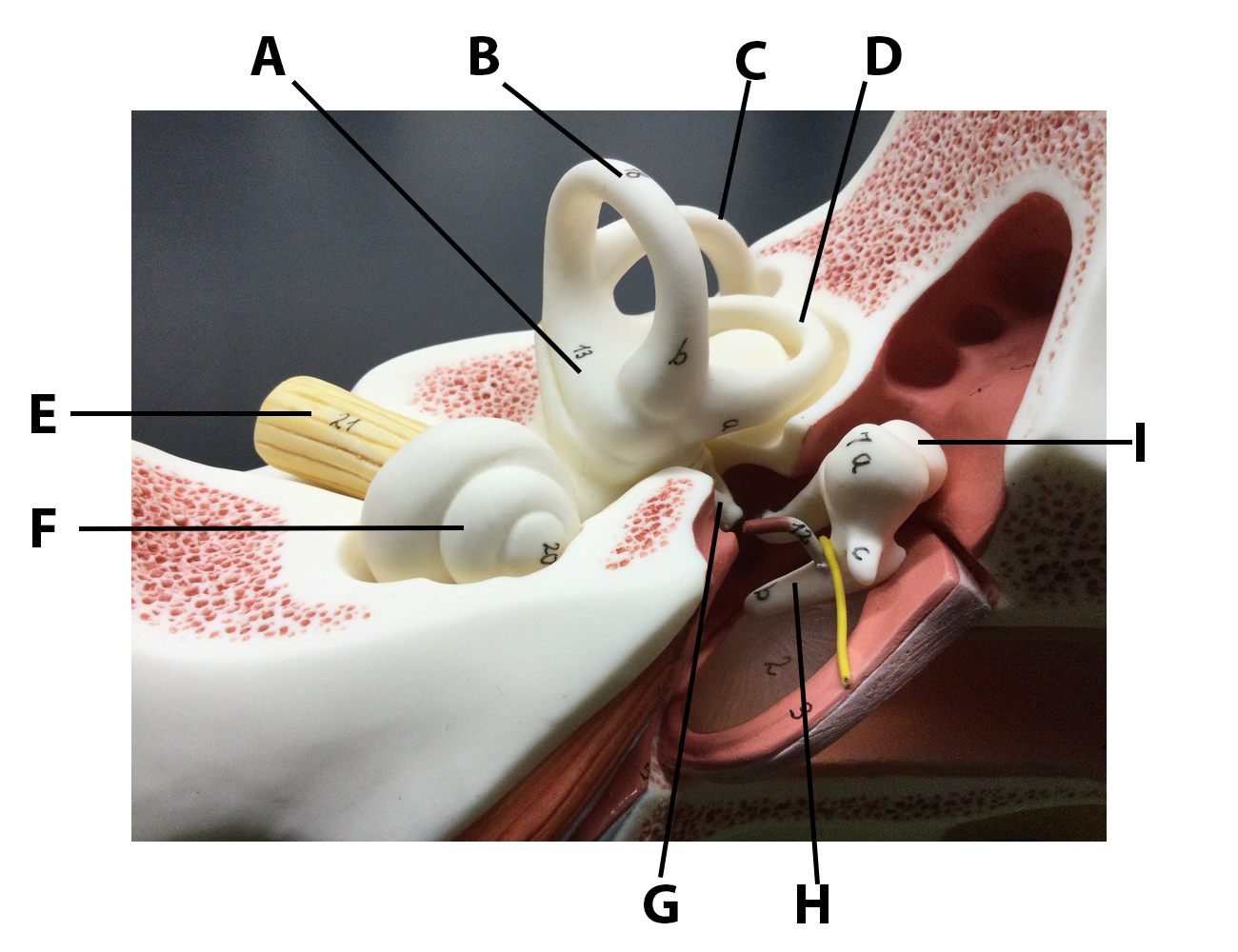
- Image Credit
-
"Middle and Inner Ear Model - Atlas" by Jennifer Lange is licensed under CC BY-NC-SA 4.0.
|
- Answer Key (click to show/hide answers)
-
A - vestibule
B - anterior semicircular canal
C - posterior semicircular canal
D - horizontal semicircular canal
E - CN VIII, vestibulocochlear nerve
F - cochlea
G - stapes
H - malleus
I - incus
|
|

- Image Credit
-
"Inner Ear Model - Atlas" by Jennifer Lange is licensed under CC BY-NC-SA 4.0.
|
- Answer Key (click to show/hide answers)
-
A - cochlea
B - vestibule
C - oval window
D - semicircular canals
|
|

- Image Credit
-
"Cochlear Model - Atlas" by Jennifer Lange is licensed under CC BY-NC-SA 4.0.
|
- Answer Key (click to show/hide answers)
-
A - basilar membrane
B - vestibular membrane
C - vestibular duct
D - cochlear duct
E - tympanic duct
F - tectorial membrane
G - hair cells
H - spiral ganglia
I - cochlear nerve (cochlear branch of CN VIII)
|
|

- Image Credit
-
"Small Inner Ear Model - Atlas" by Jennifer Lange is licensed under CC BY-NC-SA 4.0.
|
- Answer Key (click to show/hide answers)
-
A - semicircular canals
B - stapes
C - vestibule
D - cochlea
E - oval window
|
|

- Image Credit
-
"Cochlear Spiral Model - Atlas" by Jennifer Lange is licensed under CC BY-NC-SA 4.0.
|
- Answer Key (click to show/hide answers)
-
A - vestibular duct
B - vestibular membrane
C - cochlear duct
D - tectorial membrane
E - dendrites of bipolar neurons
F - hair cells
G - basilar membrane
H - tympanic duct
|
|

- Image Credit
-
"Cochlear Histology - Atlas" by Jennifer Lange is licensed under CC BY-NC-SA 4.0, slide provided by the Regents of the University of Michigan Medical School © 2022.
|
- Answer Key (click to show/hide answers)
-
A - vestibular membrane
B - basilar membrane
C - hair cells
D - vestibular duct
E - cochlear duct
F - tympanic duct
G - cochlear branch of vestibulocochlear nerve (CN VIII)
H - spiral ganglion
I - spiral organ (organ of Corti)
J - tectorial membrane
|
|

- Image Credit
-
"Spiral Organ Histology - Atlas" by Jennifer Lange is licensed under CC BY-NC-SA 4.0, slide provided by the Regents of the University of Michigan Medical School © 2022.
|
- Answer Key (click to show/hide answers)
-
A - vestibular duct
B - cochlear duct
C - tympanic duct
D - vestibular membrane
E - tectorial membrane
F - hair cells
G - basilar membrane
|
|

- Image Credit
-
"Eye in Orbit Model Anterior View- Atlas" by Jennifer Lange is licensed under CC BY-NC-SA 4.0.
|
- Answer Key (click to show/hide answers)
-
A - cornea
B - sclera
C - levator palpebrae superioris muscle
D - pupil
E - iris
F - inferior oblique muscle
G - superior oblique muscle
H - medial rectus muscle
|
|

- Image Credit
-
"Eye in Orbit Model Superior Lateral View - Atlas" by Jennifer Lange is licensed under CC BY-NC-SA 4.0.
|
- Answer Key (click to show/hide answers)
-
A - optic nerve; CN II
B - superior oblique muscle
C - medial rectus muscle
D - inferior rectus muscle
E - lateral rectus muscle
F - choroid
G - sclera
H - cornea
|
|
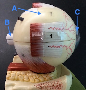
- Image Credit
-
"Lateral View of Eye Model - Atlas" by Jennifer Lange is licensed under CC BY-NC-SA 4.0.
|
- Answer Key (click to show/hide answers)
-
A - sclera
B - optic nerve; CN II
C - cornea
|
|

- Image Credit
-
"Anterior View of Eye Model - Atlas" by Jennifer Lange is licensed under CC BY-NC-SA 4.0.
|
- Answer Key (click to show/hide answers)
-
A - pupil
B - iris
C - inferior rectus
D - sclera
E - cornea
|
|
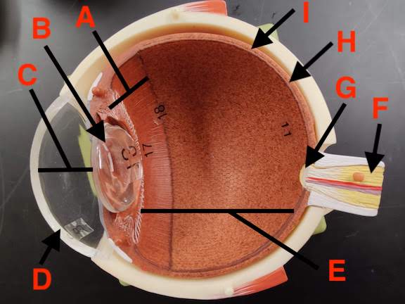
- Image Credit
-
"Interior View of Eye Model - Atlas" by Jennifer Lange is licensed under CC BY-NC-SA 4.0.
|
- Answer Key (click to show/hide answers)
-
A - ciliary body
B - lens
C - anterior cavity
D - cornea
E - posterior cavity
F - optic nerve (CN II)
G - optic disc
H - retina
I - choroid
|
|

- Image Credit
-
"Human Eye Histology - Atlas" by Jennifer Lange is licensed under CC BY-NC-SA 4.0, modification of "eye human", license: CC BY-NC-SA.
|
- Answer Key (click to show/hide answers)
-
A - sclera
B - posterior cavity
C - retina
D - choroid
E - lens
F - iris
G - cornea
H - pupil
I - anterior cavity
J - ciliary muscle (body)
|
|

- Image Credit
-
"Eye Wall Histology - Atlas" by Jennifer Lange is licensed under CC BY-NC-SA 4.0, slide provided by the Regents of the University of Michigan Medical School © 2022.
|
- Answer Key (click to show/hide answers)
-
A - sclera
B - choroid
C - retina
D - neural retina
E - photoreceptor layer - rods and cones
F - bipolar cell layer
G - ganglion cell layer
H - pigmented epithelium
I - posterior cavity
|
|

- Image Credit
-
"Retina Model - Atlas" by Jennifer Lange is licensed under CC BY-NC-SA 4.0.
|
- Answer Key (click to show/hide answers)
-
A - ganglion cell layer
B - bipolar cell layer
C - photoreceptor layer
D - pigmented epithelium
E - choroid
F - sclera
G - rod
H - cone
|
|

- Image Credit
-
"Cow Eye Dissected - Exterior - Atlas" by Jennifer Lange is licensed under CC BY-NC-SA 4.0.
|
- Answer Key (click to show/hide answers)
-
A - cornea
B - sclera
C - optic nerve (CN II)
|
|

- Image Credit
-
"Cow Eye Dissected - Interior, Anterior - Atlas" by Jennifer Lange is licensed under CC BY-NC-SA 4.0.
|
- Answer Key (click to show/hide answers)
-
A - sclera
B - iris
C - ciliary body
D - cornea
E - pupil
|
|

- Image Credit
-
"Cow Eye Dissected - Interior, Posterior - Atlas" by Jennifer Lange is licensed under CC BY-NC-SA 4.0.
|
- Answer Key (click to show/hide answers)
-
A - optic disc
B - sclera
C - tapetum lucidum
D - retina
E - choroid
|