|

- Image credit
-
“Head Model Midline - Digestive Organs Atlas" by Jennifer Lange, is licensed under CC BY-NC-SA 4.0.
|
- Answer Key (click to show/hide answers)
-
A - pharynx
B - oral cavity
C - teeth
D - tongue
E - esophagus
|
|

- Image credit
-
“Head Model Lateral - Digestive Organs Atlas" by Jennifer Lange, is licensed under CC BY-NC-SA 4.0.
|
- Answer Key (click to show/hide answers)
-
A - parotid gland
|
|

- Image credit
-
“Torso Model Head and Neck - Digestive Organs Atlas" by Jennifer Lange, is licensed under CC BY-NC-SA 4.0.
|
- Answer Key (click to show/hide answers)
-
A - pharynx
B - oral cavity
C - teeth
D - tongue
E - sublingual gland
F - submandibular gland
|
|

- Image credit
-
“Stomach and Surrounding Organs Model - Atlas" by Jennifer Lange, is licensed under CC BY-NC-SA 4.0.
|
- Answer Key (click to show/hide answers)
-
A - liver
B - gallbladder
C - pancreas
D - esophagus
E - inner oblique layer of muscularis
F - middle circular layer of muscularis
G - outer longitudinal layer of muscularis
H - serosa
|
|

- Image credit
-
“Stomach Model Close-up - Atlas" by Jennifer Lange, is licensed under CC BY-NC-SA 4.0.
|
- Answer Key (click to show/hide answers)
-
A - lower esophageal sphincter
B - gastric folds (rugae)
C - submucosa
D - muscularis (externa)
E - mucosa
|
|
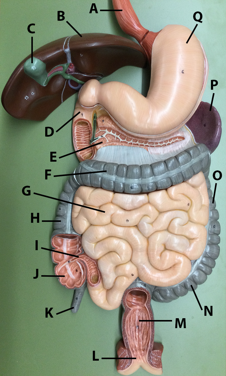
- Image credit
-
“Flat Digestive System Model - Atlas" by Jennifer Lange, is licensed under CC BY-NC-SA 4.0.
|
- Answer Key (click to show/hide answers)
-
A - esophagus
B - liver
C - gallbladder
D - duodenum (of small intestine)
E - pancreas
F - transverse colon (of large intestine)
G - small intestine (jejunum)
H - ascending colon (of large intestine)
I - iliocecal valve
J - cecum (of large intestine)
K - appendix
L - anal canal
M - rectum
N - sigmoid colon (of large intestine)
O - descending colon (of large intestine)
P - spleen
Q - stomach
|
|

- Image credit
-
“Flat Digestive System Model - Biliary Ducts Atlas" by Jennifer Lange, is licensed under CC BY-NC-SA 4.0.
|
- Answer Key (click to show/hide answers)
-
A - gallbladder
B - right hepatic duct
C - left hepatic duct
D - common hepatic duct
E - common bile duct
F - cystic duct
G - duodenum (of small intestine)
H - pancreatic duct (main)
I - pancreas
J - stomach
K - pyloric sphincter
|
|
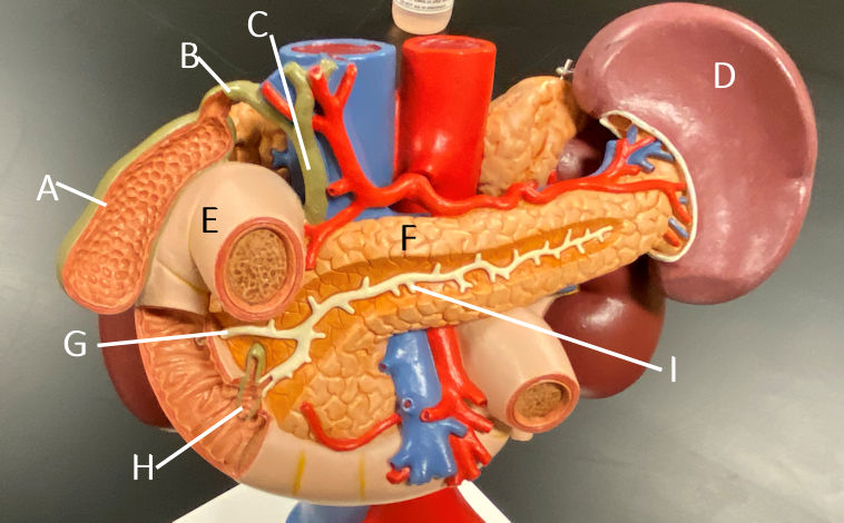
- Image credit
-
“Upper Abdominal Organs Model - Atlas" by John Polos is licensed under CC BY-NC-SA 4.0.
|
- Answer Key (click to show/hide answers)
-
A - gallbladder
B - cystic duct
C - common bile duct
D - spleen
E - duodenum (of small intestine)
F - pancreas
G - accessory pancreatic duct
H - ampulla
I - main pancreatic duct
|
 |
- Answer Key (click to show/hide answers)
A - Mucosal layer
A1 - Lacteal
A2 - Villi
A3 - Crypt
B - Submucosal layer
B1 - Peyer's patch (lymph nodule)
B2 - Mucus gland
C - Muscularis (externa)
C1 - Circular muscularis
C2 - Longitudinal muscularis
D - Serosa (serosal layer)
|
|

- Image credit
-
“Torso Model Abdominal Region - Digestive Organs Atlas" by Jennifer Lange, is licensed under CC BY-NC-SA 4.0.
|
- Answer Key (click to show/hide answers)

|
 |
- Answer Key (click to show/hide answers)
A - Serosal layer
B - Muscularis (externa)
B1 - Circular muscularis
B2 - Longitudinal muscularis
C - Submucosa
C1 - Muscularis mucosae
D - Mucosa
D1 - Gastric glands (pits) in stomach
D2 - Villi in small intestines
D3 - Crypts in large intestine (colon)
|
 |
- Answer Key (click to show/hide answers)
-
Esophagus @ 100x
A - muscularis (externa)
B - submucosa
C - mucosa
D - muscularis mucosa
E - lamina propria
F - stratified squamous epithelial tissue
|
|

- Image credit
-
“Stomach Histology @ 100x" slide provided by the Regents of the University of Michigan Medical School © 2022 CC BY-NC-SA 3.0, labeling by Jennifer Lange.
|
- Answer Key (click to show/hide answers)
-
Stomach @ 100x
A - mucosa
B - muscularis mucosa
C - MALT (mucus-associated lymphoid tissue)
D - lumen
E - gastric pits
F - gastric glands
G - submucosa
H - muscularis (externa)
|
|

- Image credit
-
“Stomach Histology @ 40x" slide provided by the Regents of the University of Michigan Medical School © 2022 under CC BY-NC-SA 3.0, labeling by Jennifer Lange.
|
- Answer Key (click to show/hide answers)
-
Stomach @ 40x
A - gastric fold
B - gastric glands
C - muscularis - inner oblique layer
D - muscularis - middle circular layer
E - muscularis - outer longitudinal layer
F - muscularis (externa)
G - submucosa
H - mucosa
I - gastric pits
|
|

- Image credit
-
“Stomach Histology @ 10x - atlas" slide provided by the Regents of the University of Michigan Medical School © 2022 under CC BY-NC-SA 3.0, labeling by Jennifer Lange.
|
- Answer Key (click to show/hide answers)
-
Stomach @ 10x
A - gastric fold
B - MALT - mucus-associated lymphoid tissue
C - mucosa
D - submucosa
E - muscularis
|
|

- Image credit
-
“Small Intestine Histology @ 200x - atlas" slide provided by the Regents of the University of Michigan Medical School © 2022 under CC BY-NC-SA 3.0, labeling by Jennifer Lange.
|
- Answer Key (click to show/hide answers)
-
Small intestine @ 200x
A - simple columnar epithelium
B - lamina propria
C - goblet cells
D - intestinal crypts
E - submucosa
F - muscularis mucosa
G - villus
|
|

- Image credit
-
“Small Intestine Histology @ 100x - atlas" slide provided by the Regents of the University of Michigan Medical School © 2022 under CC BY-NC-SA 3.0, labeling by Jennifer Lange.
|
- Answer Key (click to show/hide answers)
-
Small intestine @ 100x
A - mucosa
B - submucosa
C - muscularis mucosa
D - circular fold
E - simple columnar epithelial tissue with goblet cells
F - villus
G - lamina propria
|
|

- Image credit
-
“Small Intestine Histology @ 40x - atlas" slide provided by the Regents of the University of Michigan Medical School © 2022 under CC BY-NC-SA 3.0, labeling by Jennifer Lange.
|
- Answer Key (click to show/hide answers)
-
Small Intestine @ 40x
A - villus
B - simple columnar epithelium with goblet cells
C - mucosa
D - submucosa
E - muscularis (externa)
F - circular fold
G - lamina propria
H - muscularis mucosa
|
|

- Image credit
-
“Pancreas Histology @ 400x - atlas" micrograph provided by Berkshire Community College Bioscience Image Library is in the Public Domain, labeling by Jennifer Lange.
|
- Answer Key (click to show/hide answers)
-
Pancreas @ 400x
A - pancreatic islet
B - pancreatic acini (acinar cells)
|
|

- Image credit
-
“Liver (Human) Histology @ 100x - atlas" slide provided by the Regents of the University of Michigan Medical School © 2022 under CC BY-NC-SA 3.0, labeling by Jennifer Lange.
|
- Answer Key (click to show/hide answers)
-
Liver @ 100x
A - lobule
B - central vein
|
|

- Image credit
-
“Liver (Human) Histology @ 400x Masson Stain - atlas" slide provided by the Regents of the University of Michigan Medical School © 2022 under CC BY-NC-SA 3.0, labeling by Jennifer Lange.
|
- Answer Key (click to show/hide answers)
-
Liver @ 400x
A - central vein
B - interlobular septum
C - hepatic portal triad
|
|

- Image credit
-
"Half Head" by John Polos. All cadaver images from BlueLink at the University of Michigan and are made available by the regents under CC 4.0
For larger images, right click(PC)/command click (Mac) and click open image in new window.
|
- Answer Key (click to show/hide answers)
-
A - hard palate
B - soft palate
C - uvula
D - pharynx
E - epiglottis
|
|

- Image credit
-
“Liver" by John Polos. All cadaver images from BlueLink at the University of Michigan and are made available by the regents under CC 4.0
For larger images, right click(PC)/command click (Mac) and click open image in new window.
|
- Answer Key (click to show/hide answers)
-
A - caudate lobe
B - right lobe
C - quadrate lobe
D - gallbladder
E - left lobe
|
|

- Image credit
-
“Abdominal organs" by John Polos. All cadaver images from BlueLink at the University of Michigan and are made available by the regents under CC 4.0
For larger images, right click(PC)/command click (Mac) and click open image in new window.
|
- Answer Key (click to show/hide answers)
-
A - duodenum (of small intestine)
B - pancreas
C - ampulla
D - main pancreatic duct
|
|

- Image credit
-
“Abdominal cavity" by John Polos. All cadaver images from BlueLink at the University of Michigan and are made available by the regents under CC 4.0
For larger images, right click(PC)/command click (Mac) and click open image in new window.
|
- Answer Key (click to show/hide answers)
-
A - liver
B - stomach
C - transverse colon (of large intestine)
D - ascending colon (of large intestine)
E - small intestine (ileum)
F - descending colon (of large intestine)
G -cecum (of large intestine)
H -rectum
J -sigmoid colon (of large intestine)
K -appendix
|
|

- Image credit
-
“Abdominal cavity" by John Polos. All cadaver images from BlueLink at the University of Michigan and are made available by the regents under CC 4.0
For larger images, right click(PC)/command click (Mac) and click open image in new window.
|
- Answer Key (click to show/hide answers)
-
A - liver
B - stomach
C - transverse colon (of large intestine)
D - ascending colon (of large intestine)
E - small intestine (ileum)
F - descending colon (of large intestine)
G -cecum (of large intestine)
H -rectum
J -sigmoid colon (of large intestine)
K -appendix
|