Opening the book…
Most muscles in our body operate voluntarily. Some muscles are involuntary like swallowing and breathing (controlled without conscious thought). For the purposes of our learning here, we will only discuss a few muscles that are voluntary skeletal muscles – the muscles that move your joints. There are different types of muscles, and these include power muscles, rotator cuff muscles, foot, leg, and ankle muscles, calf muscles, oblique muscles, hip to knee muscles, hip muscles, deep rotators in the hip, inward rotators in the hip. There are more than listed below, but this reasonable scope for our learning material will provide a succinct look at some of the main muscles we use as dancers.
Power Muscles
- Pectoralis Major
- Deltoid Group
- Latissimus Dorsi

(OpenStax College. (2017, Nov 23). File:1119 Muscles that Move the Humerus b.png. https://commons.wikimedia.org/wiki/File:1119_Muscles_that_Move_the_Humerus_b.png)

Rotator Cuff Muscles
- Supraspinatus
- Teres minor
- Infraspinatus
- Subscapularis
Foot, Leg, Ankle Muscles
The anterior muscles that dorsiflex the foot/ankle include the extensor digitorum longus. The extensor digitorum longus is important when a dancer wishes to achieve a winged, or beveled foot, which is considered correct and aesthetically pleasing in techniques such as ballet and jazz dance.

(Polygon data were generated by Database Center for Life Science (DBCLS). (2019, Nov 27). Extensor Digitorum Longus muscle. https://commons.wikimedia.org/wiki/File:Extensor_digitorum_longus_muscle_-_anteriror_view.png)

Calf Muscles
The posterior muscles that plantar flex the foot/ankle include the gastrocnemius (superficial) and the soleus (inner, deep calf muscle). Pointing the foot and going into relevé are examples of plantar flexion.
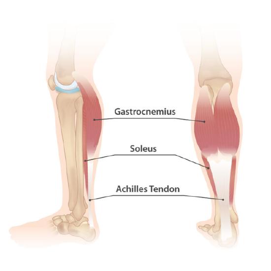
(Source: Injurymap; https://injurymap.com/free-human-anatomy-illustrations/)
Oblique Muscles
Your oblique muscles are a key component of the abdomen as they let you bend sideways and twist your trunk, moving in the transverse plane. They also help somewhat with breathing. The top of the obliques connects with the ribs while the lower end connects with the hip bone.
Hip to Knee Muscles
The Quadricep muscle group is anterior (in the front). The Hamstring muscle group is posterior (in the back). The Sartorius is the longest muscle in the human body. It wraps around the femur and attaches to the Anterior Tibia. The knee only moves on the Sagittal Plane.

(BodyParts3D/Anatomography. (2014, July 7). Rotating image of the anatomy of muscles of the upper leg. https://commons.wikimedia.org/wiki/File:Sartorius_3D.gif)

Hip Muscles
- The Iliopsoas is comprised of the psoas and the iliacus muscles.
- The psoas starts in the spine and meets at the iliacus on the crest of the ilium.
- The iliopsoas is the only hip flexor muscle that is deep in the abdominal.
Deep Rotators in the Hip
6 deep outward rotators
- Piriformis
- Gemellus inferior
- Gemellus superior
- Obturator internus
- Obturator externus
- Quadratus femoris


Inward Rotators of the Hip
- Gluteus Medius
- Gluteus Minimus
- Tensor Fasciae Latae (TFL)
This page titled 8.7: Muscles was last modified on Thu, 15 Oct 2026 21:24:48 GMT and is shared under a CC BY 4.0 license and was authored, remixed, and/or curated by Debra Worth.
The book could not be loaded just now.