Opening the book…
Search Fundamentals of Biochemistry
(Learning goals written by Claude, Sonnet 4.6, Anthropic)
Chemical Logic of Pyruvate Oxidative Decarboxylation
- Explain why α-keto acids like pyruvate cannot be decarboxylated by simple chemical means, contrasting the absence of an electron sink in α-keto acid decarboxylation with the built-in carbonyl electron sink available in β-keto acid decarboxylation, and describe how thiamine pyrophosphate (TPP) solves this problem by providing an enzymatic electron sink through its ylide carbanion.
- Explain why the thioester bond in acetyl-CoA is thermodynamically reactive relative to its hydrolysis products — including the roles of C–S bond length, reduced resonance stabilization, and diminished p-orbital overlap — and connect this to acetyl-CoA's role as a high-energy acetyl carrier in subsequent biosynthetic and catabolic reactions.
Structure and Mechanism of the Pyruvate Dehydrogenase Complex
- Describe the three-enzyme composition of the pyruvate dehydrogenase complex (E1 pyruvate dehydrogenase, E2 dihydrolipoyl acetyltransferase, and E3 dihydrolipoyl dehydrogenase), identify the five vitamin-derived cofactors (TPP, lipoamide, FAD, CoASH, and NAD⁺), and specify which cofactors are covalently attached to which subunits and why tight binding of FAD to E3 is essential for controlled catalysis.
- Trace the flow of electrons and carbon through the complete PDC reaction sequence: TPP-mediated oxidative decarboxylation of pyruvate (E1), reductive acetyl transfer from acetyl-lipoamide to CoASH (E2), and sequential reoxidation of reduced lipoamide by FAD (E3) and of FADH₂ by NAD⁺ (E3), writing the net reaction and identifying the oxidation state change at each carbon.
- Describe the quaternary organization of the PDC — including the pentagonal dodecahedral 60-mer core of E2 subunits in mammals and the peripheral arrangement of E1 and E3 — and explain why the enormous size and multi-enzyme architecture of the complex provides a kinetic and mechanistic advantage through substrate channeling via the swinging lipoamide arm
Introduction
Let's briefly review the metabolic processes for energy extraction from glucose oxidation (i.e., glycolysis). Glycolysis is a universal pathway for anaerobic energy extraction from glucose. In this linear pathway, 2-3 carbon molecules of pyruvate are produced as glucose is cleaved and converted to two molecules of glyceraldehyde-3-phosphate via an oxidation reaction using the oxidizing agent NAD+. As glycolysis continues, NADH builds up. Using lactate dehydrogenase, pyruvate, the end product of glycolysis, can be converted to lactate, regenerating NAD+, so the pathway can continue. The reactions are illustrated in Figure \(\PageIndex{1}\).

Figure \(\PageIndex{1}\): Anaerobic production of pyruvate and lactate
A careful glance at the structure of the 3-carbon pyruvate molecule reveals that much more energy could be extracted from it, presumably through oxidative decarboxylation reactions, which convert the carbons into 3 CO2 molecules. A problem arises immediately when examining pyruvate. It is an α-ketoacid, and there is no easy route to decarboxylate it, as an electron "sink" is not available to receive the electrons and stabilize the transition state and intermediate. In contrast, the decarboxylation of β-keto acids has a built-in electron "sink": an electronegative carbonyl carbon that can accept the electrons. This is illustrated in Figure \(\PageIndex{2}\).

Figure \(\PageIndex{2}\): Comparison of the decarboxylation of α and β keto acids
To begin the complete oxidation of the remnants of glucose, pyruvate enters the mitochondria and starts the process of oxidative decarboxylation by interacting with the pyruvate dehydrogenase complex (PHC). This catalyzes a complex reaction in which an electron "sink" beta is attached to the carboxylate, which is subsequently released as CO2. The end products of the PHC oxidative decarboxylation reaction are the two-carbon acetyl-CoAs, NADH, and CO2. The acetyl-CoA then enters a cyclic, non-linear pathway called the citric acid cycle, tricarboxylic acid (TCA) cycle, or Krebs cycle, named after Hans Krebs, who discovered it. We'll talk about that in section 16.2.
At a glance, we have taken glucose a small fraction of the way toward completely oxidizing every carbon to CO2 and H2O. Complete oxidation happens under aerobic conditions in the Krebs cycle. Pyruvate formed in glycolysis enters the mitochondrial matrix, where it is oxidatively decarboxylated and reacts with a small thiol, Coenzyme A (abbreviated as CoA or CoASH), to form a 2-carbon "activated acetate" acetyl group, linked through a thioester to CoASH, forming acetyl-CoA. Coenzyme A is often abbreviated as CoASH to emphasize that it has a free nucleophile thiol (-SH) group.
The third carbon from pyruvate is released as CO2. The enzyme pyruvate dehydrogenase complex (PDC) catalyzes the reaction.
Pyruvate Dehydrogenase Mechanism
This enzyme complex is enormous. The E. coli complex has a molecular weight of almost 4 million, with at least 16 chains, each containing three different enzymes that catalyze part of the reaction. The components are pyruvate dehydrogenase (E1), dihydrolipoamide dehydrogenase (E2), and dihydrolipoamide dehydrogenase (E3). The bovine complex has a molecular weight of almost 8 million and contains 22 E1, 60 E2, and 6 E3 subunits. Nature often uses the same solution for identical problems. For example, many proteases have an active-site nucleophilic serine that works with histidine and aspartate to cleave peptide bonds. Many organisms have three α-ketoacid dehydrogenase complexes. Each has a common E3 but specific E1 and E2 enzymes. Figure \(\PageIndex{3}\) shows an image of the structure so you can get an overview before we dive into the activity of each of the substrates.

Figure \(\PageIndex{3}\): View of pyruvate dehydrogenase. https://electron.med.ubc.ca/2018/07/...dehydrogenase/
The E3 subunit is not readily seen in the image above. Why has nature produced such a monstrous enzyme complex to catalyze the oxidative decarboxylation of a small three-carbon molecule? We will explore that at the end of this section.
The complex also employs five substrates/cofactors derived from vitamins collectively.
- Thiamine in the form of thiamine pyrophosphate (TPP), which is covalently attached to E1
- lipoic acid, in the form of lipoamide, which is covalently attached to a lysine side chain in E2
- riboflavin in the form of flavin adenine dinucleotide (FAD/FADH2), which is bound very tightly (and not released) to E3
- pantothenic acid, incorporated into the structure of CoASH/Acetyl-CoA, a substrate/product pair for the reaction
- niacin, nicotinic acid, in the form of NAD+/NADH, a substrate/product pair for the reaction
The structures for the five are shown in Figure \(\PageIndex{4}\), along with some additional descriptions that summarize some of the chemistry of these molecules.

Figure \(\PageIndex{4}\): Structure of the cofactors in pyruvate dehydrogenase
Figure \(\PageIndex{5}\) shows a schematic of the overall reaction.

Figure \(\PageIndex{5}\): Overall reactions catalyzed by pyruvate dehydrogenase
The net reaction is
\[\ce{pyruvate + CoASH + NAD^{+} -> Acetyl-CoA + CO2 + NADH + H^{+}} \nonumber \]
Part 1: Oxidative Decarboxylation - pyruvate dehydrogenase (E1p)
So let it begin. We need to eliminate one carbon as CO2 and transfer the other two carbons of pyruvate to CoASH to form acetyl-CoA, a CoASH thioester. Thioesters are "high energy" with respect to their hydrolysis products, as the thioester is destabilized compared to a normal carboxylic acid ester. (Remember, there is no such thing as a "high energy" bond.) Since the sulfur atom is larger than the O in the C-S and C-O bonds in their respective esters, the thioester as a reactant can not be stabilized well, as the C-S single bond length is longer, as shown in Table \(\PageIndex{1}\) below.
| bond | length (Angstroms) |
|---|---|
| C-O | 1.43 |
| C=O | 1.21 |
| C-S | 1.82 |
| C=S | 1.56 |
Table \(\PageIndex{1}\): Bond lengths of carbon-oxygen and carbon-sulfur single and double bonds
This minimizes resonance stabilization compared to the carboxylic acid ester, as shown in the figure below. The hydrolysis products of both a carboxylic and a thiol ester have comparable energies. Hence, only the thioester is relatively destabilized compared to its hydrolysis product, with the ΔG0 hydrolysis = -7.5 kcal/mol (-31 kJ/mol), the same as for the hydrolysis of a phosphoanhydride bond of ATP. Additionally, a resonance structure shows a positive charge on the carbonyl carbon and a negative charge on the oxygen, making the carbonyl carbon more electrophilic. Another, more sophisticated reason for the relative destabilization of the thiol ester is that the overlap between the carbonyl carbon p orbital and the larger sulfur p orbital is less, hindering the electron delocalization needed to stabilize the thiol ester.
Figure \(\PageIndex{6}\) illustrates these points.

Figure \(\PageIndex{6}\): Comparison of resonance stabilization of carboxylic esters and thioesters
Now, we can explore the mechanism of CO2 release and acetyl-CoA production by E1. The carbon atom between the N and S in the thiamine ring has a reduced pKa, allowing deprotonation and carbanion formation. The negative charge can't be stabilized by resonance, but the positively charged nitrogen can. This zwitterion is called a ylide: a net-neutral species with a positive charge (usually on nitrogen, phosphorus, or sulfur) and a negative charge (usually on carbon) on adjacent atoms.
The carbanion on the ylide attacks the electrophilic C=O of pyruvate, forming a TPP intermediate with an excellent electron sink (N+) beta to the carboxyl carbonyl C. This is the essence of the entire reaction, as this enables the decarboxylation event. The remaining reactions catalyzed by E2 and E3 release the other two carbon atoms of pyruvate as acetyl-CoA (E2) and return the enzymes to their original state (E2 and E3).
Figure \(\PageIndex{7}\) shows the reaction mechanism of E1.

Figure \(\PageIndex{7}\): Reaction mechanism of E1 of pyruvate dehydrogenase
Note that the carbonyl C in pyruvate has two single bonds to two other carbon atoms, while in the final covalently attached form, it has one bond to carbon and one to sulfur. Sulfur is under oxygen in the periodic table. By analogy, replacing a C-C bond with a C-S bond is an oxidation reaction that requires an oxidizing agent. The covalently attached ring of the lipoamide with an S-S bond similar to that of a disulfide bond, an oxidized form of sulfur, is the oxidizing agent. On the formation of acetyl-lipoamide, the S-S bond is cleaved, and a thioester is formed. The other sulfur is a free reduced thiol.
The bovine PDC has 22 E1 subunits. Here is an iCn3D model of one human pyruvate dehydrogenase E1 component complex with TPnP (TDP acetyl phosphonate, a TPP analog, covalently attached (PDB ID: 6CFO). One E1 subunit is an α2β2 heterodimer. The two alpha chains are cyan, while the beta chains are dark blue. Orient the model to view along the C2 rotational symmetry axes shown.
Figure \(\PageIndex{8}\) shows an interactive iCn3D model of the human pyruvate dehydrogenase E1 covalently bound to TDP acetyl phosphonate (TpnP), a TPP analog (6CFO)

 Figure \(\PageIndex{8}\): Human pyruvate dehydrogenase E1 covalently bound to TDP acetyl phosphonate (TpnP), a TPP analog (6CFO). (Copyright; author via source). Click the image for a popup or use this external link: https://structure.ncbi.nlm.nih.gov/i...7FJ2GE47tvS6g7
A heterotetramer consisting of two α (cyan) and two β chains (dark blue) is shown.
Figure \(\PageIndex{9}\) shows an interactive iCn3D model of the TPP analog covalently bound to E1 of pyruvate dehydrogenase (6CFO)

Figure \(\PageIndex{9}\): TPP analog covalently bound to E1 of pyruvate dehydrogenase (6CFO) . (Copyright; author via source). Click the image for a popup or use this external link: https://structure.ncbi.nlm.nih.gov/i...SNf5sC7SqmQkc8
The dotted lines show the interactions between the TPP analog, color-coded as shown in the legend below.

Parts 2 and 3: Acetyl-CoA (E2) Formation and Regeneration of the Active Complex.
The next part of the reaction produces acetyl-CoA (E2). After that, the enzyme is "dead" as it no longer has an oxidized form of lipoamide to serve as an oxidizing agent (which gets reduced) in another round of catalysis. To regenerate enzyme activity, the reduced lipoamide, after the release of the attached acetyl group, must be reoxidized by another oxidizing agent. The oxidizing agent is FAD, which is covalently bound to E3 and is reduced to FADH2. It must be reoxidized back to FAD to restore enzyme activity. The final oxidizing agent used is NAD+, which the enzyme releases as a product. So it's a bit complicated. PDH uses three oxidizing agents. Two are covalently attached to the enzyme (oxidized lipoamide on E2 and oxidized FAD on E3).
Figure \(\PageIndex{10}\) shows the transacetylation reaction and formation of reduced lipoamide

Figure \(\PageIndex{10}\): Transacetylation reaction and formation of reduced lipoamide by pyruvate dehydrogenase E2
The reaction of E3 follows to restore the fully catalytic enzyme, as shown in Figure \(\PageIndex{11}\).

Figure \(\PageIndex{11}\): Regeneration of oxidized lipoamide by pyruvate dehydrogenase E3.
Let's look in greater detail at the structures of both E2 and E3.
E2: dihydrolipoyl acetyltransferase -
In the mammalian complex, 60 E2 subunits arrange into a pentagonal dodecahedron. Most gram-negative bacteria's E2 subunits arrange into a cube of 24 monomers. Figure \(\PageIndex{12}\) shows a simple view of a pentagonal dodecahedron with 12 equivalent faces.
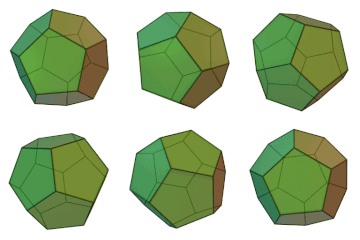
Figure \(\PageIndex{12}\): Pentagonal dodecahedron. 1. Rotating dodecahedron: https://commons.wikimedia.org/wiki/F...decahedron.gif. User Cyp on en.Wikipedia, CC BY-SA 3.0 <http://creativecommons.org/licenses/by-sa/3.0/>, via Wikimedia Commons

First, let's consider a single E2 monomer. It has a longer disulfide redox domain followed by a smaller dimerization domain, which allows multiple subunits to assemble into the dodecahedron. In greater detail, the monomer consists of two lipoyl domains, a small domain that facilitates binding to E1, and a C-terminal catalytic domain.
Figure \(\PageIndex{12}\) shows an interactive iCn3D model of E2 inner core 60-mer of human pyruvate dehydrogenase (pdb 6CT0). Symmetry axes are not shown.

Figure \(\PageIndex{12}\): E2 inner core 60-mer of human pyruvate dehydrogenase (pdb 6CT0). (Copyright; author via source). Click the image for a popup or use this external: https://structure.ncbi.nlm.nih.gov/i...zs6pwfKNuJ2VS6
Each of the 60 subunits is shown in light cyan. To see the C symmetry axes:
- select the menu =
- Choose/Check in order: Analysis, Symmetry, From PDB, 1(global), apply
- When you see just a single monomeric chain, choose Clear
The symmetry axes will then appear.
E3: dihydrolipoyl dehydrogenase
The sole function of this subunit is the reoxidation of the now-reduced lipoamide with the free sulfhydryl to the cyclic disulfide form, so the enzyme can engage in further catalysis. FAD covalently bound to the E3 subunit is the oxidizing agent. This is our first encounter with FAD. Similar to NAD+, this dinucleotide gains a hydride (:H-) but also, in contrast to NAD+, a proton to form FADH2.
Another way that the FAD/FADH2 differs from NAD+/NADH is that the FAD/FADH2 or their mononucleotide analog (FMN/FMNH2) pairs are either covalently attached (in about 10% of flavoproteins) or bound with such a low KD (often in the nanomolar range) that they don't dissociate from the enzyme during catalysis. Hence, after oxidizing a bound substrate, the reduced FADH2 must be reoxidized by another oxidizing agent, often NAD+, which can diffuse into the active site to perform its role and then dissociate from the complex as NADH, leaving the enzyme competent for another round of catalysis. (DOI: 10.1002/chem.201704622)
Figure \(\PageIndex{13}\) shows an interactive iCn3D model of E3 bound to both FAD (noncovalently) and NADH (NAI) (1ZMD) in the B chain of E3. Symmetry axes are not shown.

Figure \(\PageIndex{13}\): E3 bound to both FAD (noncovalently) and NADH (NAI) (1ZMD). (Copyright; author via source). Click the image for a popup or use this external: https://structure.ncbi.nlm.nih.gov/i...LAMqAS3Tx9SPE8.
Figure \(\PageIndex{14}\) shows an interactive iCn3D model highlighting the noncovalent interactions stabilizing bound FAD and NADH (NAI) in the E3 subunit of pyruvate dehydrogenase (1ZMD).

Figure \(\PageIndex{14}\): Noncovalent interactions stabilizing bound FAD and NADH (NAI) in the E3 subunit of pyruvate dehydrogenase (1ZMD). (Copyright; author via source). Click the image for a popup or use this external:https://structure.ncbi.nlm.nih.gov/i...gUED1ZrJVjKcD8
Let's put it all together! Figure \(\PageIndex{15}\) shows a video of the pyruvate dehydrogenase complex from the HHMI.
Figure \(\PageIndex{15}\): Video of the pyruvate dehydrogenase complex
Summary
(Summary written by Claude, Sonnet 4.6, Anthropic)
This chapter describes the oxidative decarboxylation of pyruvate to acetyl-CoA, the essential metabolic bridge between glycolysis and the citric acid cycle, focusing on the reaction's chemical logic and the elaborate multi-enzyme machinery—the pyruvate dehydrogenase complex (PDC)—that catalyzes it.
The chapter opens by establishing why pyruvate, a three-carbon α-keto acid, cannot be straightforwardly decarboxylated by the same mechanism used for β-keto acids. In β-keto acid decarboxylation, the carbonyl carbon adjacent to the carboxyl group serves as an electron sink, accepting the electrons released when CO₂ departs and stabilizing the resulting carbanion intermediate. Pyruvate lacks this built-in electron sink: its carbonyl carbon is adjacent to the carboxylate, not to a β-carbonyl, and no stable intermediate can form upon decarboxylation without enzymatic assistance. The PDC resolves this problem by using thiamine pyrophosphate (TPP), whose thiazolium ring contains a C2 carbon of uniquely low pKa. Deprotonation of this carbon generates a zwitterionic carbanion (the ylide), which attacks the electrophilic carbonyl carbon of pyruvate. The resulting adduct places an excellent electron sink — the positively charged ring nitrogen of the thiazolium — in the β-position relative to the scissile C–C bond, enabling decarboxylation with release of CO₂ and formation of a stabilized hydroxyethyl-TPP intermediate.
The chapter then describes the thermodynamic basis for acetyl-CoA's reactivity. The thioester linkage in acetyl-CoA is destabilized relative to its hydrolysis products (acetic acid and CoASH) because the C–S bond is longer (1.82 Å vs. 1.43 Å for C–O) and the lone pairs on the larger sulfur atom overlap poorly with the carbonyl carbon's p orbital, preventing the resonance delocalization that stabilizes conventional carboxylic acid esters. As a result, the thioester reactant is relatively high in energy compared to its products, with ΔG°' of hydrolysis = −7.5 kcal/mol (−31 kJ/mol) — equivalent to that of ATP phosphoanhydride hydrolysis. This thermodynamic activation makes acetyl-CoA a potent acyl group donor in biosynthetic and catabolic reactions.
The PDC is one of the largest known enzyme complexes — ~4 × 10⁶ Da in E. coli and ~8 × 10⁶ Da in bovine mitochondria — and employs three distinct enzymes (E1, E2, E3) and five vitamin-derived cofactors. E1 (pyruvate dehydrogenase) contains covalently bound TPP and catalyzes the oxidative decarboxylation described above, transferring the resulting two-carbon acetyl fragment to the oxidized lipoamide arm of E2. E2 (dihydrolipoyl acetyltransferase) contains lipoamide covalently tethered via an amide linkage to a lysine side chain; this flexible "swinging arm" (approximately 14 Å long) receives the acetyl group from TPP as a thioester, simultaneously reducing the disulfide of the lipoamide ring. E2 then catalyzes transacetylation — transfer of the acetyl group to CoASH — to form acetyl-CoA and a fully reduced dihydrolipoamide. The 60 E2 subunits assemble into a pentagonal dodecahedral core in the mammalian complex, providing a scaffold around which E1 and E3 are organized.
E3 (dihydrolipoyl dehydrogenase) then reoxidizes the reduced dihydrolipoamide in two sequential steps. First, the reduced lipoamide is reoxidized by the FAD that is tightly but noncovalently bound to E3, forming FADH₂ and restoring the cyclic disulfide of the lipoamide. Second, FADH₂ is reoxidized by NAD⁺ — which diffuses into the E3 active site and is released as NADH — regenerating the fully oxidized enzyme. This sequential use of three oxidizing agents (lipoamide, FAD, and NAD⁺) is required because only NAD⁺/NADH can diffuse freely into and out of the complex; lipoamide and FAD must remain enzyme-bound to prevent their reduction products from reacting with cellular oxidants, and their tight binding ensures that electron flow is channeled exclusively toward NAD⁺ reduction. The swinging lipoamide arm is the key architectural feature that enables substrate channeling among all three active sites without releasing intermediates into solution — a structural explanation for why evolution has produced such a massive complex to catalyze the conversion of a simple three-carbon molecule to a two-carbon thioester.
This page titled 16.1: Production of Acetyl-CoA (Activated Acetate) was last modified on Mon, 14 Sep 2026 11:31:36 GMT and is shared under a not declared license and was authored, remixed, and/or curated by Henry Jakubowski and Patricia Flatt.
The book could not be loaded just now.