Opening the book…
Search Fundamentals of Biochemistry
(Learning goals written by Claude, Sonnet 4.6, Anthropic)
The Urea Cycle: Logic, Reactions, and Mechanisms
- Explain the logic of the urea cycle as a cyclic pathway — using Krebs's experimental reasoning with ornithine to establish cyclicity — and identify the sources of the two nitrogen atoms and the carbon atom in urea, accounting for the three ATP equivalents consumed per cycle.
- Describe the mechanism of carbamoyl phosphate synthase I (CPS I), including the roles of two sequential ATP-driven phosphorylations in activating bicarbonate and ammonia, the function of the internal tunnel that channels the reactive mixed anhydride intermediate between active sites, and the allosteric role of N-acetylglutamate (NAG) in opening the tunnel and activating the enzyme.
- Explain how arginosuccinate synthase (ASS1) incorporates the second nitrogen atom from aspartate into the urea precursor at the cost of one ATP (cleaved to AMP + PPᵢ), and describe the β-elimination mechanism by which arginosuccinate lyase (ASL) releases arginine and fumarate.
- Describe the mechanism of arginase I, explaining how a dinuclear Mn²⁺ cluster lowers the pKa of a bound water molecule to generate a nucleophilic hydroxide that attacks the guanidino group of arginine, forming a tetrahedral intermediate that collapses to produce ornithine and urea with assistance from Asp128 as a general acid.
Regulation of the Urea Cycle
- Explain the short-term allosteric regulation of the urea cycle through NAG — including how arginine activates NAG synthase to produce NAG, which in turn allosterically activates CPS I by inducing a conformational change that forms a complete substrate tunnel — and describe how long-term regulation occurs through transcriptional upregulation of urea cycle enzymes in response to high-protein diets.
- Explain the clinical consequences of ornithine transcarbamoylase (OTC) deficiency as an X-linked disorder, and describe why supplemental arginine is given to patients with urea cycle disorders in terms of its effect on NAG synthase and NAG levels.
Integration of the Urea Cycle with the TCA Cycle and Nitrogen Transport
- Describe the metabolic connections between the urea cycle and the TCA cycle — specifically how fumarate produced in the cytoplasm re-enters the TCA cycle via the aspartate-argininosuccinate shunt, and how aspartate (providing the second urea nitrogen) is regenerated through transamination of oxaloacetate — and explain how the malate-aspartate shuttle simultaneously coordinates these cycles and transports cytoplasmic NADH equivalents into the mitochondria for oxidative phosphorylation.
- Explain why the urea cycle is energetically expensive (four phosphoanhydride bonds per urea produced), how this cost is partially offset by the ATP generated from fumarate processing through the linked TCA cycle, and connect this to the broader metabolic principle that toxic waste excretion requires a thermodynamic investment.
The Urea Cycle
Now, we can see what happens to excess NH3/NH4+ that accumulates in the liver mitochondria. The ammonia formed in the liver by oxidative deamination via glutamate dehydrogenase is converted to urea. Alternatively, it can be added to glutamic acid to form glutamine, which can be transported to the kidney. There, after the sequential actions of glutaminase (a deamidation reaction) and glutamate dehydrogenase, ammonia is produced for direct excretion in the urine. If your diet is high in protein and hence amino acids, they can't be stored as proteins per se, so they are metabolized for energy, and the metabolic products can be converted to fat or carbohydrates. That leaves excess nitrogen, which is mainly excreted as urea.
Urea is a highly water-soluble, non-toxic molecule that biochemists use in the lab to chemically denature proteins (at concentrations of 3-6 M). Clinical blood urea concentrations are expressed as Blood Urea Nitrogen (BUN), which ranges from 7 to 20 mg/dL N, equivalent to 2.5 to 7.1 mM urea. Serum levels depend predominantly on the balance between urea synthesis in the liver and its elimination by the kidney. Normal cellular urea concentrations should be similar.
Urea is produced through a cyclic pathway known as the urea cycle. You are familiar with perhaps the most favorite biochemical cycle, the tricarboxylic (citric or Krebs) cycle. Krebs also discovered the urea cycle before conceiving the citric acid cycle. He concluded that the urea formation pathway was a cycle, using the same logic he had applied to the citric acid cycle. After your first view of the full urea cycle, we'll explain how, as shown in Figure \(\PageIndex{1}\).

Figure \(\PageIndex{1}\): The Urea Cycle
The enzyme names are:
- CPS1: carbamoyl phosphate synthase I
- OTC: ornithine transcarbamoylase
- ASS1: arginosuccinate synthase 1
- ASL: arginosuccinase lysase (aka arginosuccinase)
- ARG1: arginase
It is color-coded to designate the sources of the C and N atoms. One N comes from ammonia (or indirectly from the amido N of glutamine), while the C atom comes from carbonate. The other comes from aspartate's amine group. Note that the cycle uses three ATPs. Also note that the guanidino group of arginine (NH(NH2+)NH2) is the immediate donor of the 2 N atoms in the step that produces urea. Knowing that one amino acid was the immediate donor of the N atoms in urea, you would easily predict that.
Let's start with the first step, the cleavage of arginine to produce ornithine and urea. What happens to the ornithine derived as a "waste" product of urea formation? Experimentally, one might add more ornithine to a system running the urea formation pathway and ask what happens to the rate of urea synthesis. If it were a one-step process, you might expect, based on mass action, that adding ornithine, a product, would slow that reaction rate by driving it backward. Krebs found the opposite: adding more ornithine increased the rate of urea formation, but in a way that did not alter the ornithine concentration. Hence, ornithine was not consumed per se in a subsequent step. Ornithine appeared to act as a traditional catalyst, speeding up the reaction without being consumed. To do so, it must generate more arginine. The way to account for this was a cyclic pathway, not a linear or branched one. In a cycle, none of the participants is consumed overall.
Now, let's examine some of the steps, starting with those powered by ATP, which also incorporate nitrogen into the urea precursors.
Carbamoyl phosphate synthase I (CPS I)
This enzyme catalyzes the first "committed" step of the pathway and makes carbamoyl phosphate, the reactant that enters the cycle. It requires a specific cofactor, N-acetylglutamate (NAG), produced by acetylation of glutamate with acetyl-CoA via NAG synthase, which is activated by arginine. The reaction is not part of the cycle but provides input.
A cytosolic version of this enzyme, CPS II, synthesizes arginine and pyrimidine nucleotides using glutamine as a donor of NH4+, which reacts with carboxyphosphate to produce carbamoyl phosphate. NAG does not regulate it.
CPS I provides a way to form a (C=O)NH2 unit in urea by condensing bicarbonate and ammonia to form a carbamate, which contains a high-energy "motif" relative to its hydrolysis product. (Note: This does not imply that the broken bond is high-energy, a misnomer found in many books.) Hence, the reaction is powered by 2 ATP molecules. Urea is a molecule that "carries" both NH3/NH4+ and carbonate.
A reaction mechanism for carbamoyl phosphate synthase is shown in Figure \(\PageIndex{2}\).

Figure \(\PageIndex{2}\): Reaction mechanism for carbamoyl phosphate synthase
The two high-energy motif molecules (again with respect to their hydrolysis products) are protected from nonspecific hydrolysis as the mixed anhydride passes along a sequestered tunnel to a more distal phosphorylation site in the multimeric enzyme.
Figure \(\PageIndex{3}\) shows an interactive iCn3D model of the human carbamoyl phosphate synthetase I with bound ADP and N-acetyl-glutamate (5DOU).

 Figure \(\PageIndex{3}\): Human carbamoyl phosphate synthetase I with bound ADP and N-acetyl-glutamate (5DOU). (Copyright; author via source). Click the image for a popup or use this external link: https://structure.ncbi.nlm.nih.gov/i...yV8fW3mW9idy6A
The model shows one chain of the multimer. The two ADPs and a single N-acetylglutamate (NLG) are shown in spacefill and labeled. Additional bound ions are shown (unlabeled).
The protein exists in a monomer-dimer equilibrium mixture. Each monomer has 1 NAG and 2 ADPs in the two binding sites required for the two phosphorylation steps, as shown in the reaction above. A tunnel connects the two phosphorylation sites, allowing the mixed anhydride to reach the carbamate phosphorylation site. The NAG is an allosteric modifier as it is not bound near the phosphorylation sites. On binding to the apo form of the enzyme, it elicits a large conformational change, allowing a competent enzyme conformation with a complete tunnel to predominate.
An image of the tunnel for one monomer of the human carbamoyl phosphate synthetase I is shown in Figure \(\PageIndex{4}\).
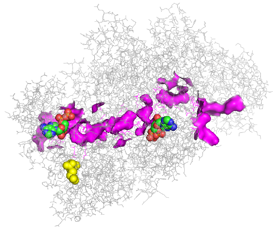
Figure \(\PageIndex{4}\): Tunnel for one monomer of the human carbamoyl phosphate synthetase I enzyme.
Two ADPs are shown below in spacefill with CPK color. The tunnel between the two ADP sites is highlighted in magenta. The yellow molecule is NAG, which again exerts its activation effect by binding to an allosteric site, effectively opening up the tunnel.
Figure \(\PageIndex{5}\) shows the enzyme monomer in a different orientation with a tunnel made using ChExVis: a tool for molecular channel extraction and visualization. The large red and green spheres show the surface openings.
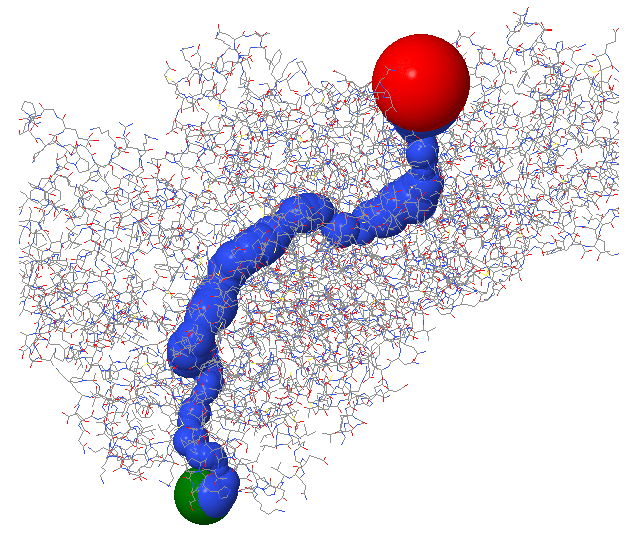
Figure \(\PageIndex{5}\): Second view of the tunnel in human carbamoyl phosphate synthetase I
The top panel in Figure \(\PageIndex{6}\) shows the radius in Angstroms of the tunnel going from the Red sphere to the Green sphere in the image above. The bottom panel shows the hydrophobicity (red)// hydrophilicity (blue) of the amino acids surrounding the tunnel.
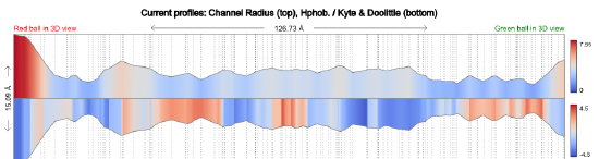
Figure \(\PageIndex{6}\): Tunnel size and polarity profile in human carbamoyl phosphate synthetase I
ASS1: ArginoSuccinate Synthase 1
Another ATP is cleaved to convert citrulline to arginosuccinate, as shown in Figure \(\PageIndex{7}\). The product could easily be cleaved in the next step to form the amino acid arginine.

Figure \(\PageIndex{7}\): ATP cleavage drives the conversion of citrulline to arginosuccinate
ASL: arginosuccinase lyase (aka arginosuccinase)
This enzyme catalyzes a beta-elimination reaction that may proceed via an E2 or E1 mechanism. A general but very abbreviated mechanism showing a two-step (E1) elimination proceeding through a carbanion intermediate is shown in Figure \(\PageIndex{8}\) below.
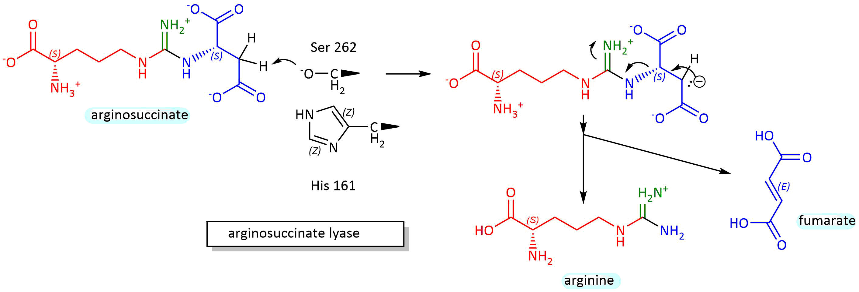
Figure \(\PageIndex{8}\): Mechanism for conversion of arginosuccinate to arginine and fumarate
Figure \(\PageIndex{9}\) shows an interactive iCn3D model of the tetrameric arginosuccinate lyase (ASL) from Mycobacterium tuberculosis (6IEN).

Figure \(\PageIndex{9}\): Tetrameric arginosuccinate lyase (ASL) from Mycobacterium tuberculosis (6IEN). (Copyright; author via source). Click the image for a popup or use this external link: https://structure.ncbi.nlm.nih.gov/i...32EuouGQ6xMVk6
Each of the four monomers is shown in a different color. Three arginosuccinates (substrates) and fumarate, as well as arginine (products), are shown in spacefill with CPK colors and labeled. Each monomer has N-, M-, and C-domains. The four binding sites are formed by residues from three monomers. The products are in the fourth site. Catalytic residues probably include a serine and a histidine acting as general acids/bases with a deprotonated lysine, making the serine a general base.
Ornithine Transcarbamoylase
The reaction mechanism is quite simple here. The carbamoyl phosphate is an activated electrophile in which a deprotonated amino group of ornithine attacks the carbonyl C to produce citrulline. The gene is found on the X chromosome in humans, so mutations in males lead to a "hyperammonemia coma" and death. Heterozygous females are either asymptomatic or have problems arising from defects in pyrimidine biosynthesis, which a low-protein diet can alleviate with the addition of arginine.
Arginase
This enzyme has two general variants. Arginase I is a cytosolic enzyme expressed in the liver that participates in urea synthesis. Arginase II is a mitochondrial enzyme involved in arginine metabolism. Arginase I converts arginine to ornithine and urea. The enzyme has 2 Mn2+ ions in the active site. A hydroxide ion bridges the two Mn ions and acts as a nucleophile, attacking the arginine guanidino group and forming a tetrahedral intermediate. An active site aspartate (Asp 128) acts as a general acid, protonating the amino leaving group to form ornithine and urea. Water then re-adds to the Mn dinuclear cluster and ionizes, donating a proton to the solvent via an intermediary His 41 as the enzyme reforms.
The model below depicts the trimeric human arginase I (3KV2) in complex with the inhibitor N(omega)-hydroxy-nor-L-arginine, in which one guanidino nitrogen is replaced with an OH group. The two gray spheres are Mn ions.
Figure \(\PageIndex{10}\) shows an interactive iCn3D model of the trimeric human arginase I in complex with the inhibitor N(omega)-hydroxy-nor-L-arginine (3KV2).

Figure \(\PageIndex{10}\): Trimeric human arginase I in complex with the inhibitor N(omega)-hydroxy-nor-L-arginine (3KV2). (Copyright; author via source). Click the image for a popup or use this external link: https://structure.ncbi.nlm.nih.gov/i...QaAQpt5t2KKic6
Figure \(\PageIndex{11}\) show an active site water (#480) near the Mn ions.
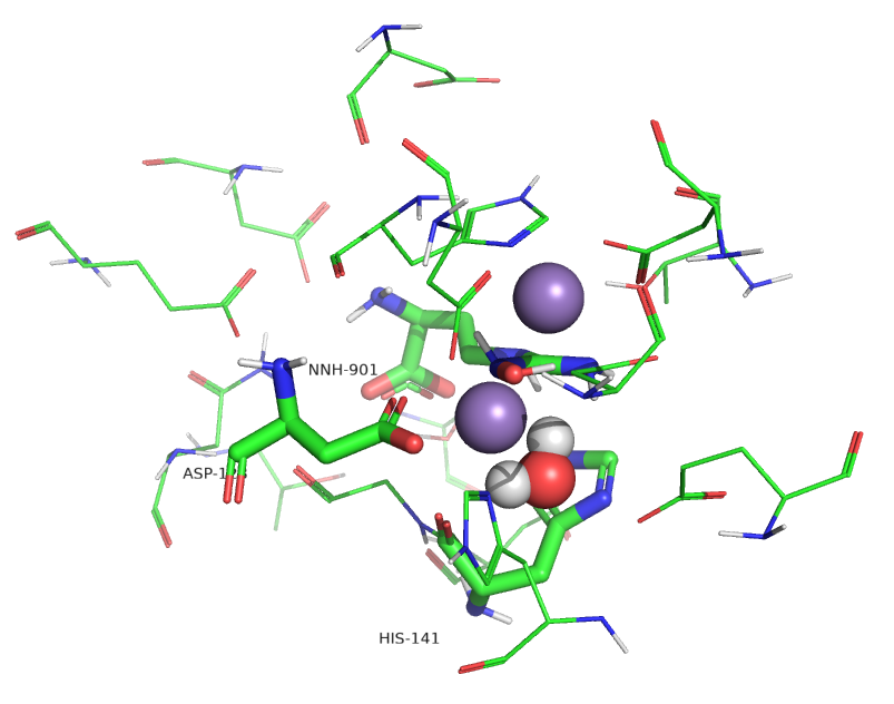
Figure \(\PageIndex{11}\): Active site of human arginase I with an active site water
Water molecules can act as ligands and bind transition-state metal ions through a coordinate covalent bond. The pKa of the bound water shifts to a lower value, making it more likely to deprotonate to form OH-, which is both a better base and nucleophile. Water 480 in the active site appears to form a coordinate covalent bond to the Mn ion, forming an active site hydroxide that attacks the guanidino group of arginine, leading to the formation of urea and ornithine.
Regulation
If excess amino acids/proteins are consumed, gene expression can change to increase levels of the enzymes in the urea cycle. In the short term, the enzyme that provides access to the cycle, carbamoyl phosphate synthase, is regulated by N-acetylglutamic acid and catalyzes the rate-limiting step. As shown above, the enzyme is functionally inactive without the allosteric regulator, NAG. The levels of NAG are determined by the enzyme NAG synthase, which is regulated by free arginine, the immediate precursor of urea in the cycle. Supplemental arginine is given to patients with urea cycle disorders, and its effect is likely mediated by NAG synthase regulation.
The link between the urea cycle and the TCA cycle
Two major pathways you have explored, the TCA and urea cycles, are not linear but cyclic. What advantages do "circular" pathways offer over linear ones? A comparison to economic pathways may offer a clue. In a linear economy, raw materials are used to produce a product, which is eventually discarded as trash. Unfortunately, linear pathways dominate our world at a great cost to our environment. To reduce the inefficiencies in a linear economy, make it more viable, and decrease environmental damage and associated climate change, the world should move to a circular economy. A circular economy is characterized by the 3Rs: reduce, reuse, and recycle. This saves resources and energy. These characteristics also apply to circular biochemical pathways, where metabolites are reused and recycled.
Given this, it should not be surprising that the urea and TCA cycles are linked, especially given that fumaric acid is a product of the urea cycle and a cyclic metabolite of the TCA cycle. In addition, aspartic acid is one transamination step away from oxaloacetate. The interconnections between the urea and TCA cycles are shown in Figure \(\PageIndex{12}\), along with two additions, the malate aspartate shuttle and the aspartate arginosuccinate shunt.

Figure \(\PageIndex{12}\): Urea and TCA cycles linked by the malate aspartate shuttle and the aspartate arginosuccinate shunt.
One of the nitrogens in urea comes from the amine of aspartate, which can be formed through the transamination of oxaloacetate from the TCA cycle to form aspartate. Since the TCA cycle is a cycle, an equivalent of one oxaloacetate must return to the cycle. It does so indirectly from fumarate, a TCA intermediate produced as a by-product of the urea cycle, but the fumarate is produced in the cytoplasm. There, it is converted to malate, which can be transported into the mitochondria by the mitochondrial 2-oxoglutarate/malate transporter, which is part of yet another cycle shown in the figure above: the aspartate-argininosuccinate shunt. Fumarate enters the shuttle through the aspartate-argininosuccinate shunt, as shown in the figure above. Once malate enters the mitochondrial matrix, it can be directly converted to oxaloacetate.
These linked cycles require both mitochondrial and cytoplasmic forms of several enzymes and a balance between the two key amino acids, glutamate and aspartate, across the mitochondrial divide. One key enzyme is aspartate aminotransferase (AST), also known as glutamate oxaloacetate transaminase (GOT), named for the reverse reaction: aspartate + α-ketoglutarate ↔ oxaloacetate + glutamate. As mentioned in the previous chapter section, this key enzyme can be detected in the blood if the liver is damaged, so its levels are routinely used in medical tests to assess impaired liver function.
Another key function of the malate-aspartate shuttle is to bring into the mitochondria reduced "equivalents" of NADH produced in the cytoplasm from glycolysis and fatty acid oxidation. No membrane transporter carries NAD+ or NADH. Instead, cytoplasmic malate, produced by NADH's cytosolic reduction of oxaloacetate, can be transported into the matrix, where it can be reconverted to oxaloacetate, reforming mitochondrial NADH. NADH can then power ATP production via mitochondrial electron transport/oxidative phosphorylation.
Energetically, four phosphoanhydride bonds in 3 ATP molecules are used to produce one urea molecule. This large energy expenditure is partially compensated for by ATP generated from the "fumarate equivalents" that enter the mitochondria via the aspartate arginosuccinate and aspartate arginosuccinate shunts, and by the passage of NADH through oxidative phosphorylation.
Summary
(Summary written by Claude, Sonnet 4.6, Anthropic)
This chapter describes the urea cycle — the primary mechanism by which vertebrates convert toxic NH₄⁺ produced by amino acid catabolism into non-toxic, water-soluble urea for excretion — and examines its integration with the TCA cycle through shared metabolic intermediates and shuttle systems.
Krebs's discovery of the urea cycle followed the same experimental logic he later applied to the TCA cycle: adding ornithine to liver preparations accelerated urea synthesis without net ornithine consumption, indicating a cyclic pathway in which ornithine is regenerated. The cycle accepts two nitrogen atoms — one from NH₄⁺ (derived from glutamate dehydrogenase activity on glutamate in the mitochondrial matrix) and one from the α-amino group of aspartate in the cytoplasm — and one carbon from bicarbonate, packaging them into the highly water-soluble, non-toxic urea molecule. Three ATP molecules (four phosphoanhydride bonds total) are consumed per urea produced, reflecting the thermodynamic cost of detoxification.
The first and committed step, catalyzed by carbamoyl phosphate synthase I (CPS I) in the mitochondrial matrix, condenses bicarbonate and NH₄⁺ to form carbamoyl phosphate in two sequential ATP-dependent phosphorylation steps. The first ATP activates bicarbonate to carbonylphosphate, which reacts with NH₃ to form carbamate; the second ATP phosphorylates carbamate to form the high-energy (relative to its hydrolysis products) carbamoyl phosphate. These reactive intermediates are channeled through an internal tunnel within the multimeric enzyme, preventing their hydrolysis by bulk solvent. CPS I is absolutely dependent on N-acetylglutamate (NAG) as an allosteric activator: NAG binding to a distal site induces a large conformational change that creates a complete, competent tunnel between the two active sites. NAG is synthesized by NAG synthase from glutamate and acetyl-CoA, and NAG synthase is itself activated by arginine — creating a feedback loop in which the arginine produced by the cycle signals for increased flux.
Ornithine transcarbamoylase (OTC) in the mitochondrial matrix transfers the carbamoyl group from carbamoyl phosphate to ornithine, forming citrulline by nucleophilic attack of ornithine's deprotonated amino group on the carbamoyl carbonyl. OTC is encoded on the X chromosome; males with loss-of-function mutations develop hyperammonemia, which is often fatal, while heterozygous females may be asymptomatic or develop pyrimidine biosynthesis defects (since CPS II, the cytoplasmic isozyme, uses glutamine to produce carbamoyl phosphate for pyrimidine synthesis). Citrulline exits the mitochondria and enters the cytoplasm, where arginosuccinate synthase (ASS1) condenses it with aspartate using ATP (cleaved to AMP + PPᵢ) to form arginosuccinate, incorporating the second nitrogen that will ultimately appear in urea. Arginosuccinate lyase (ASL) then performs a β-elimination reaction, releasing arginine and fumarate; the reaction likely proceeds through an enamine/carbanion intermediate assisted by a Ser-His-Lys catalytic system, with residues contributed from three of the four subunits in the tetrameric enzyme. Finally, cytoplasmic arginase I cleaves the guanidino group of arginine to produce ornithine (which returns to the mitochondria to complete the cycle) and urea. The active site contains a dinuclear Mn²⁺ cluster that coordinates a bridging water molecule, lowering its pKa to generate a nucleophilic hydroxide. Asp128 acts as a general acid to protonate the leaving amino group, and His141 facilitates proton transfer from the regenerating water. The two Mn²⁺ ions are essential for catalysis and are coordinated by active-site His and Asp residues.
The urea cycle is elegantly integrated with the TCA cycle through two metabolic connections. The first is direct: fumarate, the byproduct released by ASL in the cytoplasm, is converted to malate by cytoplasmic fumarase and then transported into the mitochondrial matrix, where malate dehydrogenase reforms oxaloacetate — a TCA cycle intermediate. Oxaloacetate can then be transaminated to aspartate (regenerating the nitrogen donor for the next turn of the urea cycle) or enter the TCA cycle directly. The second connection is the malate-aspartate shuttle, which is coupled to the aspartate-argininosuccinate shunt: cytoplasmic malate enters the mitochondria via the 2-oxoglutarate/malate transporter, carrying the reducing equivalent of cytoplasmic NADH (produced in glycolysis) into the matrix for oxidative phosphorylation. The key enzyme at the interface of these systems is aspartate aminotransferase (AST/GOT), which interconverts aspartate and oxaloacetate in both compartments; elevated serum AST is a clinical marker of liver damage. While four phosphoanhydride bonds are consumed per urea produced, the energy cost is partially offset by ATP generated from the oxidative phosphorylation of NADH derived from fumarate equivalents processed through the linked TCA cycle — illustrating how cyclic biochemical pathways, like circular economies, achieve resource efficiency through reuse and recycling of intermediates.
This page titled 18.3: Nitrogen Excretion and the Urea Cycle was last modified on Mon, 14 Sep 2026 14:31:57 GMT and is shared under a not declared license and was authored, remixed, and/or curated by Henry Jakubowski and Patricia Flatt.
The book could not be loaded just now.