Opening the book…
Search Fundamentals of Biochemistry
(Learning goals written by Claude, Sonnet 4.6, Anthropic)
Electron Flow from Plastoquinone to NADPH
- Trace the complete linear electron flow pathway from reduced plastoquinol (PQH₂) through the cytochrome b₆f complex (via the Q cycle with bifurcated high- and low-potential electron pathways), through the mobile carrier plastocyanin, to Photosystem I, and finally through ferredoxin and ferredoxin-NADP⁺ reductase (FNR) to produce NADPH, identifying structural and mechanistic parallels to mitochondrial Complex III and Complex I at each step.
- Explain the Q cycle in cytochrome b₆f, describing how two successive oxidations of PQH₂ at the Qp site bifurcate electrons — one through the high-potential Fe₂S₂/cytochrome f path to plastocyanin and PSI, and one through the low-potential heme bL/bH/ci path to regenerate PQH₂ at the Qn site — and explain how this doubles the number of protons translocated into the lumen per electron delivered to plastocyanin.
- Explain why Photosystem I has a quantum efficiency approaching 1 for NADPH production, contrast the spectral characteristics of PSI (favoring far-red light absorption, important for plants in low-canopy positions) with those of PSII, and describe the role of the four peripheral LHCI proteins (Lhca1–4) in extending PSI's light-harvesting capacity.
Linear vs. Cyclic Electron Flow and Regulation
- Distinguish linear photophosphorylation from cyclic photophosphorylation, explaining how cyclic electron flow through the NDH complex (returning reduced ferredoxin electrons to plastoquinone without producing NADPH) continues to generate a proton gradient and ATP while uncoupling NADPH and ATP synthesis, and describe three physiological conditions that favor cyclic over linear flow.
- Describe the structural and functional homology between chloroplast NDH and mitochondrial Complex I — including the conserved antiporter subunits (NdhB, D, F/ND2, ND4, ND5) for proton translocation and the conserved Q-binding architecture — and explain the key difference that NDH accepts electrons from ferredoxin rather than NADH, positioning it at the intersection of cyclic electron flow and the NDH–PSI–LHCI-2 supercomplex.
- Explain how the redox state of the plastoquinone pool regulates state 1/state 2 transitions through LHCII kinase (STN7/Stt7): high PQH₂ activates the kinase to phosphorylate LHCII trimers, driving their migration from PSII to PSI (state 2), while high PQ and high-light conditions inactivate the kinase via stromal thioredoxin, returning LHCII to PSII (state 1).
Chloroplast ATP Synthase and Light-Dark Regulation
- Describe the structure and mechanism of chloroplast ATP synthase (CF₁F_o), identifying its structural similarity to mitochondrial F₁F_o ATP synthase and explaining the unique redox switch in its γ subunit — in which a disulfide bond between two cysteines in the oxidized state stabilizes β-hairpin structures that constrain γ rotation and reduce ATP synthesis activity by ~80% — and how thioredoxin-mediated reduction of the disulfide upon illumination relieves this constraint to fully activate the enzyme.
- Explain the daily regulatory cycle of chloroplast ATP synthase activity — tracing the sequence from nighttime oxidative inactivation (preventing ATP hydrolysis), through sunrise-induced proton gradient formation and partial activation of the oxidized enzyme, to full thioredoxin-mediated reduction and maximum ATP synthesis activity during the day, and back to sunset-triggered re-oxidation — and connect thioredoxin reduction to the ferredoxin produced by PSI electron transport.
Introduction
In the previous sections, we studied light absorption by chlorophylls, energy transfer to the reaction center of photosystem II, the oxidation of H2O by the oxygen-evolving complex (OEC), and the transfer of electrons from these events to the lipophilic electron carrier plastoquinone. Now, we are ready to see how the process continues as electrons are passed on from reduced plastoquinone to the cytochrome b6f complex, through photosystem I (which has no OEC), and on to the terminal electron acceptor NADP+, which forms NADPH. This is used for the reductive biosynthesis of glucose after atmospheric CO2 is fixed by ribulose bisphosphate carboxylase (RuBisCo). As in mitochondrial electron transport, electron passage is accompanied by proton movement from the lumen to the stroma, with the proton gradient ultimately collapsing back into the lumen through a rotary ATP synthase, forming the ATP required for reductive biosynthesis. Figure \(\PageIndex{1}\) reviews the light reactions of photosynthesis again.
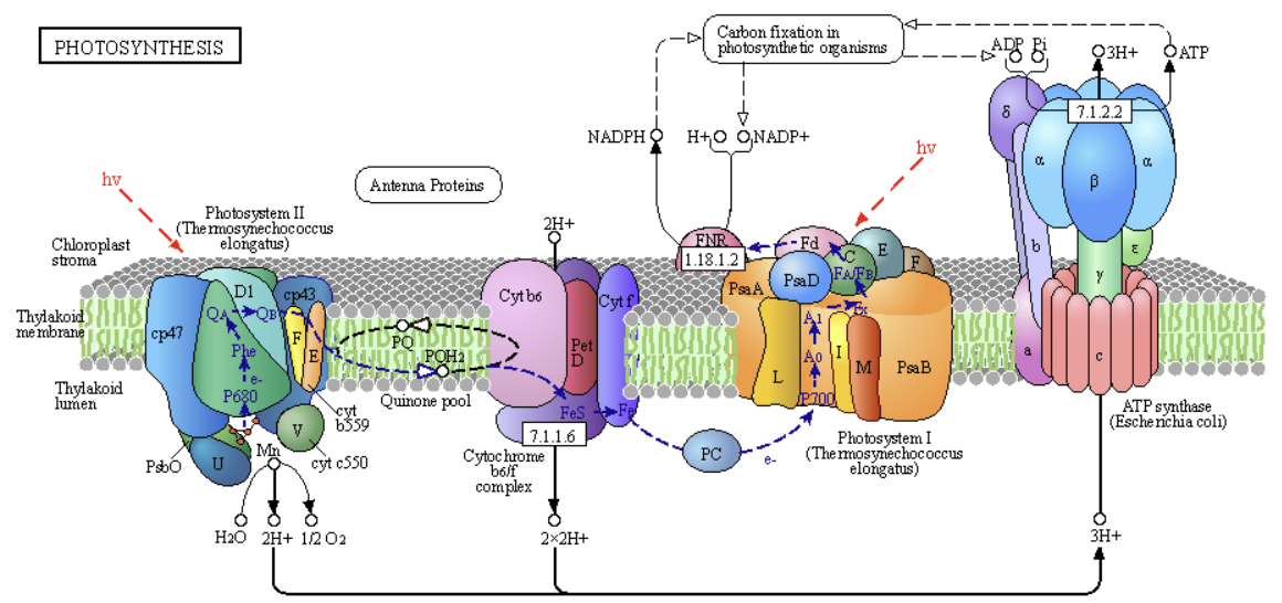
The pathway shows a "linear" sequential flow of electrons from:
- PSII to a mobile electron carrier, plastoquinone (PQ/PQH2), then to
- the membrane complex Cytochrome b6f complex to the mobile carrier plastocyanin (PC), then to
- PSI to the mobile carrier flavodoxin (Fd), then to
- FNR (Ferredoxin-NADP⁺ Reductase (FNR), a peripheral membrane protein, passes electrons from bound reduced flavodoxin to the mobile electron acceptor, NADP+.
This linear electron flow is accompanied by a linear proton (H+) flow, forming a proton gradient that collapses through ATP synthase to produce ATP. Hence, these linear flows of electrons and protons couple NADPH and ATP synthesis. This sequence of events is also called linear photophosphorylation and leads to an ATP/NADPH ratio ≈ of 1.3.
Below, we'll see an alternative "circular" electron flow and the advantages of each.
Cytochrome b6f
This complex moves electrons from the mobile lipophilic electron carrier reduced plastoquinol (PQH2), an isoprenoid quinone, to the mobile Cu-containing protein plastocyanin, which plays an analogous role to the mobile protein carrier in mitochondrial electron transport, cytochrome C. It catalyzes the rate-limiting step in electron transport in the light reactions. Figure \(\PageIndex{2}\) shows an interactive iCn3D model of the spinach plastocyanin (1AG6)
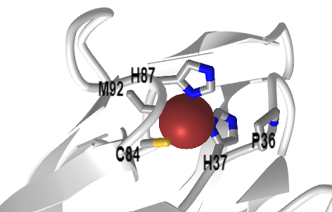
 Figure \(\PageIndex{2}\): Spinach plastocyanin (1AG6). (Copyright; author via source). Click the image for a popup or use this external link: https://structure.ncbi.nlm.nih.gov/i...DwSDDrYMUb9Xn7
The complex that mediates electron transfer between reduced plastoquinone and the protein plastocyanin (cytochrome b6f) is centrally positioned between the two photosystems. It also moves 2 H+ from the stroma into the lumen. These are joined by 2 H+s from water oxidation by the OEC (on the luminal side of PSII) to create a transmembrane proton gradient, which powers ATP synthesis.
Electron transfer with cytochrome b6f occurs through the quinol (Q) cycle, similar to Complex III in mitochondrial electron transport; therefore, we will not delve into the details here. Figure \(\PageIndex{3}\) shows a summary diagram with electron and proton flow.
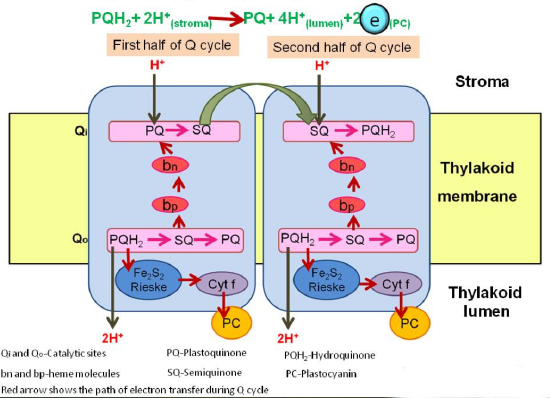
Figure \(\PageIndex{4}\) shows another version of the Q cycle as an alternative representation.

The left box shows the b6f complex transferring two protons (green arrows) per electron transferred (blue arrows) along high- (Fe2S2 cluster, cytochrome f) and low-potential chains (bl, bh, ci hemes). It also shows Quinol (QH2) oxidation at Qo site and Quinone (Q) reduction at Qi site. The right structure depicts haems b (purple), ci and f (red), Fe2S2 cluster (yellow and orange ball-and-stick model), cytochrome b6 (cyan), subunit IV (blue), Rieske subunit (yellow), cytochrome f (red), PetG, L, M and N subunits (green). Magnification of Qi site comprising bh and ci hemes.
Both cytochrome bc1 and cytb6f are dimeric complexes with 2 Fe2S2 clusters, two cytochrome bs, and a cytochrome c. The cytb6f complex also contains 9-cis β-carotene and an additional c1 heme. The electrons move from PQH2 through the complex in a similar fashion to that of the bc1 complex.
PQH2 is oxidized at the Qp site with a bifurcation of electrons:
- One electron moves through the high-potential Fe2S2 center and cyt f pathway (bottom left in pathway diagram of Figure 3), with the electron moving to the soluble peripheral protein plastocyanin and one to photosystem I.
- The other moves through the low-potential bl, bh, ci hemes pathway (top left in pathway diagram of Figure 3), with the electron moving to a plastoquinone at the Qn site near the stroma. A second round of oxidation of PQH2 at the Qp site eventually donates another electron to a plastoquinone-. which regenerates PQH2 after addition of 2 H+ from the stroma.
Two successive oxidations of PQH2 result in twice as many protons moving into the lumen. 2 H+ ions move into the lumen. Cytochrome b6f also senses the redox state of the plastoquinol/plastoquinone pool.
Figure \(\PageIndex{5}\) shows an interactive iCn3D model of the spinach cytochrome b6f complex (6RQF)

Figure \(\PageIndex{5}\): Spinach cytochrome b6f complex (6RQF). (Copyright; author via source). Click the image for a popup or use this external link: https://structure.ncbi.nlm.nih.gov/i...tgUhjekB4Tndw6
The region away from the red leaflet represents the lumen side of the complex. The FeS cluster and heme C are in the luminal domains. The coloring scheme is as follows:
- cytochrome bcf subunits 4, 5, 6, 7, and 8: light gray
- cytochrome b6: dark gray
- cytochrome b6 FeS subunit: plum
- cytochrome F: cyan
- chlorophylls: orange spacefill, labeled
- FeS clusters: CPK spacefill, labeled
- HEC - Heme C: CPK spacefill, labeled
- heme - porphyrin IX containing Fe: yellow spacefill
Cytochrome b6f is the rate-limiting step for electron flow, but what is its role in regulating the photosynthetic pathway (light reaction plus the dark reaction of carbon metabolism? Data suggest that the complex regulates electron transport in low-light conditions but promotes a switch to carbon metabolism under saturating light. We'll see that below when we discuss circular electron flow.
Plant photosystem I-LHCI super-complex
If the goal of the photosystem complexes is to transduce light energy delivered by photons into electrons that can be used for reductive biosynthesis of glucose from atmospheric carbon capture of CO2, then PSI is quite amazing. It has a "quantum efficiency" close to 1, which implies that one absorbed photon produces 1 electron that can be used to reduce NADP+. This occurs because the transfer of photon energy to other molecules in PSI is so rapid compared to non-radiative decay processes for the excited-state chlorophylls.
The same excitation and electron (charge) transfer process we saw in PSII occurs in PSI, with the reaction-center chlorophyll involved in charge transfers. PSI's light-absorbing molecules enable it to absorb light at the far-red end of the spectrum. The complex contains 16 proteins, 155 chlorophyll molecules, and 35 carotene derivatives. As with PSII, it has a core complex similar to cyanobacterial PSI. The supercomplex also has, on one side (looking down), light-harvesting complex I proteins surrounding it, comprising four LHCI proteins (Lhca 1-4), which increase light absorption. They have 57 chlorophylls and 13 carotene derivatives.
The apoproteins that bind the chlorophylls, including the LHCI proteins, have similar but slightly different topologies, allowing for tuning the absorbance spectra of the bound chromophores. Seven of the chlorophylls, when bound, have local environments that allow the absorption of far-red light. These are not found in PSII. Far-red light would be more abundant in the lower parts of the plant canopies, as photons of lower wavelengths would be more filtered out by upper leaves in the canopy.
Because of PSI's complexity, we provide several iCn3D models to illustrate the features of the photosystem I-LHCI supercomplex. As with PSII, PSI has a core surrounded by a light-harvesting complex (LHCI). Figure \(\PageIndex{6}\) shows an interactive iCn3D model of the Plant (pea) photosystem I-LHCI super-complex (4XK8)

Figure \(\PageIndex{6}\): C. (Copyright; author via source). Click the image for a popup or use this external link: https://structure.ncbi.nlm.nih.gov/i...uWBzFrVBUDHHQ6 (long load time)
Again, the red leaflet represents the lumen side of the complex, while the gray represents the chlorophyll and other lipid molecules in the membrane.
Figure \(\PageInde.x{7}\) shows an interactive iCn3D model highlighting the chlorophylls, beta-carotene derivatives, and FeS clusters of the plant photosystem I-LHCI super-complex (4XK8)

Figure \(\PageIndex{7}\): Chlorophylls, beta-carotene derivatives, and FeS clusters of Plant photosystem I-LHCI super-complex (4XK8). (Copyright; author via source). Click the image for a popup or use this external link: https://www.ncbi.nlm.nih.gov/Structu...BecS9cgNqdr8J6 (long load time)
The proteins in the complex are shown in gray cartoons. Chlorophylls A and B are shown in green and teal spacefill, respectively. Beta-carotene derivatives are shown in orange spacefill.
Figure \(\PageIndex{8}\) shows an interactive iCn3D model of the ApoA1 protein with surrounding chlorophyll and other components from plant photosystem I-LHCI super-complex (4XK8)

Figure \(\PageIndex{8}\): ApoA1 protein with surrounding chlorophyll and other components from plant photosystem I-LHCI super-complex (4XK8). (Copyright; author via source). Click the image for a popup or use this external link: https://structure.ncbi.nlm.nih.gov/i...L6tsJ3oMDZ2YQA (long load time)
ApoA1 is shown in a cartoon, colored by hydrophobicity. The chlorophylls, carotenes, and other components within 5 Å are shown in red. Rotate the image to see how the chlorophyll and other lipids encircle the membrane protein components.
Figure \(\PageIndex{9}\) shows an interactive iCn3D model of the Plant (pea) photosystem I-LHCI super-complex, highlighting LHCIs (4XK8)

Figure \(\PageIndex{9}\): Plant (pea) photosystem I-LHCI super-complex highlighting LHCIs (4XK8). (Copyright; author via source). Click the image for a popup or use this external link: https://structure.ncbi.nlm.nih.gov/i...9vHynkRTLxAct7 (long load time)
The LHCI subunits (Lhca 1-4) are shown in alternating magenta and cyan cartoon structures surrounding one side of the core complex. The model above is viewed from above.
We discussed in Chapter 20.1 that PSI absorbance is tilted toward the red end of the spectrum, with the effective absorbance ratio over a broad wavelength range for the two photosystems, PSI/(PSI+PSII), deviating from around 0.5 at the red/far red end of the spectrum (670-730 nm), where the ratio is close to 1. When exposed to far-red light, the systems move to state I. In this state, the major mobile antenna proteins (LHCIIs) move to PSII to restore a "photoabsorption" balance. When exposed to light depleted in the high end of the visible spectrum, the system moves to state II, where mobile LHCIIs move to PSI.
LHC location is regulated by phosphorylation, which is controlled by plastoquinone levels, making excellent biological sense. When the concentration of plastoquinones in the reduced state, PQH2 (plastoquinol), is high, it would be optimal to increase the activity of PSI to relieve the high concentration of the substrate for cyto b6f and shift the system to higher PSI activity and continue electron flow. This regulation is mediated by the phosphorylation of LHCs by LHCII kinase (a Ser/Thr kinase), which is activated by high concentrations of reduced PQH2. Phosphorylation of LHCs leads to their movement from PSII to PSI. When the oxidized form of plastoquinone is high, LHCII kinase is inactivated by dephosphorylation, causing the mobile LHC to move back to PSII to increase output (PQH2) from PSII. These events occur in low light. In high-light conditions, when the system is functioning at a high level, stromal thioredoxin inhibits the LHCII kinase. Specifically, the phosphorylation status of Lhcb1 and Lhcb2 in LHCII homo- or heterotrimers determines the movement between PSII and PSI. The chloroplastic serine/threonine-protein kinase (STT7, also known as STN7) is another LHCII kinase.
Recent Updates: 4.7.25
Linear vs Cyclic Flow of Electrons in Photosynthesis
Sometimes, electrons from PSI do not flow through ferredoxin to NADP+ via Ferredoxin-NADP⁺ Reductase (FNR). Instead, the electrons are repeatedly shunted repeatedly from reduced ferredoxin (FdH2) back to PSI through cytochrome b6f in a circular flow, which is often called cyclic photophosphorylation. If this happens, NADPH is not made. Yet protons continue to flow across the membrane, increasing ATP synthesis. Here are several reasons this occurs:
- NADPH is in excess, and it's not needed
- NADP+ is low and must be regenerated
- Prevention of damage to the photosystems by excess light
This cyclic flow essentially uncouples NADPH and ATP production, enabling continuous ATP production.
The actual return of electrons in this circular flow occurs through a thylakoid membrane enzyme called NAD(P)H dehydrogenase (Ndh). This enzyme is structurally very similar to mitochondrial Complex I, also known as NADH:ubiquinone oxidoreductase or Type I NADH dehydrogenase. As the name implies, Complex I binds NADH and passes electrons from it in a dehydrogenation reaction to another mobile electron carrier, ubiquinone (UQ), which forms ubiquinol (UQH2).
NADH + ubiquinone (UQ) ↔ NAD+ + UQH2
The chloroplastic Ndh, however, does not bind NADPH, which is confusing given the name. Rather, it binds reduced ferredoxin from PSI and transfers electrons to plastoquinone (PQ), which, when reduced, can pass electrons to Cyto b6f and then to PSI. Ferredoxin is considered the principal electron donor. The redox state does not change in net. Note that the cyclic electron pathway, once started, does not need PSII, so water oxidation is not needed. Hence, Ndh binds ferredoxin and plastoquinones/ols at distinct sites.
Essentially, FNR makes NADPH and ATP for biosynthesis, while Ndh boosts ATP and helps modulate the NADPH/ATP ratio.
Spinach NDH has many subunits. Ndh A-G are in the membrane arm and are similar to subunits (ND1-ND6) in the analogous arm of mitochondrial complex I. Ndh H-K in the peripheral arm are similar to the Q (ubiquinone) reduction part of complex I. Instead of having a binding site for NADH, it interacts with ferredoxin. Other subunits include NdhL-O and S-V. NdhC, I, J, L, M, O, and U form the plastoquinone-binding site. Other subunits (PnsL1–PnsL5 and PnsB1–PnsB5) are found in the spinach chloroplast NDH but not in cyanobacteria.
NDH used for cyclic electron flow is part of a much larger NDH–PSI–LHCI-2 supercomplex. The structure of a larger supercomplex in spinach, composed of NDH, PSI, and LHCI-2, determined by cryo-electron microscopy, is shown in Figure \(\PageIndex{10}\) below.

Figure \(\PageIndex{10}\): Cryo-EM structure of the NDH–PSI–LHCI-2 supercomplex from S. oleracea. Introini, B., Hahn, A. & Kühlbrandt, W. Cryo-EM structure of the NDH–PSI–LHCI supercomplex from Spinacia oleracea. Nat Struct Mol Biol (2025). https://doi.org/10.1038/s41594-024-01478-1. Creative Commons Attribution 4.0 International License. http://creativecommons.org/licenses/by/4.0/.
Lateral (a,b) views of the cryo-EM map of the supercomplex (contour level: 0.38). Shades of cyan (module SubA, NdhH–NdhO) and magenta (module SubE, NdhU) indicate the NDH peripheral arm. The membrane arm is drawn in shades of blue (module SubM, NdhA–NdhG). Lumenal SubL subunits (PnsL1–PnsL5) are shown in shades of purple, and subunits of the stromal SubB (PnsB1–PnsB5) are in shades of gray. PSI–LHC-2 is colored in shades of green. Cryo-EM densities of lipids are salmon (PG), violet (MGDG), pink (SQDG) or green (DGDG). Carotenoids are orange, and chlorophylls are bright green. The detergent belt is transparent gray.
Figure \(\PageIndex{11}\) below shows a proton flow model for the spinach complex—Subunits A-G form transmembrane modules. B, D, and F form antiporter subunits for H+ translocation, and each has two discontinuous helices, TMH7 and TMH12. These homologous antiporter structures are also found in mitochondrial Complex I. Subunits A, C, E, and G form the plastoquinone binding site and the early part of the proton translocation machinery.

Figure \(\PageIndex{11}\): Proposed proton transfer path in the membrane arm of S. oleracea NDH. Introini, B. et al., ibid.
Side view of NDH. From right to left, NdhA helices and NdhH and NdhK loops contributing to the formation of the platoquinon (PQ)-binding pocket (gray surface), NdhC TMH1–TMH2, NdhG TMH3, NdhE TMH2–TMH3, and discontinuous TMHs of NdhB, NdhD and NdhF are shown as solid cylinders. Other subunits are shown as outlines. The 4Fe–4S N2 cluster (upper right) is indicated. Charged residues of NdhA–NdhG and residues involved in proton translocation that are conserved between spinach NDH and A. thaliana complex I are shown as stick models and are color-coded. Positively charged residues are blue, negatively charged residues are red and key aromatic residues are white. PQ pocket, voids, channels, and depressions within NDH are likely to be relevant for proton translocation and are shown as gray surfaces. The light-blue arrow delineates a continuous hydrophilic path from the Q-binding site to NdhF. Stromal half-channels above NdhB, NdhD, and NdhF are indicated as dotted cyan arrows and labeled 1–3; the channel between NdhB and NdhD (magenta) is labeled 4.
Figure \(\PageIndex{12}\) shows an interactive iCn3D model the complex of NDH and the PSI-LHCI-2 supercomplex from S. oleracea (9GRX).

Figure \(\PageIndex{12}\): NDH–PSI–LHCI-2 supercomplex from S. oleracea (9GRX) (Copyright; author via source). Click the image for a popup or use this external link: ttps://www.ncbi.nlm.nih.gov/Structur...kJXW3YGGrM9sQA
NdhP, D, and B are the antiporters involved in H+ translocation. NdhH and NdhK show the PQ-binding pocket region. Yellow highlights show FeS complexes (spacefill)
The charged side chains in Complex I involved in proton flow are also found in NDH, supporting the idea that NDH in chloroplasts and Complex I in mitochondria have identical functions resulting from homologous structures.
Electronic regulation of an enzyme complex
- Click here
-
Johnson and Berry have analyzed electron flow with carbon metabolism in a fashion analogous to transistors in a circuit board, as illustrated in the figure below.

Electron transport system as an electrical circuit (and associated text below). Johnson and Berry. Photosynthesis Research (2021) 148:101–136. Creative Commons Attribution 4.0 International License. http:// creativecommons.org/ licenses/by/4.0/. They define a transistor as a regulated circuit element that uses variable conductance to control current flow. The linear flow of electrons from water to the reductant is viewed as a light-driven current, controlled by many regulatory feedback loops stemming from carbon metabolism. In limiting light, Cyt b6f presents maximal conductance to flow, and feedback from carbon metabolism adjusts the excitation of PS I and PS II in such a way as to balance the relative rates of linear and cyclic electron flow to the NADPH, Fd, and ATP requirements of the sinks. When the light becomes saturating, feedback from carbon metabolism also decreases the apparent conductance of Cyt b6f, controlling the linear flow of electrons through the plastoquinone pool and the associated flow of protons into the thylakoid lumen. In this way, Cyt b6f regulation simultaneously permits efficient photosynthesis and protects the system from photodamage.
This model is organized around the idea that the distribution of excitation between PS II and PS I is regulated to minimize losses of absorbed light and maximize potential electron transport through Cyt b6f. The expression for the potential electron transport rate has the form of a Michaelis-Menten expression for a single substrate (i.e., light). Still, it describes the kinetic behavior of the entire electron transport chain (i.e., including both photochemical and biochemical reactions). It predicts that electron transport has a hyperbolic dependence on irradiance, with maximum efficiency realized at the limit where absorbed irradiance approaches zero and maximum speed realized at the limit where absorbed irradiance approaches infinity.
The trade-off between the speed and efficiency of potential electron transport is driven by the need for the supplies of reduced plastoquinone and oxidized plastocyanin to be balanced to sustain Cyt b6f turnover at the maximum catalytic rate. This causes progressive closure of the PS II and PS I reaction centers, with PS II accumulating in a reduced state and PS I in an oxidized state. As the excitation pressure on PS II and PS I increases, the closure of the reaction centers causes the photochemical yields of PS II and PS I, as well as the absorbed quantum yield, to decrease as the potential electron flow through Cyt b6f and the potential photosynthetic rate increase.
The key prediction of the expression for the potential electron transport rate is that the maximum activity of Cyt b6f defines the upper limit for the theoretical maximum speed of electron transport. The expressions describing feedback control of Cyt b6f activity are based on the idea that Cyt b6f functions like a transistor, i.e., an electrical component that uses variable conductance to control the flow of current.
The fact that Cyt b6f can modulate its conductance to linear electron flow within milliseconds of a perturbation in light suggests that photosynthetic control is the first line of defense against overexcitation, protecting the acceptor side of PS I from being flooded with highly reduced intermediates.
In response to a sustained increase in light, the induction of photosynthetic control is followed by the induction of PQN. As NPQ alleviates the electron overpressure in the PQ pool, photosynthetic control progressively relaxes. The two forms of regulation gradually settle into a steady state at the new light intensity. This interaction appears to enable electron transport to proceed at the Cyt b6f-limited rate under low light intensities, then smoothly transitions to the Rubisco-limited rate once the light intensity is high enough to become saturating. It also appears to enable photosynthesis to operate safely and efficiently in a wide range of biochemical environments, from those characteristic of natural variation in photosynthetic capacity (with balanced electron transport and carbon metabolism) to those resulting from genetic manipulations (with imbalances in electron transport and carbon metabolism).
In this framework, the excitation balance of PS II and PS I, as well as the maximum activities of Cyt b6f and Rubisco, emerge as key limits on system dynamics. For example, the trade-off between the speed and efficiency of electron transport is shown to be controlled by the excitation balance of PS II and PS I, as well as the maximum activity of Cyt b6f. The development of PQN is shown to be controlled by the excitation balance of PSII and PSI, as well as the demand for linear electron flow (LEF) through the light reactions and circular electron flow (CEF) around PSI. The onset of photosynthetic control depends on the maximum activities of Cyt b6f and Rubisco.
Chloroplast ATP synthase - CF1FOATPase
Finally, let's look at chloroplast ATP synthase. It is structurally and functionally similar to mitochondrial FoF1ATPase, so we won't spend much time on its mechanism. Like its mitochondrial counterpart, it is a rotary enzyme that transduces the free energy of a proton gradient collapse into chemical energy in the form of ATP. One difference is that light levels should regulate the rotary enzyme, decreasing its activity at night. This is accomplished in higher plants by conversion between a reduced and oxidized state, an effective redox switch, in one subunit (γ) of the ATP synthase. Deactivation is important at night, as if the rotary enzyme runs in reverse, ATP hydrolysis will occur.
Figure \(\PageIndex{13}\) shows an interactive iCn3D model of the reduced R1 state of chloroplast ATP synthase (R1, CF1FO) (6VON). It should look familiar to you, given its similarity to mitochondrial ATP synthase.

Figure \(\PageIndex{13}\): Reduced R1 state of chloroplast ATP synthase (R1, CF1FO) (6VON) (Copyright; author via source). Click the image for a popup or use this external link: https://www.ncbi.nlm.nih.gov/Structu...HT8YZefdWN5XM7 (long load)
- alpha - light green
- beta - dark green
- gamma - yellow
- gamma - crimson
- epsilon - indigo
- b - blue
- b' - light blue
- a - light pink
- c ring - purple
- ATP - spacefill, cyan
- TENTOXIN (TTX) - space fill gray
- ADP - spacefill magenta
In the oxidized state, a disulfide in the γ subunit inhibits torsion by stabilizing two β hairpins. Reduction relieves this constraint and enhances rotation. In the reduced structure, tentoxin, an uncompetitive inhibitor, is present, which allowed the structure to be determined.
Material below from Yang, JH., Williams, D., Kandiah, E. et al. Structural basis of redox modulation on chloroplast ATP synthase. Commun Biol 3, 482 (2020). https://doi.org/10.1038/s42003-020-01221-8. Creative Commons Attribution 4.0 International License. http://creativecommons.org/licenses/by/4.0/.
At night, the enzyme is in the low-activity form. When light becomes available during the day, the collapse of the pH gradient releases tightly bound ATP from the enzyme in its oxidized state. PSI can engage in photon-induced electron transfer to ferredoxin. This binds to NADP reductase, forming NADPH. These processes are illustrated in Figure \(\PageIndex{14}\).
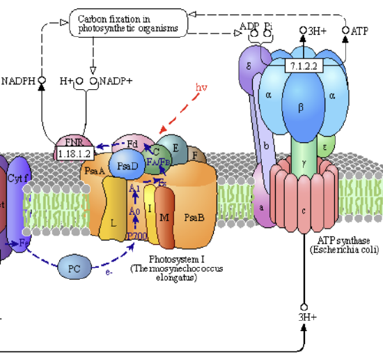
Increased reduced ferredoxin leads to the formation of reduced thioredoxin, a small redox protein involved in redox signaling and protection of cells against oxidative stress. A similar process activates the enzyme sedoheptulose-1,7-bisphosphatase (SBPase), which is involved in the dark reaction of the Calvin cycle in photosynthesis. Its activation is illustrated in Figure \(\PageIndex{15}\).
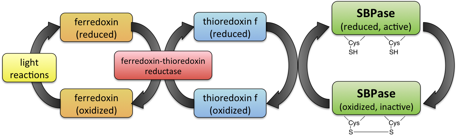

Reduced thioredoxin both activates and inhibits chloroplastic ATP synthase.
Figure \(\PageIndex{16}\) the activities and structures of the ATP synthase (CF1FO)

Panel a: For the experiments, purified CF1FO was reconstituted into a liposome (orange) mixed with phosphatidylcholine and phosphatidic acid lipids. The pH gradient across the membrane drove the reconstituted CF1FO to synthesize ATP, which was detected using a luciferin/luciferase assay (green). Val indicates valinomycin.
Pane b: The activity of the enzyme is shown in the reduced state (in the presence of dithiothreitol - DTT, blue curve), oxidized state (in the presence of iodosobenzoate - IBZ, orange curve, and with no additive (green curve). Note that the activity is reduced by about 80% in the oxidized state.
Panel c: The images are from Cryo-EM density maps of the oxidized and reduced forms of the CF1FO. The color codes are as follows: α (light green), β (dark green), δ (yellow), bb' (blue and light blue), γ (crimson), ε (indigo), a (light pink), and c ring (purple). R indicates a reduced state, and O indicates an oxidized state. The three-dimensional (3D) reconstructions are categorized into three rotary states (1, 2, and 3). The upper insets are the density maps of the F1 domains.
Figure \(\PageIndex{17}\) shows the conformation changes in the γ subunit between the oxidized and reduced forms.

Panel a: Structures of the reduced (light blue) and oxidized (orange) γ subunits. Two β-hairpin structures (from γGlu238 to γLeu282) are shown in light green, and the two cysteines of the redox switch are shown in yellow in circular, enlarged views. The diagram on the right shows the topology of the two β hairpin structures.
Panel b: Superposition of the reduced and oxidized γ subunits (RMSD 1.016 Å). The two β hairpins are shown in light blue and orange for the reduced and oxidized forms, respectively. Other regions are shown in white.
Panel c: Interaction networks of the β hairpin 2 and βDELSEED motif. The left and right panels are the reduced (γ subunit in light blue) and oxidized (γ subunit in orange) forms. Light green represents the β hairpin 2, dark green represents the β subunit, and yellow represents the βDELSEED motif. The distances connecting the residues of the γ coiled-coil (γArg73, γGln76, and γGlu77) with the βGlu412 are labeled.
Panel d: Interaction of the EDE motif with the γ subunit. The EDE motif (yellow) does not interact with any part of the reduced γ subunit but forms an extensive interaction network with its neighborhood when the γ subunit is oxidized.
Figure \(\PageIndex{18}\) shows a cartoon model illustrating the structures of the overall oxidized and reduced states of ATP synthase

Panel a: This cartoon schematic shows redox modulation. The upper and lower models are the reduced and oxidized states, respectively. Color codes are the same as in Fig. 1c, and the β hairpin structures of the γ subunit are shown in light green. The two redox states are aligned in the same view.
Panel b: At night, no energy input from light is available for the photosynthetic electron transport chain, and thus, no electrochemical potential (ΔΨ) and proton gradient (ΔpH) are generated. The oxidized γ subunit prevents CF1FO from hydrolyzing ATP. During the day, light induces charge separation, developing an electrochemical potential across the membrane. Although the CF1FO begins to synthesize ATP molecules, the γ subunit is still oxidized while ΔΨ is small. ATP synthesis is not yet at its maximum rate. At sunrise, thioredoxin subsequently reduces the γ subunit, fully activating CF1FO. The molecular motor, consisting of the γ-ε central shaft and the c14-ring, can rotate at full speed to maximize its ATP synthesis activity. Three ATP molecules per rotation of the c14 ring are produced. At sunset, the membrane becomes de-energized, resulting in small ΔΨ and ΔpH, and ATP hydrolysis occurs. To prevent ATP loss from excess ATP hydrolysis, the γ subunit is then oxidized again. Light regulation and redox modulation on the CF1FO will cycle daily.
Summary
(Summary written by Claude, Sonnet 4.6, Anthropic)
This chapter completes the description of the light reactions of photosynthesis, tracing the flow of electrons from reduced plastoquinol through cytochrome b₆f, Photosystem I, ferredoxin, and ferredoxin-NADP⁺ reductase to produce NADPH, describing the structural and mechanistic parallels to mitochondrial electron transport at each step, and presenting the regulatory mechanisms that balance linear and cyclic electron flow while controlling chloroplast ATP synthase activity in a light-dependent manner.
After water oxidation by the OEC of PSII, electrons are transferred through the reaction center to pheophytin A, plastoquinone A, and then to the mobile lipophilic carrier plastoquinol (PQH₂) in the thylakoid membrane. PQH₂ is the substrate for the cytochrome b₆f complex, the rate-limiting step in light-reaction electron transport and the structural and mechanistic analog of mitochondrial Complex III (cytochrome bc₁). Like Complex III, cytochrome b₆f operates through a Q cycle in which oxidation of PQH₂ at the luminal Qp site bifurcates two electrons along two distinct pathways. One electron takes the high-potential path through the Rieske Fe₂S₂ cluster and cytochrome f to the mobile electron carrier plastocyanin (a soluble copper-containing protein analogous to cytochrome C in mitochondria), which delivers its electron to Photosystem I. The other electron takes the low-potential path through hemes bL, bH, and ci to reduce a plastoquinone at the stromal Qn site; after two complete rounds of this cycle, the two electrons delivered to the Qn site convert one oxidized PQ to PQH₂, consuming two protons from the stroma. The net result is that for every two electrons delivered to plastocyanin/PSI, four protons are released into the lumen — two from the Qp site oxidations and two incorporated into PQH₂ at the Qn site from the stroma — doubling the proton gradient contribution per electron relative to a simple two-electron transfer. Cytochrome b₆f also contains unique cofactors not found in Complex III: a 9-cis β-carotene and an additional ci heme.
Photosystem I is remarkable for its near-unity quantum efficiency: essentially every absorbed photon results in a productive electron transfer. The PSI-LHCI supercomplex contains 16 proteins, 155 chlorophyll molecules, and 35 carotenoid derivatives, organized as a core complex (homologous to cyanobacterial PSI) flanked on one side by four LHCI proteins (Lhca1–4) that extend PSI's antenna system with an additional 57 chlorophylls. Seven chlorophylls in PSI bind in environments that shift their absorption to the far-red region, giving PSI a preferential spectral response at 670–730 nm relative to PSII — advantageous in the lower parts of plant canopies where shorter-wavelength photons are filtered by upper leaves. Charge separation at the PSI reaction center (P700) generates a strong reductant that is transferred through a series of Fe/S clusters to ferredoxin, an iron-sulfur protein. Ferredoxin-NADP⁺ reductase (FNR), a peripheral membrane flavoprotein, then reduces NADP⁺ to NADPH using electrons from reduced ferredoxin. This completes linear photophosphorylation, which produces NADPH and ATP in an approximate 1:1.3 ratio.
An alternative pathway, cyclic photophosphorylation, occurs when NADPH is in excess, NADP⁺ is depleted, or excess light must be safely dissipated. In this pathway, electrons from reduced ferredoxin are returned to the plastoquinone pool via the chloroplastic NDH complex (NAD(P)H dehydrogenase) rather than being used to reduce NADP⁺. The NDH complex is structurally homologous to mitochondrial Complex I: its membrane arm subunits NdhA–G correspond to the ND1–ND6 subunits of Complex I, and the antiporter subunits NdhB, NdhD, and NdhF (containing discontinuous transmembrane helices TM7 and TM12) carry out proton translocation by the same mechanism as their mitochondrial counterparts. The key mechanistic difference is that NDH accepts electrons from ferredoxin rather than NADH. The entire cyclic electron flow apparatus is organized as a massive NDH–PSI–LHCI-2 supercomplex, recently solved at high resolution by cryo-EM, in which NDH is physically coupled to PSI to enable efficient electron recycling. Cyclic flow maintains proton gradient formation and ATP synthesis without NADPH production, effectively uncoupling the two and allowing the ATP/NADPH ratio to adjust to biosynthetic demands. FNR primarily drives NADPH and ATP synthesis in linear mode, while NDH boosts ATP and modulates the NADPH/ATP balance in cyclic mode.
The redox state of the plastoquinone pool dynamically regulates the distribution of mobile LHCII trimers between PSII and PSI, creating state 1/state 2 transitions that balance light absorption between the two photosystems. When PQH₂ accumulates (indicating excess PSII activity), the LHCII kinase STN7/Stt7 is activated, phosphorylating LHCII proteins and triggering their migration from PSII grana stacks to PSI-containing single-layer thylakoids — shifting the system to state 2 and increasing PSI activity to consume the PQH₂ substrate. When PQ is oxidized or light levels are high, STN7 is inactivated by stromal thioredoxin, unphosphorylated LHCII trimers return to PSII, and the system reverts to state 1. This elegant regulatory circuit uses the redox state of the mobile electron carrier as a direct signal to balance throughput across the two photosystems.
Chloroplast ATP synthase (CF₁F_o) is structurally homologous to mitochondrial F₁F_o ATP synthase and operates by the same proton-driven rotary mechanism, synthesizing three ATP per c-ring revolution (14 c-subunits in the spinach enzyme). However, it possesses a unique light-dependent regulatory mechanism not found in its mitochondrial counterpart: a disulfide bond between two cysteines in the γ subunit that functions as a redox switch controlling enzyme activity. In the oxidized state (nighttime), the disulfide stabilizes two β-hairpin structures in the γ subunit, restricting rotation and reducing ATP synthesis activity by approximately 80%. This oxidative inactivation is functionally critical — it prevents the enzyme from running in reverse and hydrolyzing cellular ATP in the dark when no proton gradient is available to drive synthesis. In the reduced state (daytime), thioredoxin — itself reduced by ferredoxin generated by light-driven PSI electron transport — breaks the γ-subunit disulfide, relieving the conformational constraints on rotation and allowing full-speed ATP synthesis as the proton gradient builds during illumination. This daily regulatory cycle, mediated by the ferredoxin-thioredoxin system, couples ATP synthase activity to the photosynthetic electron transport chain, ensuring that the enzyme operates only when light is available to generate the proton-motive force that powers it.
This page titled 20.3: Plant Electron Transport and ATP Synthesis was last modified on Mon, 14 Sep 2026 17:08:31 GMT and is shared under a not declared license and was authored, remixed, and/or curated by Henry Jakubowski and Patricia Flatt.
The book could not be loaded just now.