From LibreTexts, under CC BY-NC 4.0, as the source states it; its equations are written in TeX, which the PDF shows typeset. This page at LibreTexts.
9.1: Using Microscopes
Opening the book…
The Microscope
Dissecting Microscope
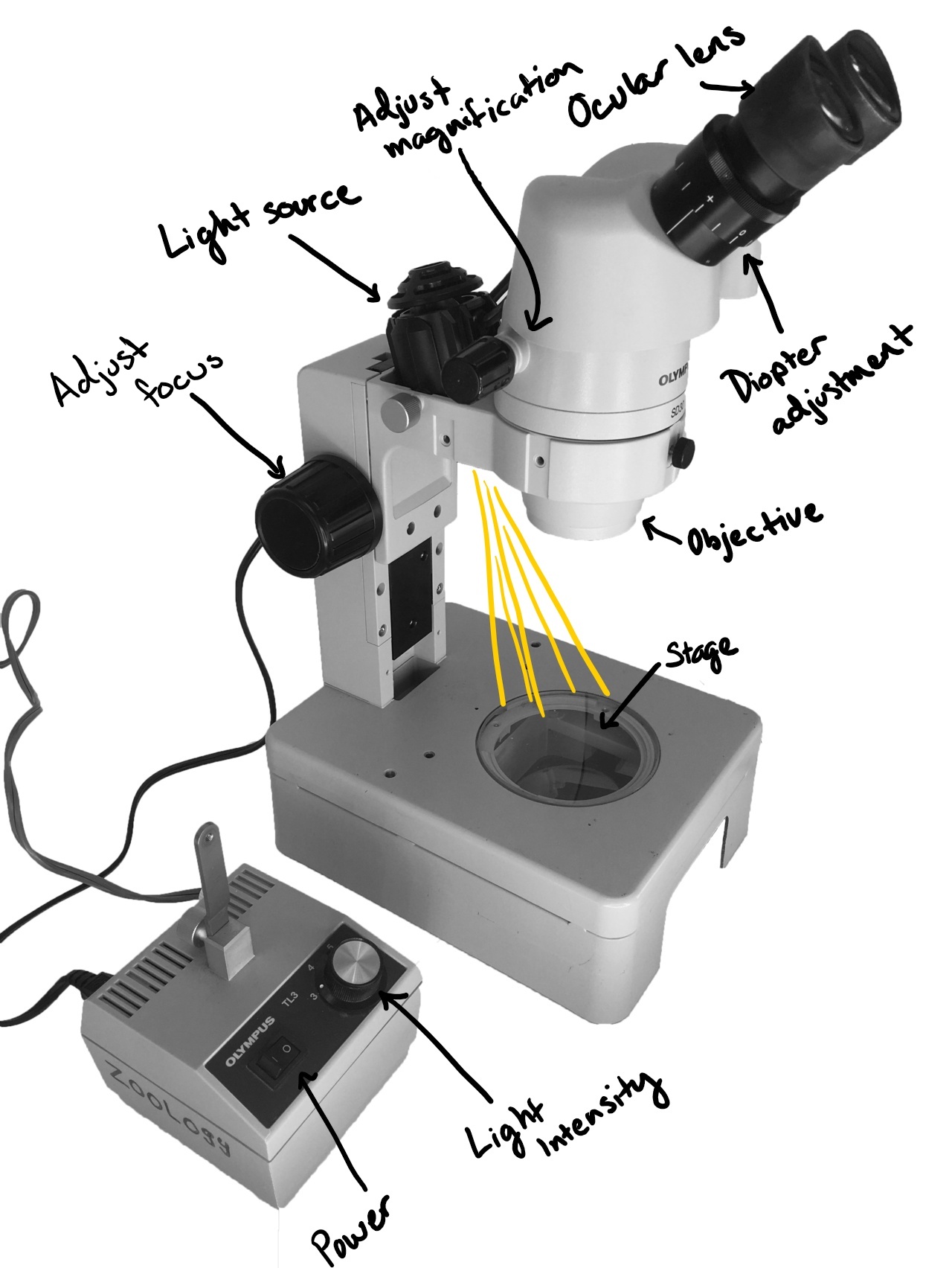
A dissecting microscope generally has lower magnification and uses incident light, meaning the light shines onto (not through) the specimen to view it.
Figure \(\PageIndex{1}\): A labeled dissecting microscope. Two ocular lenses extend from the top with a diopter adjustment around the base of one. These are at the top of the stereo head. At the base of the head is the objective. On the side of the head is the magnification adjustment. A light source goes through the head and shines light onto the stage. The head is connected to the base by an arm where the focus adjustment is located. The light is connected by a cord to a separate device with a power switch and a knob to adjust light intensity. Photo by Sarah Greenwood via Wikimedia Commons with labels added by Maria Morrow. CC BY 4.0
Compound Microscope
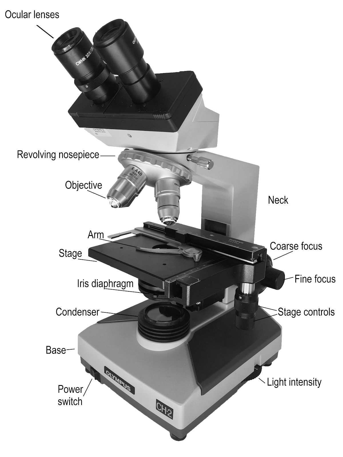
A compound microscope is used for viewing small samples or pieces of a larger specimen at higher magnification. This type of microscope uses transmitted light, where the light must pass through the specimen to view it.
Figure \(\PageIndex{2}\): A labeled compound microscope, with the stage for the specimen located between the lenses and the light source (transmitted light). From top to bottom on the left side of the image, the labels read: Ocular lenses, revolving nosepiece, objective, arm, stage, iris diaphragm, condenser, base, power switch.From top to bottom on the right side of the image: neck, coarse focus, fine focus, stage controls, light intensity. Adapted from Compound Microscope by Sarah Greenwood via Wikimedia Commons. CC BY 4.0
Making and Staining Slides
There are many ways to make a slide. Preparation method and technique will vary, depending on your specimen and what you need to see. Below are some basic guidelines, but you should experiment with what works best for you and your specimen.
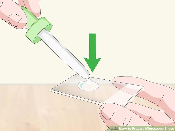
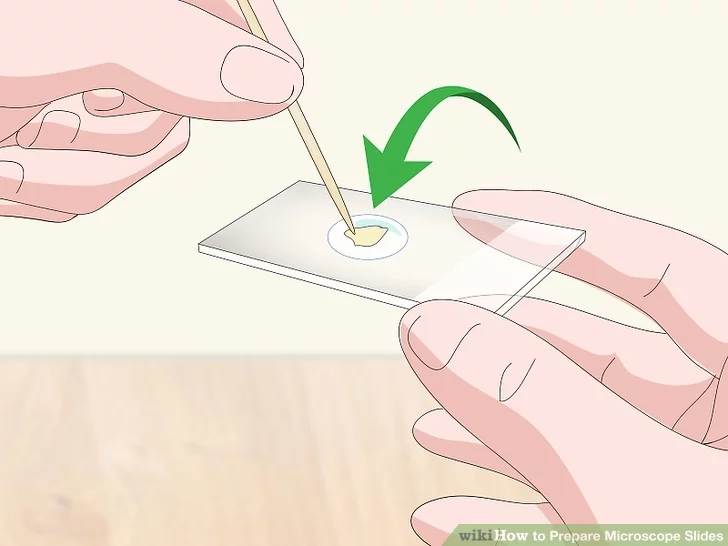
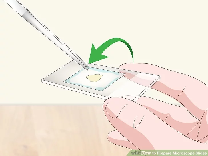
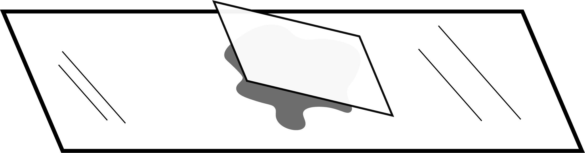
Figure \(\PageIndex{3}\): Preparing a wet mount. Add a few drops of liquid to the slide. Put your specimen onto the droplet of liquid. Add a cover slip. The best method for placing the cover slip is to touch it to the slide at an angle (see bottom image) and slowly bring it toward the droplet of liquid. Once the cover slip touches the liquid, lower it gently until flat. This method reduces the amount of air bubbles in your sample. First three images from WikiHow, CC-BY-NC-SA. Bottom image by Sarah Greenwood, CC BY 4.0, via Wikimedia Commons.
With compound microscopes, the light must pass through the specimen. For this to work, your prepared sample must be quite thin. Many prepared slides are made using a machine called a microtome to achieve the extremely thin, even sections needed to see many anatomical features. As a human trying to perform this process, you'll need as much practice as possible to get a good thin section.
Video \(\PageIndex{1}\): This video provides a demonstration of making thin cross-sections by hand for plant material such as stems and roots; includes a demonstration of staining the material using TBO (toluidine blue O), a metachromatic stain useful for many plant materials. [See Episode 6 for how to prepare TBO.] Sourced from YouTube.
Focusing with a Compound Microscope
Video \(\PageIndex{2}\): Dr. Patrick demonstrates the steps in focusing a compound light microscope from 10X to 100X. She shows you how the field of view changes with each lens. The use of immersion oil for the 100X lens is specifically shown. Sourced from YouTube.
Measuring Your Specimen
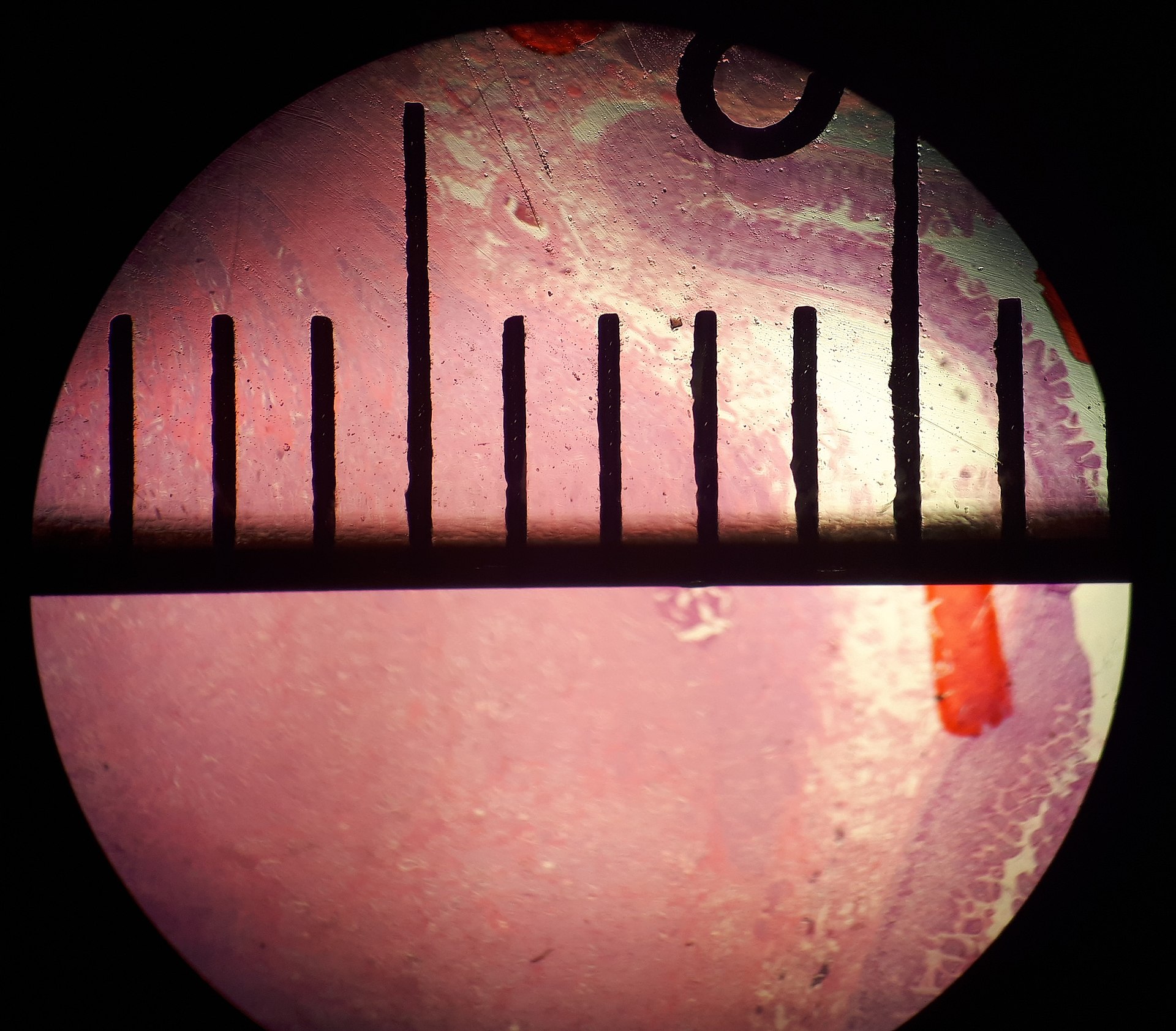
Figure \(\PageIndex{4}\): This image shows the field of view through a compound microscope. A ruler or reticle can be used to measure the field of view and/or the specimen you are viewing. The marks on the ruler in this image are millimeters. The diameter of the field of view at this magnification is approximately 11 mm. Photo by Mikael Häggström, M.D. - Author info - Reusing imagesConsent from the patient or patient's relatives is regarded as redundant, because of absence of identifiable features (List of HIPAA identifiers) in the media and case information (See also HIPAA case reports guidance)., CC0, via Wikimedia Commons.