From LibreTexts, under CC BY 4.0, as the source states it; its equations are written in TeX, which the PDF shows typeset. This page at LibreTexts.
10.2.1: The -enchymas
Opening the book…
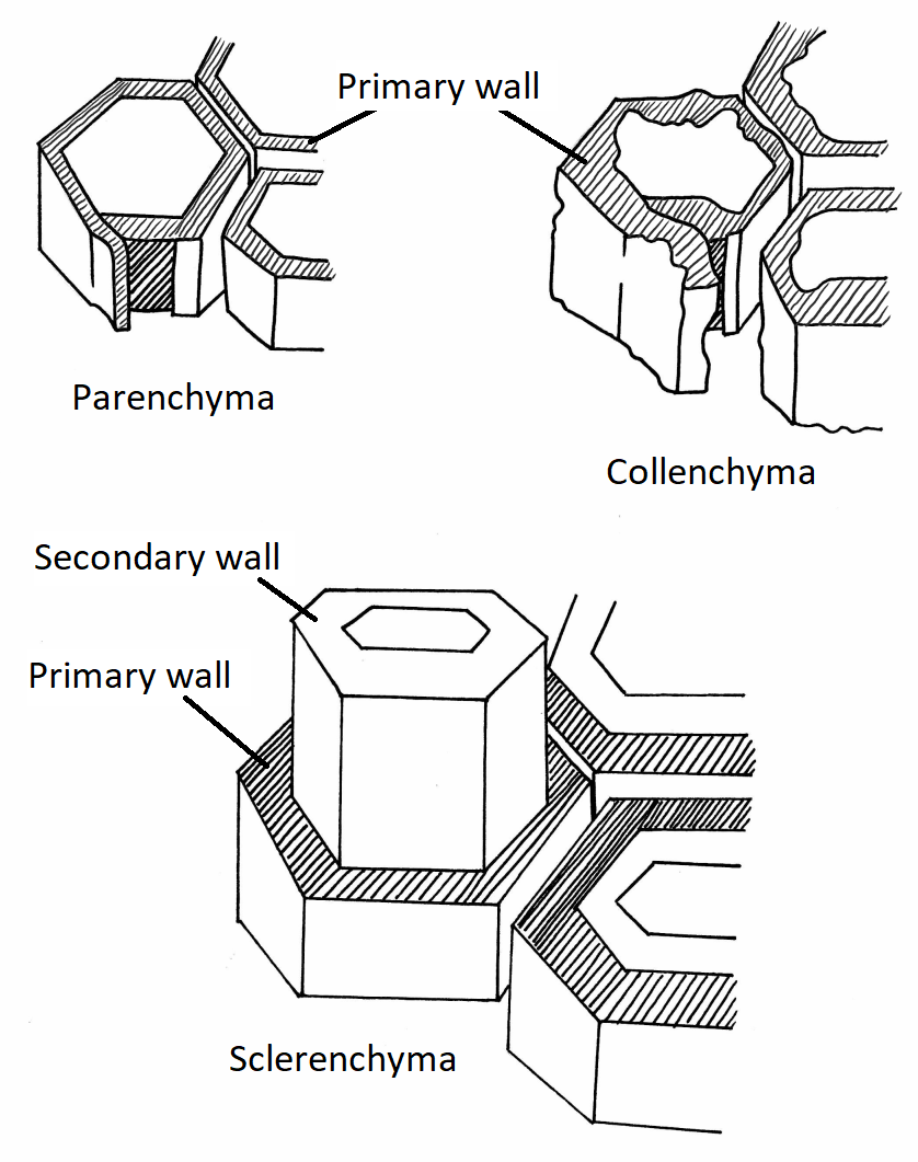
There are three major cell types in plants that we can distinguish by differences in their cell walls. These three cell types are parenchyma, collenchyma, and sclerenchyma.
Figure \(\PageIndex{1}\): This diagram shows the three major cell types found in plants. In the upper left, parenchyma cells have an evenly thickened primary wall (shaded with stripes in the illustration). The primary wall of parenchyma cells is quite thin, though it has been widened in the diagram to make it easier to see. To the right of the parenchyma, collenchyma cells also have only a primary wall, but it is unevenly thickened, giving them a chunky appearance. Unlike parenchyma cells, the primary wall of a collenchyma cell is often quite thick. On the bottom half of the diagram, sclerenchyma cells are shown with a thick secondary wall (unshaded) that develops within the primary wall (shaded). Eventually, the primary wall of the sclerenchyma cells will die away. Artwork by Nikki Harris, CC-BY 4.0 with labels added by Maria Morrow.
Parenchyma
Parenchyma cells are characterized by an even, relatively thin primary wall.
Figure \(\PageIndex{2}\): A cross section of a Ranunculus root showing parenchyma cells filling the cortex. Note the evenly thin cell walls. These cortex cells contain many starch grains. Photo by Berkshire Community College Bioscience Image Library, CC0, via Wikimedia Commons.
Collenchyma
Collenchyma cells are characterized by an uneven, relatively thick primary wall.
Figure \(\PageIndex{3}\): Collenchyma cells. Left: Angular collenchyma is a supportive tissue found in peppermint and other species like celery. 200X magnification. Taken in the Plant Lab at CU:Boulder. Photo by Spencer Llerandi, CC BY 4.0, via Wikimedia Commons with an added arrow by Maria Morrow. Right: Collenchyma tissue from celery. Photo from Clematis, CC BY-SA 3.0, via Wikimedia Commons.
Sclerenchyma
Sclerenchyma cells are characterized by the formation of a secondary wall composed of lignin. This secondary wall forms within the primary wall and eventually leads to the death of the cell. Sclerenchyma cells are dead at functional maturity.
Figure \(\PageIndex{4}\): The first image is a cross section through a cluster of fiber cells (sclerenchyma). These cells have relatively evenly thick secondary walls that have stained red due to lignin. In the second image, there is a single fiber cell. The reddish donut is formed by its secondary wall. There are several gaps in this wall called pits, two are indicated by arrows. These are pits where plasmodesmata used to traverse the cell walls. The center (L) is the lumen, a now empty region where the cytoplasm used to be. First image: Snowman frosty at en.Wikipedia, Public domain, via Wikimedia Commons. Second image: Roland Gromes, CC BY-SA 4.0, via Wikimedia Commons with labels added by Maria Morrow.Figure \(\PageIndex{5}\): Sclereids are sclerenchyma cells found in pears. These woody cells give pears a gritty texture. In the first photo, the sclereids are stained a dark pink color and clustered into large groups, surrounded by parenchyma cells. In the second photo, a single sclereid is visible. It has a thick secondary wall with the channels of former plasmodesmata visible running through it, forming pits. First: Berkshire Community College Bioscience Image Library, CC0, via Wikimedia Commons. Second: Snowman frosty at en.Wikipedia, Public domain, via Wikimedia Commons.