Opening the book…
Overview
Inspection of the skin, hair, and nails is a significant part of a physical examination. Dermatology is the scientific study and medical treatment of the integumentary system. Skin is a complex organ. An average square inch of skin contains 650 sweat glands, 20 blood vessels, and more than 1,000 nerve endings. Despite being just a few millimeters thick, the skin makes up around one-seventh of a person’s body weight.
Did you know your skin sheds about 40,000 cells every hour? Imagine the renewal process happening without you even noticing! Would you like to explore more about how this renewal helps in healing wounds?
Human skin is the first anatomical site a medical professional examines when seeing a patient, using it as an indicator of general health such as oxygenation, nutritional status, and injury. Skin color is mostly based on the pigmentation or melanin found in the keratinocytes in the epidermis of skin. In addition, the blood supply to the surface (redness), injury (bruising), and jaundice (yellowing) can affect skin color.
Ready to peel back the layers of your body's protective shield? Want me to unpack that step-by-step? Although you may not typically think of the skin as an organ, it is in fact made of tissues that work together as a single structure to perform unique and critical functions. The skin is made of multiple layers of cells and tissues, which are held to underlying structures by connective tissue. The deeper layer of skin is well vascularized (has numerous blood vessels). It also has numerous sensory, autonomic and sympathetic nerve fibers ensuring communication to and from the brain. The skin and its accessory structures make up the integumentary system, let's explore its key functions:
- Physical protection - the integumentary system is the covering of the human body and it is most apparent function is physical protection: skin - a tightly knit network of cells, with each layer contributing to its strength. The epidermis has an outermost layer created by layers of dead keratin that can withstand wear and tear of the outer environment, the dermis provides the epidermis with blood supply and has nerves that bring danger to attention amongst other functions. Hypodermis (subcutaneous layer) provides physical cushioning to any mechanical trauma through adipose storage; cutaneous glands secrete protective films; nails protect the digits; hairs throughout the body filter harmful particles from entering the eyes, ears, nose, etc.
- Immunity - the skin is the body’s first line of defense, acting as a physical barrier preventing direct entry of pathogens. Resident immune cells are present in the skin, and some, Langerhans cells or dermal dendritic cells, can travel to the periphery and activate the greater immune system.
- Wound healing - when our body undergoes trauma with a resulting injury, the integumentary system orchestrates the wound healing process through hemostasis, inflammation, proliferation, and remodeling.
- Thermoregulation - the skin has a large surface area that is highly vascularized, which allows it to conserve and release heat through vasoconstriction and vasodilation, respectively.
- Vitamin D synthesis - the primary sources of vitamin D are sun exposure and oral intake (crucial for bone health).
- Sensation - skin innervation is by various types of sensory nerve endings that discriminate pain, temperature, touch, and vibration. Each type of receptor and nerve fiber varies in its adaptive and conductive speeds, leading to a wide range of signals that can be integrated to create an understanding of the external environment and help the body to react appropriately.
Layers of the Integument
The skin consists of the epidermis and the dermis. The epidermis is composed of keratinized, stratified squamous epithelium. It is made of four or five layers of epithelial cells, depending on its location in the body. It does not have any blood vessels within it (i.e., it is avascular). The layers of epidermis tissue are described below from deep to superficial:
- Stratum basale - This deepest layer of the epidermis is composed mainly of one layer of proliferating and non-proliferating keratinocytes (keratin-containing cells), attached to the basement membrane by hemidesmosomes. Melanocytes are present in this layer (melanin containing cells also known as pigment cells), connected to numerous keratinocytes through dendrites. Merkel cells are also found in the stratum basale (large numbers of touch-sensitive receptors are found in fingertips and lips).
- Stratum spinosum - Keratinocytes become connected through desmosomes and start to produce lamellar bodies, from within the Golgi, enriched in polar lipids, glycosphingolipids, free sterols, phospholipids, and catabolic enzymes. Langerhans cells (immunologically active cells) and melanin are found in the middle of this layer.
- Stratum granulosum - A layer in which keratinocytes lose their nuclei and their cytoplasm appears granular. Lipids, contained in those keratinocytes within lamellar bodies, are released into the extracellular space through exocytosis to form a lipid barrier.
- Stratum lucidum - A clear, translucent layer of dead keratinocytes, only present in thick skin.
- Stratum corneum - Superficial layer of cells, composed of 10 to 30 layers anucleated keratinocyte, with the palms and soles having the most layers. Most of the barrier functions of the epidermis localize to this layer.
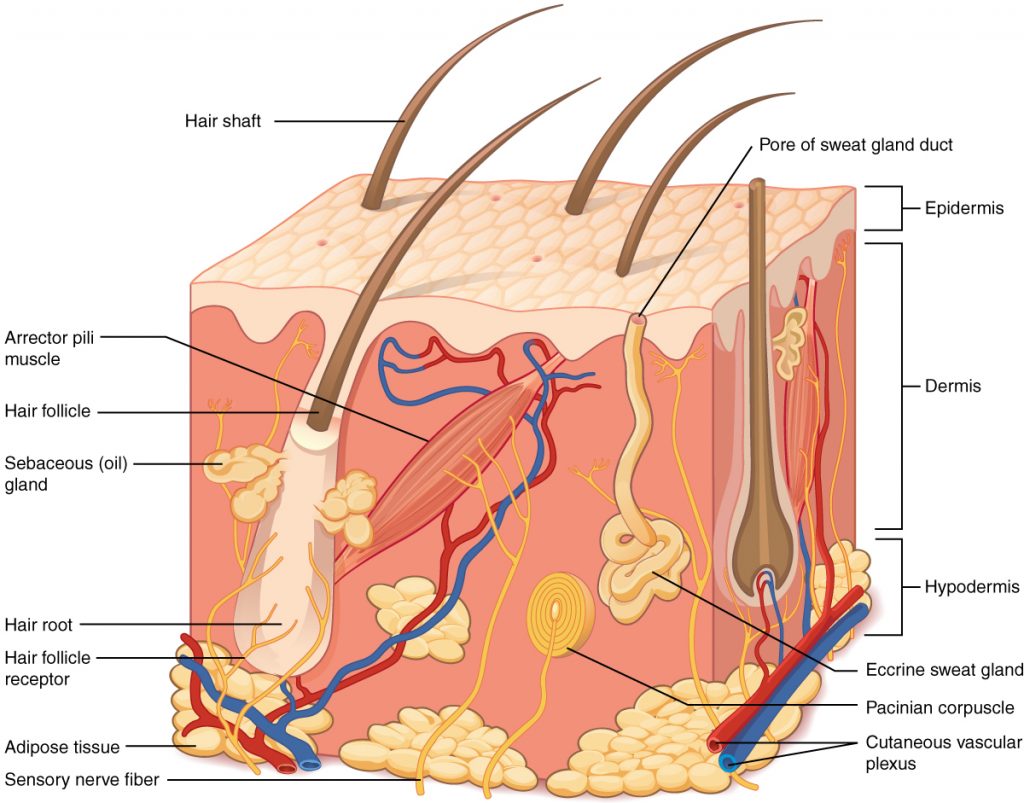
Figure \(\PageIndex{1}\) cross-section illustration of skin, highlighting the epidermis (stratified squamous epithelium), dermis (dense irregular connective tissue containing hair follicles, sweat glands, and blood vessels), and hypodermis (adipose tissue). Each part is labeled to help identify its function and location:
Layers of the Skin:
- Epidermis: The outermost layer, providing a protective barrier.
- Dermis: The middle layer, containing blood vessels, nerves, and glands.
- Hypodermis: The deepest layer, made up of adipose tissue (fat) for insulation and cushioning.
Structures Within the Skin:
- Hair Shaft: The visible part of the hair above the skin surface.
- Hair Root: The part of the hair embedded in the skin.
- Hair Follicle: The structure surrounding the hair root.
- Hair Follicle Receptor: Detects movement of the hair.
- Arrector Pili Muscle: A small muscle attached to the hair follicle that causes hair to stand up (goosebumps).
- Sebaceous (Oil) Gland: Produces oil to lubricate the skin and hair.
- Eccrine Sweat Gland: Produces sweat for temperature regulation.
- Pore of Sweat Gland Duct: The opening on the skin surface where sweat is released.
- Sensory Nerve Fiber: Transmits sensory information like touch and pain.
- Pacinian Corpuscle: A deep pressure receptor.
- Cutaneous Vascular Plexus: A network of blood vessels that supplies the skin.

Figure \(\PageIndex{2}\) is a microscopic view of thick skin epidermis, illustrating the strata basale (with Merkel cell, melanocyte, and sensory neuron), spinosum, granulosum (lamellar granules), lucidum, and corneum (keratinized cells). It shows the layers of the epidermis from top to bottom, along with key cell types and structures.
- Layers of the Epidermis (top to bottom):
- Stratum Corneum - Outermost layer contains dead cells filled with keratin, providing a tough, protective barrier.
- Stratum Lucidum - A thin, clear layer found only in thick skin (like palms and soles).
- Stratum Granulosum - Contains lamellar granules, which help form a waterproof barrier.
- Stratum Spinosum - Contains keratinocytes, which produce keratin for skin strength and resilience.
- Stratum Basale - Deepest layer of the epidermis.Contains:Merkel cells, which are connected to sensory neurons and help detect touch.
- Melanocytes - Produce melanin, the pigment responsible for skin color.
- Dermis:
- Located beneath the epidermis.
- Supports the epidermis and contains blood vessels, nerves, and connective tissue.
The dermis contains blood and lymphatic vessels, nerves, and other structures, such as hair follicles and sweat glands. The epidermis is avascular and the cells of this layer must get their oxygen and nutrients from capillaries in the dermis. The dermis is made of two layers of connective tissue that compose an interconnected mesh of elastin and collagenous fibers, produced by fibroblasts. The more superficial papillary layer (made of loose, areolar connective tissue) serves as an anchor point for the epidermis above and is intimately connected to the deeper reticular layer (composed of dense, irregular connective tissue, is well vascularized and has a rich sensory and sympathetic nerve supply).
Figure \(\PageIndex{3}\) shows a histological section of human skin, focusing on the dermis, which is divided into two layers:
Papillary Layer
- Located just beneath the epidermis.
- Composed of loose connective tissue with fine collagen fibers and capillaries.
- Supports the epidermis and aids in nutrient exchange and sensory function.
Reticular Layer
- Found below the papillary layer.
- Made of dense connective tissue with thicker collagen fibers.
- Provides structural strength and elasticity.
- Contains sweat glands, hair follicles, and other deep dermal structures.
The hypodermis (also called the subcutaneous layer or superficial fascia) is a layer directly below the dermis and serves to connect the skin to the underlying fascia (fibrous tissue) surrounding the muscles. It is not strictly a part of the skin, although the border between the hypodermis and dermis can be difficult to distinguish. The hypodermis consists of well-vascularized, loose areolar connective tissue and abundant adipose tissue, which functions as a mode of fat storage and provides insulation and cushioning for the integument. Fascia is a thick connective tissue wrapping that surrounds skeletal muscles, anchoring them to surrounding tissues and investing groups of muscles.
Pigmentation
The color of skin is influenced by a number of pigments, including melanin, carotene, and hemoglobin. Recall that melanin is produced by cells called melanocytes, which are found scattered throughout the stratum basale of the epidermis. The melanin is transferred into the keratinocytes via a cellular vesicle called a melanosome.

Figure \(\PageIndex{4}\) shows a side-by-side cellular comparison of dark and light skin, divided into two labeled sections:
- Left: "Dark"
- Right: "Light"
Skin Layers (Top to Bottom):
- Surface – Outermost skin layer
- Upper Keratinocytes – Contain melanin
- Melanosomes – Pigment organelles within keratinocytes
- Basal Keratinocytes – Base layer skin cells
- Melanocytes – Pigment-producing cells
Key Differences:
- Dark Skin has more melanin in keratinocytes and melanosomes. Melanocytes are more active.
- Light Skin has less melanin and fewer melanosomes. Melanocytes produce less pigment
Dark-skinned individuals produce more melanin than those with pale skin. Exposure to the UV rays of the sun or a tanning salon causes melanin to be manufactured and built up in keratinocytes, as sun exposure stimulates keratinocytes to secrete chemicals that stimulate melanocytes. The accumulation of melanin in keratinocytes results in the darkening of the skin, or a tan. This increased melanin accumulation protects the DNA of epidermal cells from UV ray damage and the breakdown of folic acid, a nutrient necessary for our health and well-being. In contrast, too much melanin can interfere with the production of vitamin D, an important nutrient involved in calcium absorption. Thus, the amount of melanin present in our skin is dependent on a balance between available sunlight and folic acid destruction, and protection from UV radiation and vitamin D production. It requires about 10 days after initial sun exposure for melanin synthesis to peak, which is why pale skinned individuals tend to suffer sunburns of the epidermis initially. Dark-skinned individuals can also get sunburns, but are more protected than pale-skinned individuals. Melanosomes are temporary structures that are eventually destroyed by fusion with lysosomes; this fact, along with melanin-filled keratinocytes in the stratum corneum sloughing off, makes tanning impermanent. Too much sun exposure can eventually lead to wrinkling due to the destruction of the cellular structure of the skin, and in severe cases, can cause sufficient DNA damage to result in skin cancer. When there is an irregular accumulation of melanocytes in the skin, freckles appear. Moles are larger masses of melanocytes, and although most are benign, they should be monitored for changes that might indicate the presence of cancer.
Accessory structures
Accessory structures of the skin include hair, nails, sweat glands, and sebaceous glands. These structures embryologically originate from the epidermis and can extend down through the dermis into the hypodermis.
Hair
Hair is a keratinous filament growing out of the epidermis. It is primarily made of dead, keratinized cells. Strands of hair originate in an epidermal penetration of the dermis called the hair follicle. The hair shaft is the part of the hair not anchored to the follicle, and much of this is exposed at the skin’s surface. The rest of the hair, which is anchored in the follicle, lies below the surface of the skin and is referred to as the hair root. The hair root ends deep in the dermis at the hair bulb, and includes a layer of mitotically active basal cells called the hair matrix. The hair bulb surrounds the hair papilla, which is made of connective tissue and contains blood capillaries and nerve endings from the dermis.

Figure \(\PageIndex{5}\) illustrates sectional view of the thin skin showing hair bulb (located at the base of the root, contains active cells that produce new hair), hair papilla (containing blood vessels), hair matrix (region within the bulb where new hair cells divide and differentiate), outer root sheath (a multilayered tissue that surrounds the hair follicle and extends from the hair bulb to the skin surface; plays a role in hair growth, sensory perception, and immune functions within the skin), inner root sheath (a specialized structure that surrounds the hair shaft within the follicle, providing support and protection, sebaceous gland (connected to the follicle, producing sebum to lubricate the hair and skin), hair shaft (the visible portion of hair, composed of three layers: the cuticle, cortex, and, in some cases, the medulla) and the Arrector pili muscle (attached to the follicle, responsible for making hair stand on end - goosebumps).
Nails
Nail has three main parts, the root, plate, and free margin. Other structures around or under the nail include the nail bed, cuticle, and nail fold. Nails grow from a deep layer of living epidermal tissue, known as the nail matrix, at the proximal end of the nail. The nail root is the portion of the nail found under the surface of the skin at the near (or proximal) end of the nail. It is where the nail begins. The free margin is the portion of the nail that protrudes beyond the distal end of the finger or toe. This is the part that is cut or filed to keep the nail trimmed.

Figure \(\PageIndex{6}\) illustrates the top diagram in this diagram shows the external, visible part of the nail plate (or the body) and the cuticle (layer of dead epithelial cells that overlaps and covers the edge of the nail plate, it helps to seal the edges of the nail to prevent infection of the underlying tissues). The bottom diagram shows nail fold (a groove in the skin in which the side edges of the nail plate are embedded), nail bed (the area of skin under the nail plate; it is pink in color, due to the presence of capillaries in
the dermis) and the nail matrix (the area at the base of the nail, under the cuticle, that is responsible for nail growth; it is a living, growing tissue that produces the nail plate).
Sweat
When the body becomes warm, sweat (sudoriferous) glands produce sweat to cool the body. Sweat glands develop from epidermal projections into the dermis and are classified as merocrine glands; that is, the secretions are excreted by exocytosis through a duct without affecting the cells of the gland. There are two types of sweat glands, each secreting slightly different products. An eccrine sweat gland is type of gland that produces a hypotonic sweat for thermoregulation. These glands are found all over the skin’s surface, but are especially abundant on the palms of the hand, the soles of the feet, and the forehead. They are coiled glands lying deep in the dermis, with the duct rising up to a pore on the skin surface, where the sweat is released. Eccrine glands are a primary component of thermoregulation in humans and thus help to maintain homeostasis.

Figure \(\PageIndex{7}\) is a cross-sectional anatomical diagram of human skin, showing multiple layers and internal structures.
Labeled Components:
- Skin Surface – Outermost visible layer
- Pores – Openings for sweat and oil release
- Eccrine Sweat Gland – Deep coiled gland that regulates body temperature
Additional Visible Structures (Unlabeled):
- Hair Follicles – Tubes housing hair roots
- Blood Vessels – Arteries and veins supplying nutrients
- Nerves – Sensory pathways for touch, temperature, and pain
- Other Glands – Likely sebaceous glands secreting oil near hair follicles
Apocrine Sweat Glands - An apocrine sweat gland is usually associated with hair follicles in densely hairy areas, such as armpits and genital regions. Apocrine sweat glands are larger than eccrine sweat glands and lie deeper in the dermis, sometimes even reaching the hypodermis, with the duct normally emptying into the hair follicle. In addition to water and salts, apocrine sweat includes organic compounds that make the sweat thicker and subject to bacterial decomposition and subsequent smell.
Sebaceous Glands
A sebaceous gland is a type of oil gland that is found all over the body and helps to lubricate and waterproof the skin and hair. Most sebaceous glands are associated with hair follicles. They generate and excrete sebum, a mixture of lipids, onto the skin surface, thereby naturally lubricating the dry and dead layer of keratinized cells of the stratum corneum, keeping it pliable. The fatty acids of sebum also have antibacterial properties, and prevent water loss from the skin in low-humidity environments. The secretion of sebum is stimulated by hormones, many of which do not become active until puberty. Thus, sebaceous glands are relatively inactive during childhood.

Figure \(\PageIndex{8}\) is a labeled cross-sectional diagram of human skin, focusing on the structure of a hair follicle.
Labeled Components:
- Hair – Strand extending from the follicle through the skin surface
- Skin Surface – Outermost layer where hair emerges
- Sebum – Oily substance near the follicle
- Follicle – Tubular structure housing the hair root
- Sebaceous Gland – Gland connected to the follicle that secretes sebum
Arrector pili - Each hair root is connected to a smooth muscle called the arrector pili Figure \(\PageIndex{5}\) that contracts in response to nerve signals from the sympathetic nervous system, making the external hair shaft “stand up.” The primary purpose for this is to trap a layer of air to add insulation. This is visible in humans as goose bumps and even more obvious in animals, such as when a frightened cat raises its fur.
Concept and Applications About Skin Conditions
Let's explore integumentary system issues, focusing on albinism, burn classifications (including the Rule of Nines), and the concept of SPF.
- Albinism is a genetic condition resulting from a deficiency or absence of melanin, the pigment responsible for skin, hair, and eye color. This melanin deficiency leads to:
- Photosensitivity - Extreme sensitivity to sunlight, significantly increasing the risk of sunburn and skin cancer.
- Vision Problems - Melanin's role in eye development means albinism often causes vision impairments like nystagmus (involuntary eye movements) and photophobia (light sensitivity).
- Social and Psychological Impacts - The visible differences can unfortunately lead to social stigma and psychological challenges.
- Burns are tissue injuries caused by heat, chemicals, electricity, or radiation. Severity is classified by degree:
- First-degree (Superficial) - Affects only the epidermis; characterized by redness, pain, and mild swelling.
- Second-degree (Partial-Thickness) - Involves the epidermis and part of the dermis; blistering, intense pain, and swelling are common. Sub-classified as superficial partial-thickness (upper dermis) or deep partial-thickness (deeper into the dermis).
- Third-degree (Full-Thickness) - Damages the entire epidermis and dermis, often extending deeper; appears charred, white, or leathery, with potentially less pain (due to nerve damage).
- Fourth-degree - The most severe; extends through all skin layers and into underlying structures like muscle and bone.
- The Rule of Nines estimates the total body surface area (TBSA) affected by burns, vital for fluid resuscitation and treatment. The body is divided into regions representing 9% or multiples of 9% TBSA.
- Sun Protection Factor (SPF): SPF indicates a sunscreen's ability to protect against UVB radiation, a major cause of sunburn and skin cancer. An SPF of 15 means it takes 15 times longer to sunburn with the sunscreen than without. Higher SPF values offer greater protection, but even high SPF doesn't block 100% of UVB rays. While SPF focuses on UVB, UVA rays also contribute to skin aging and cancer risk. Choosing a sunscreen with broad-spectrum protection is crucial
| Anatomic structure of Adult | Surface area | Anatomic structure of Child (less than 10 years) | Surface area |
|---|---|---|---|
| Anterior head | 9% | Anterior head | 12% |
| Posterior head | 9% | Posterior head | 12% |
| Anterior torso | 18% | Anterior torso | 18% |
| Posterior torso | 18% | Posterior torso | 18% |
| Anterior leg, each | 6.75% | Anterior leg, each | 5.5% |
| Posterior leg, each | 6.75% | Posterior leg, each | 5.5% |
| Anterior arm, each | 4.5% | Anterior arm, each | 4.5% |
| Posterior arm, each | 4.5% | Posterior arm, each | 4.5% |
| Genitalia/perineum | 1% | Genitalia/perineum | 1% |
This page titled 6.1: Background Information was last modified on Tue, 01 Sep 2026 01:45:36 GMT and is shared under a CC BY-NC 4.0 license and was authored, remixed, and/or curated by Lakshmikanta Sengupta and Susmita Sengupta.
The book could not be loaded just now.