Opening the book…
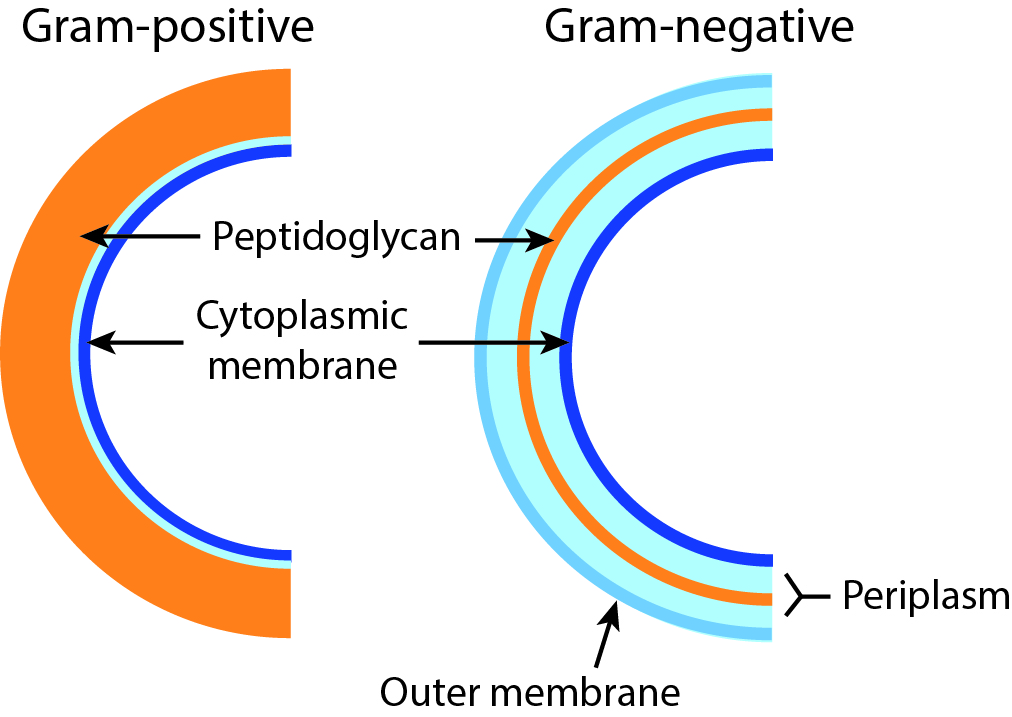
The cell walls of Bacteria have a rigid layer composed of peptidoglycan that is the main source of strength of the wall (Madigan et al., 2003). Peptidoglycan is a polymer consisting of sugars and amino acids. For some Bacteria, peptidoglycan makes up as much as 90% of the cell wall. However, for other Bacteria, it makes up only about 10% of the cell wall (Fig. \(3.3\)).
These two groups of Bacteria can be distinguished based on a staining procedure. By this method, cells are exposed to a stain, crystal violet, that binds to peptidoglycan. Cells with an abundance of peptidoglycan retain more of the stain than those that have little. The cells that retain more stain are referred to as gram-positive cells (i.e., the staining result is positive). Those that bind little stain are referred to as gram-negative cells. This difference in stain retention was traditionally used to help classify Bacteria and was originally developed in the nineteenth century by a Danish scientist, Hans Christian Gram.
Aside from the difference in peptidoglycan thickness, gram-negative cell walls differ from gram-positive cell walls in a few other ways as well. Gram-negative cell walls have an outer membrane. Unlike the cytoplasmic membrane, the outer membrane is not composed entirely of phospholipid but also contains polysaccharide and protein components and can be referred to as the lipopolysaccharide layer (LPS). Moreover, gram-negative cells also contain a thicker periplasm, a gel-like matrix between the inner cytoplasmic membrane and the outer membrane (Fig. \(3.3\)).

This page titled 3.3: Gram-positive and gram-negative bacteria was last modified on Sun, 31 Mar 2024 22:36:55 GMT and is shared under a CC BY-SA 4.0 license and was authored, remixed, and/or curated by Matthew F Kirk via source content that was edited to the style and standards of the LibreTexts platform.
The book could not be loaded just now.